

Supercharging BRD4 with NUT in carcinoma
- PMID: 33452461
- PMCID: PMC7914217
- DOI: 10.1038/s41388-020-01625-0
Supercharging BRD4 with NUT in carcinoma
Abstract
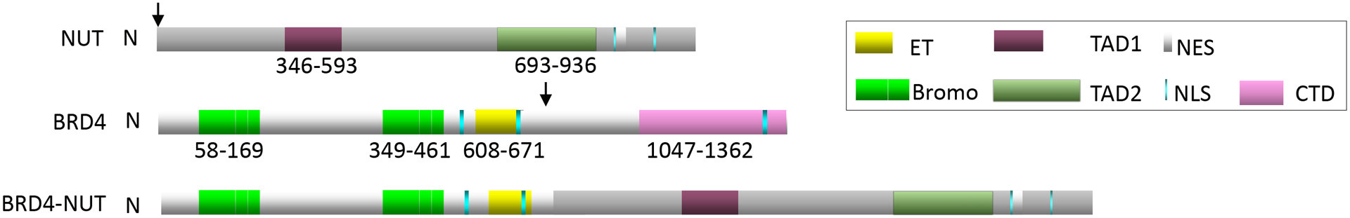
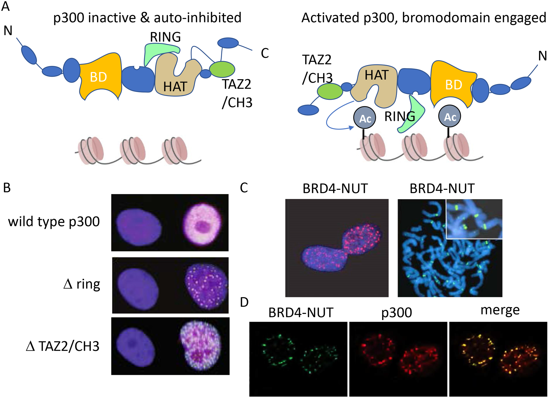
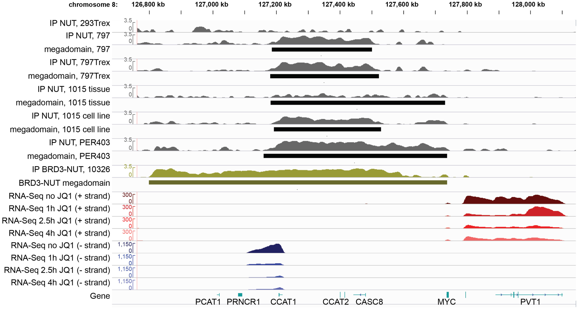
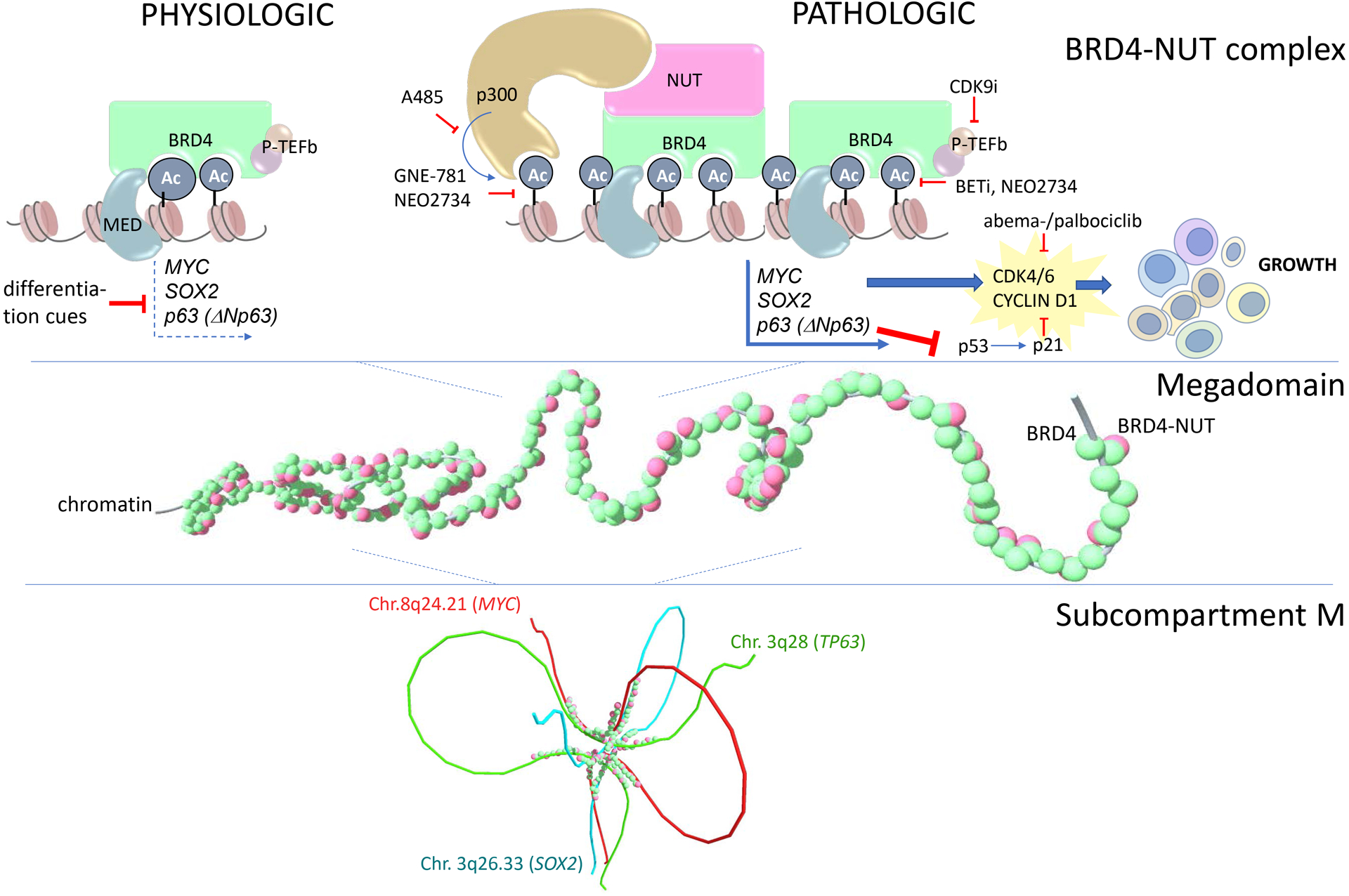
NUT carcinoma (NC) is an extremely aggressive squamous cancer with no effective therapy. NC is driven, most commonly, by the BRD4-NUT fusion oncoprotein. BRD4-NUT combines the chromatin-binding bromo- and extraterminal domain-containing (BET) protein, BRD4, with an unstructured, poorly understood protein, NUT, which recruits and activates the histone acetyltransferase p300. Recruitment of p300 to chromatin by BRD4 is believed to lead to the formation of hyperacetylated nuclear foci, as seen by immunofluorescence. BRD4-NUT nuclear foci correspond with massive contiguous regions of chromatin co-enriched with BRD4-NUT, p300, and acetylated histones, termed "megadomains" (MD). Megadomains stretch for as long as 2 MB. Proteomics has defined a BRD4-NUT chromatin complex in which members that associate with BRD4 also exist as rare NUT-fusion partners. This suggests that the common pathogenic denominator is the presence of both BRD4 and NUT, and that the function of BRD4-NUT may mimic that of wild-type BRD4. If so, then MDs may function as massive super-enhancers, activating transcription in a BET-dependent manner. Common targets of MDs across multiple NCs and tissues are three stem cell-related transcription factors frequently implicated in cancer: MYC, SOX2, and TP63. Recently, MDs were found to form a novel nuclear sub-compartment, called subcompartment M (subM), where MD-MD interactions occur both intra- and inter-chromosomally. Included in subM are MYC, SOX2, and TP63. Here we explore the possibility that if MDs are simply large super-enhancers, subM may exist in other cell systems, with broad implications for how 3D organization of the genome may function in gene regulation and maintenance of cell identity. Finally, we discuss how our knowledge of BRD4-NUT function has been leveraged for the therapeutic development of first-in-class BET inhibitors and other targeted strategies.
Figures
Similar articles
-
Therapeutic targeting of p300/CBP HAT domain for the treatment of NUT midline carcinoma.Oncogene. 2020 Jun;39(24):4770-4779. doi: 10.1038/s41388-020-1301-9. Epub 2020 May 4. Oncogene. 2020. PMID: 32366905 Free PMC article.
-
Chemical Screen Identifies Diverse and Novel Histone Deacetylase Inhibitors as Repressors of NUT Function: Implications for NUT Carcinoma Pathogenesis and Treatment.Mol Cancer Res. 2021 Nov;19(11):1818-1830. doi: 10.1158/1541-7786.MCR-21-0259. Epub 2021 Jul 20. Mol Cancer Res. 2021. PMID: 34285087 Free PMC article.
-
Mechanistic analysis of the role of bromodomain-containing protein 4 (BRD4) in BRD4-NUT oncoprotein-induced transcriptional activation.J Biol Chem. 2015 Jan 30;290(5):2744-58. doi: 10.1074/jbc.M114.600759. Epub 2014 Dec 15. J Biol Chem. 2015. PMID: 25512383 Free PMC article.
-
BRD4 and MYC: power couple in transcription and disease.FEBS J. 2023 Oct;290(20):4820-4842. doi: 10.1111/febs.16580. Epub 2022 Aug 3. FEBS J. 2023. PMID: 35866356 Free PMC article. Review.
-
NUT Is a Driver of p300-Mediated Histone Hyperacetylation: From Spermatogenesis to Cancer.Cancers (Basel). 2022 Apr 29;14(9):2234. doi: 10.3390/cancers14092234. Cancers (Basel). 2022. PMID: 35565363 Free PMC article. Review.
Cited by
-
Nuclear protein of the testis midline carcinoma of the thorax.Jpn J Clin Oncol. 2022 May 31;52(6):531-538. doi: 10.1093/jjco/hyac033. Jpn J Clin Oncol. 2022. PMID: 35325167 Free PMC article. Review.
-
Precision Targeting of BET Proteins - Navigating Disease Pathways, Inhibitor Insights, and Shaping Therapeutic Frontiers: A Comprehensive Review.Curr Drug Targets. 2025;26(3):147-166. doi: 10.2174/0113894501304747240823111337. Curr Drug Targets. 2025. PMID: 39385413 Review.
-
Nuclear protein in testis carcinoma of the lung.Transl Oncol. 2023 Apr;30:101640. doi: 10.1016/j.tranon.2023.101640. Epub 2023 Feb 11. Transl Oncol. 2023. PMID: 36780749 Free PMC article. Review.
-
A novel MXI1-NUTM2B fusion detected in an undifferentiated ovarian cancer.Gynecol Oncol Rep. 2024 Dec 6;57:101653. doi: 10.1016/j.gore.2024.101653. eCollection 2025 Feb. Gynecol Oncol Rep. 2024. PMID: 39758708 Free PMC article. No abstract available.
-
Targeting lysine acetylation readers and writers.Nat Rev Drug Discov. 2025 Feb;24(2):112-133. doi: 10.1038/s41573-024-01080-6. Epub 2024 Nov 21. Nat Rev Drug Discov. 2025. PMID: 39572658 Review.
References
-
- French CA, Kutok JL, Faquin WC, Toretsky JA, Antonescu CR, Griffin CA, et al. Midline carcinoma of children and young adults with nut rearrangement. J Clin Oncol. 2004;22(20):4135–9. - PubMed
-
- Shiota H, Barral S, Buchou T, Tan M, Coute Y, Charbonnier G, et al. Nut directs p300-dependent, genome-wide h4 hyperacetylation in male germ cells. Cell Rep. 2018;24(13):3477–87 e6. - PubMed
-
- French CA, Miyoshi I, Kubonishi I, Grier HE, Perez-Atayde AR, Fletcher JA. Brd4-nut fusion oncogene: A novel mechanism in aggressive carcinoma. Cancer Res. 2003;63(2):304–7. - PubMed
Publication types
MeSH terms
Substances
Grants and funding
LinkOut - more resources
Full Text Sources
Other Literature Sources
Research Materials
Miscellaneous
