

Automated quantitative MRI volumetry reports support diagnostic interpretation in dementia: a multi-rater, clinical accuracy study
- PMID: 33452627
- PMCID: PMC8213665
- DOI: 10.1007/s00330-020-07455-8
Automated quantitative MRI volumetry reports support diagnostic interpretation in dementia: a multi-rater, clinical accuracy study
Abstract
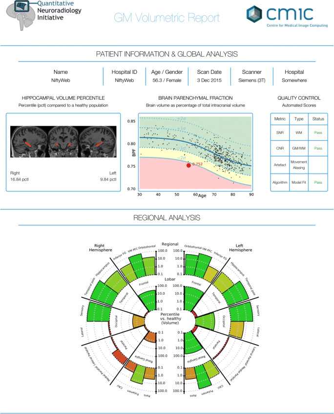
Objectives: We examined whether providing a quantitative report (QReport) of regional brain volumes improves radiologists' accuracy and confidence in detecting volume loss, and in differentiating Alzheimer's disease (AD) and frontotemporal dementia (FTD), compared with visual assessment alone.
Methods: Our forced-choice multi-rater clinical accuracy study used MRI from 16 AD patients, 14 FTD patients, and 15 healthy controls; age range 52-81. Our QReport was presented to raters with regional grey matter volumes plotted as percentiles against data from a normative population (n = 461). Nine raters with varying radiological experience (3 each: consultants, registrars, 'non-clinical image analysts') assessed each case twice (with and without the QReport). Raters were blinded to clinical and demographic information; they classified scans as 'normal' or 'abnormal' and if 'abnormal' as 'AD' or 'FTD'.
Results: The QReport improved sensitivity for detecting volume loss and AD across all raters combined (p = 0.015* and p = 0.002*, respectively). Only the consultant group's accuracy increased significantly when using the QReport (p = 0.02*). Overall, raters' agreement (Cohen's κ) with the 'gold standard' was not significantly affected by the QReport; only the consultant group improved significantly (κs 0.41➔0.55, p = 0.04*). Cronbach's alpha for interrater agreement improved from 0.886 to 0.925, corresponding to an improvement from 'good' to 'excellent'.
Conclusion: Our QReport referencing single-subject results to normative data alongside visual assessment improved sensitivity, accuracy, and interrater agreement for detecting volume loss. The QReport was most effective in the consultants, suggesting that experience is needed to fully benefit from the additional information provided by quantitative analyses.
Key points: • The use of quantitative report alongside routine visual MRI assessment improves sensitivity and accuracy for detecting volume loss and AD vs visual assessment alone. • Consultant neuroradiologists' assessment accuracy and agreement (kappa scores) significantly improved with the use of quantitative atrophy reports. • First multi-rater radiological clinical evaluation of visual quantitative MRI atrophy report for use as a diagnostic aid in dementia.
Keywords: Alzheimer’s disease; Atrophy; Frontotemporal dementia; Magnetic resonance imaging; Radiologists.
Conflict of interest statement
SH is a speaker for General Electric, a consultant for Spineart and on the imaging advisory board for the European Prevention of Alzheimer’s Disease. JC and SO are founders of Brainminer. MPW received speaker and/or consultancy fees from Bayer, Biogen, Biologix, Celgene, GeniLac, Imcyse, Medison, Merck-Serono, Novartis, Sanofi Genzyme, and Roche. FB is a board member for Neurology, Brain, Radiology, and MSJ; section editor for Neuroradiology; personal fees from Bayer, Biogen, Roche, IXICO, and GeNeuro; grants from Novartis, Teva Pharmaceuticals, Merck, Biogen, Innovative Medicines Initiative, General Electric Healthcare, UK MS Society, Dutch Foundation MS Research, NWO, and NIHR. JMS has received research funding and PET tracer from AVID Radiopharmaceuticals (a wholly owned subsidiary of Eli Lilly); has consulted for Roche, Eli Lilly, Biogen, Merck, and GE; received royalties from Oxford University Press and Henry Stewart Talks; given education lectures sponsored by Eli Lilly, Biogen, and GE; and served on a Data Safety Monitoring Committee for Axon Neuroscience SE. He is Chief Medical Officer for Alzheimer’s Research UK.
Figures

References
-
- Duchesne S, Caroli A, Geroldi C, Barillot C, Frisoni GB, Collins DL (2008) MRI-based automated computer classification of probable AD versus normal controls. IEEE Trans Med Imaging 27:509–520. 10.1109/TMI.2007.908685 - PubMed
-
- ten Kate M, Barkhof F, Boccardi M et al (2017) Clinical validity of medial temporal atrophy as a biomarker for Alzheimer’s disease in the context of a structured 5-phase development framework. Neurobiol Aging. 10.1016/j.neurobiolaging.2016.05.024 - PubMed
MeSH terms
Grants and funding
LinkOut - more resources
Full Text Sources
Other Literature Sources
Medical
