

The cerebral network of COVID-19-related encephalopathy: a longitudinal voxel-based 18F-FDG-PET study
- PMID: 33452633
- PMCID: PMC7810428
- DOI: 10.1007/s00259-020-05178-y
The cerebral network of COVID-19-related encephalopathy: a longitudinal voxel-based 18F-FDG-PET study
Erratum in
-
Correction to: The cerebral network of COVID-19-related encephalopathy: a longitudinal voxel-based 18F-FDG-PET study.Eur J Nucl Med Mol Imaging. 2022 Jul;49(9):3304. doi: 10.1007/s00259-022-05812-x. Eur J Nucl Med Mol Imaging. 2022. PMID: 35570216 Free PMC article. No abstract available.
Abstract
Purpose: Little is known about the neuronal substrates of neuropsychiatric symptoms associated with COVID-19 and their evolution during the course of the disease. We aimed at describing the longitudinal brain metabolic pattern in COVID-19-related encephalopathy using 18F-FDG-PET/CT.
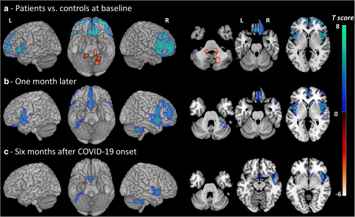
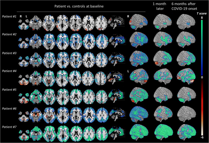
Methods: Seven patients with variable clinical presentations of COVID-19-related encephalopathy were explored thrice with brain 18F-FDG-PET/CT, once in the acute phase, 1 month later and 6 months after COVID-19 onset. PET images were analysed with voxel-wise and regions-of-interest approaches in comparison with 32 healthy controls.
Results: Patients' neurological manifestations during acute encephalopathy were heterogeneous. However, all of them presented with predominant cognitive and behavioural frontal disorders. SARS-CoV-2 RT-PCR in the CSF was negative for all patients. MRI revealed no specific abnormalities for most of the subjects. All patients had a consistent pattern of hypometabolism in a widespread cerebral network including the frontal cortex, anterior cingulate, insula and caudate nucleus. Six months after COVID-19 onset, the majority of patients clinically had improved but cognitive and emotional disorders of varying severity remained with attention/executive disabilities and anxio-depressive symptoms, and lasting prefrontal, insular and subcortical 18F-FDG-PET/CT abnormalities.
Conclusion: The implication of this widespread network could be the neural substrate of clinical features observed in patients with COVID-19, such as frontal lobe syndrome, emotional disturbances and deregulation of respiratory failure perception. This study suggests that this network remains mildly to severely impaired 6 months after disease onset.
Keywords: 18F-FDG-PET; COVID-19; Glucose metabolism; Prefrontal impairment; SARS-CoV-2.
Conflict of interest statement
The authors declare that they have no conflict of interest.
Figures
References
-
- Cani I, Barone V, D’Angelo R, Pisani L, Allegri V, Spinardi L, et al. Frontal encephalopathy related to hyperinflammation in COVID-19. J Neurol. 2020; http://link.springer.com/10.1007/s00415-020-10057-5. Accessed 11 Sept 2020. - DOI - PMC - PubMed
-
- Delorme C, Paccoud O, Kas A, Hesters A, Bombois S, Shambrook P, et al. Covid-19-related encephalopathy: a case series with brain FDG-PET/CT findings. Eur J Neurol. 2020; https://www.ncbi.nlm.nih.gov/pmc/articles/PMC7461074. Accessed 5 Sept 2020. - PMC - PubMed
MeSH terms
Substances
LinkOut - more resources
Full Text Sources
Other Literature Sources
Medical
Miscellaneous
