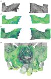
Validation of a novel method for canine eruption assessment in unilateral cleft lip and palate patients
- PMID: 33452746
- PMCID: PMC8204035
- DOI: 10.1002/cre2.397
Validation of a novel method for canine eruption assessment in unilateral cleft lip and palate patients
Abstract
Objective: The aim of this study was to propose and validate a three-dimensional (3D) methodology for the assessment of canine eruption in patients born with unilateral cleft lip and palate (UCLP) following secondary alveolar bone graft (SABG).
Methods and materials: A total of 10 patients (four females, six males; mean age: 8.8 years) with UCLP who underwent SABG were recruited. Pre- and 6-month post-operative cone-beam computed tomography (CBCT) was acquired for all patients. Post-operative data was registered onto pre-operative data utilizing voxel-based registration. Following superimposition, a segmentation process was applied to segment maxillary canine on both cleft and non-cleft side. Thereafter, translational and rotational changes in canine position were assessed for both cleft and non-cleft side by two observers.
Results: The intra-class correlation coefficient (ICC) indicated excellent reliability (≥0.90) with inter and intra-observer error of less than 0.05 mm. The overall ICC was found to be high for assessing both translational and rotational changes. The mean absolute inter- and intra-observer difference for translational and rotational changes was found to be less than 1 mm and 3°.
Conclusion: The present method was found to be reliable proving to be clinically applicable for assessing maxillary canine eruption changes in both cleft and non-cleft bone.
Keywords: 3-D imaging; cleft lip; cleft palate; cone-beam computed tomography; tooth eruption.
© 2021 The Authors. Clinical and Experimental Dental Research published by John Wiley & Sons Ltd.
Conflict of interest statement
The authors report no conflict of interest.
Figures


Similar articles
-
Validation of a 3D methodology for the evaluation and follow-up of secondary alveolar bone grafting in unilateral cleft lip and palate patients.Orthod Craniofac Res. 2022 Aug;25(3):377-383. doi: 10.1111/ocr.12546. Epub 2021 Dec 1. Orthod Craniofac Res. 2022. PMID: 34817927
-
Three-dimensional evaluation of secondary alveolar bone grafting in patients with unilateral cleft lip and palate: A 2-3 year post-operative follow-up.Orthod Craniofac Res. 2024 Jun;27 Suppl 1:100-108. doi: 10.1111/ocr.12763. Epub 2024 Feb 1. Orthod Craniofac Res. 2024. PMID: 38299981
-
A CBCT Based Assessment of Canine Eruption and Development Following Alveolar Bone Grafting in Patients Born With Unilateral Cleft lip and/or Palate.Cleft Palate Craniofac J. 2023 Apr;60(4):386-394. doi: 10.1177/10556656211064477. Epub 2021 Dec 7. Cleft Palate Craniofac J. 2023. PMID: 34873962
-
Comparison of early and conventional autogenous secondary alveolar bone graft in children with cleft lip and palate: A systematic review.Orthod Craniofac Res. 2020 Nov;23(4):385-397. doi: 10.1111/ocr.12394. Epub 2020 Jun 28. Orthod Craniofac Res. 2020. PMID: 32446283
-
Effects of Secondary Alveolar Bone Grafting on Maxillary Growth in Cleft Lip or Palate Patients: A Systematic Review and Meta-Analysis.Cleft Palate Craniofac J. 2024 Nov;61(11):1860-1872. doi: 10.1177/10556656231188600. Epub 2023 Jul 12. Cleft Palate Craniofac J. 2024. PMID: 37438927
References
-
- Almukhtar, A. , Ju, X. , Khambay, B. , McDonald, J. , & Ayoub, A. (2014). Comparison of the accuracy of voxel based registration and surface based registration for 3D assessment of surgical change following orthognathic surgery. PLoS One, 9(4), e93402. 10.1371/journal.pone.0093402 - DOI - PMC - PubMed
-
- Bergland, O. , Semb, G. , & Aabyholm, F. E. (1986). Elimination of the residual alveolar cleft by secondary bone grafting and subsequent orthodontic treatment. The Cleft Palate Journal, 23(3), 175–205. - PubMed
-
- Bianchi, A. , Muyldermans, L. , Di Martino, M. , Lancellotti, L. , Amadori, S. , Sarti, A. , & Marchetti, C. (2010). Facial soft tissue esthetic predictions: Validation in craniomaxillofacial surgery with cone beam computed tomography data. Journal of Oral and Maxillofacial Surgery, 68(7), 1471–1479. 10.1016/j.joms.2009.08.006 - DOI - PubMed
MeSH terms
LinkOut - more resources
Full Text Sources
Other Literature Sources
Medical

