. 2021 Mar 4;81(5):1074-1083.e5.
doi: 10.1016/j.molcel.2020.12.036. Epub 2021 Jan 15.

RADX controls RAD51 filament dynamics to regulate replication fork stability

Affiliations

RADX controls RAD51 filament dynamics to regulate replication fork stability

Madison B Adolph et al. Mol Cell. .

Abstract

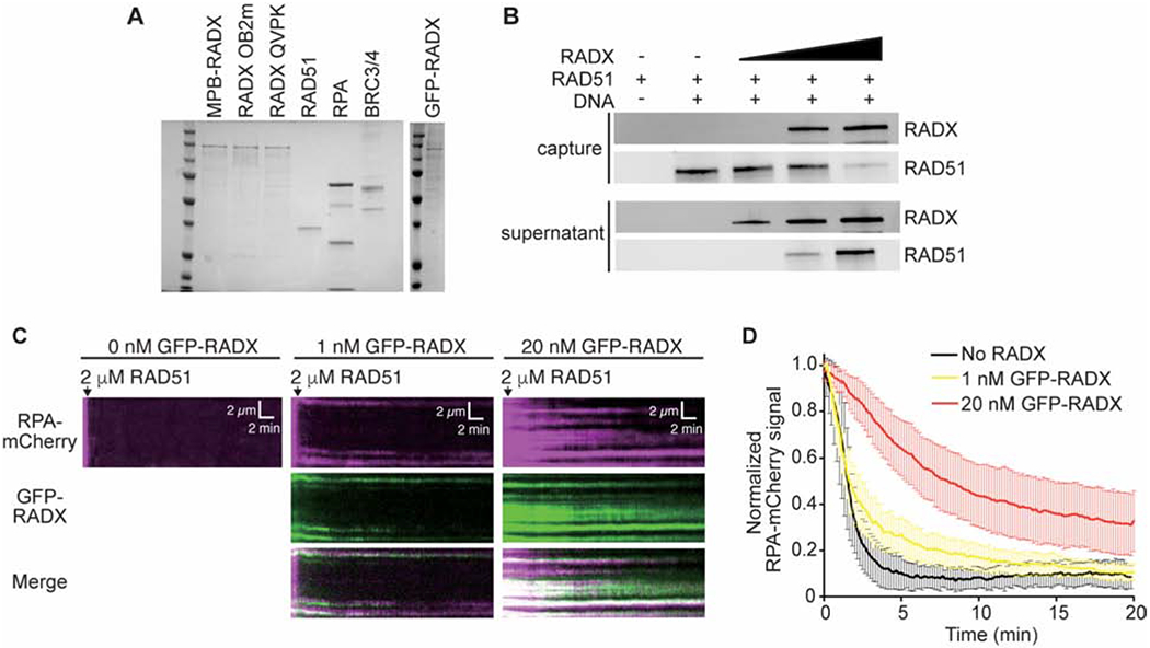
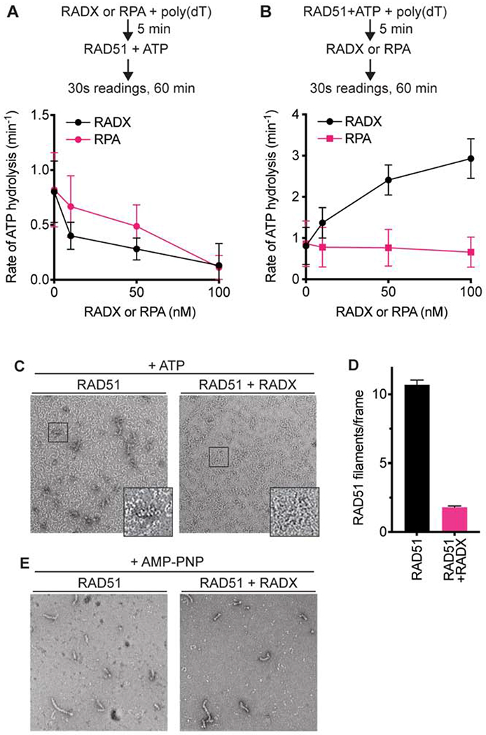
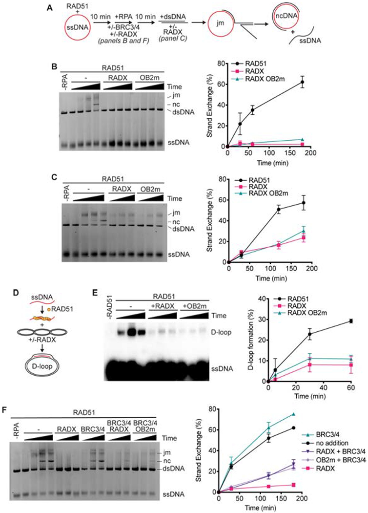
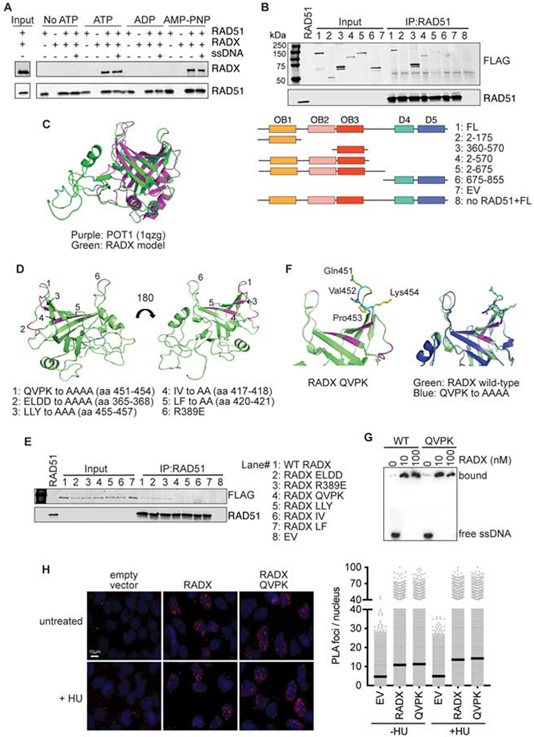
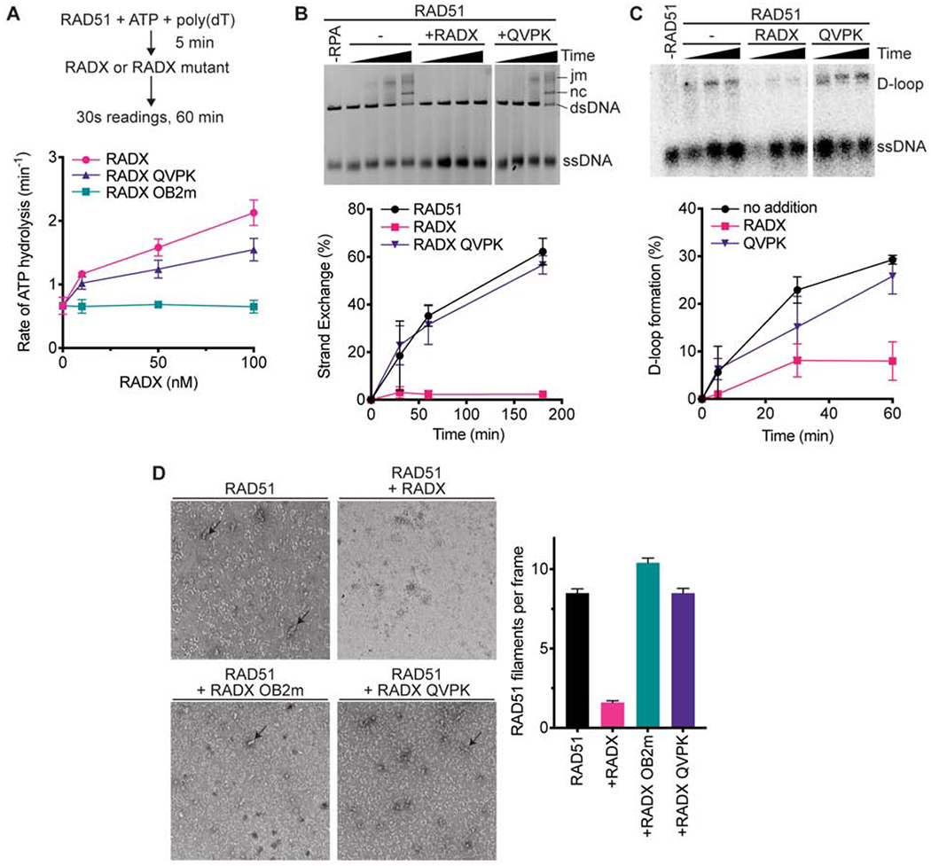
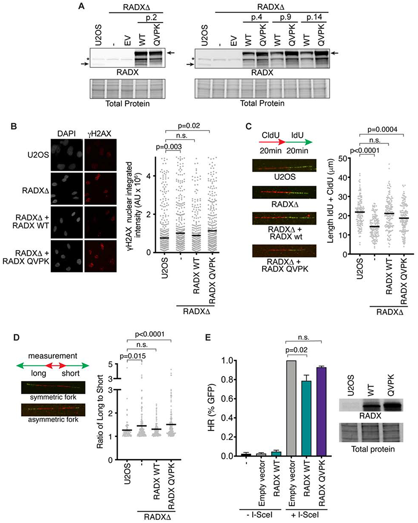
The RAD51 recombinase forms nucleoprotein filaments to promote double-strand break repair, replication fork reversal, and fork stabilization. The stability of these filaments is highly regulated, as both too little and too much RAD51 activity can cause genome instability. RADX is a single-strand DNA (ssDNA) binding protein that regulates DNA replication. Here, we define its mechanism of action. We find that RADX inhibits RAD51 strand exchange and D-loop formation activities. RADX directly and selectively interacts with ATP-bound RAD51, stimulates ATP hydrolysis, and destabilizes RAD51 nucleofilaments. The RADX interaction with RAD51, in addition to its ssDNA binding capability, is required to maintain replication fork elongation rates and fork stability. Furthermore, BRCA2 can overcome the RADX-dependent RAD51 inhibition. Thus, RADX functions in opposition to BRCA2 in regulating RAD51 nucleofilament stability to ensure the right level of RAD51 function during DNA replication.

Keywords: DNA curtain; DNA damage response; DNA repair; double-strand break; electron microscopy; fork reversal; replication stress.


Conflict of interest statement

Declaration of interests The authors declare no competing interests.

Figures

Figure 1.
Figure 1.. RADX inhibits RAD51 assembly on DNA.
(A) Coomassie stained SDS-PAGE gel showing purified proteins used in this study. Each lane contains ~200 nM of purified protein. Actual amounts used is specified in each experiment. (B) Pull-down experiments showing MBP-RADX (0.5, 1, and 2nM) outcompetes RAD51 (4μM) for binding ssDNA. (C) Kymographs showing the assembly of RAD51 on ssDNA molecules coated with RPA-mCherry and 0 nM (left), 1 nM (middle), and 20 nM GFP-RADX (right). Assembly was initiated by injecting 2 μM RAD51 together with 2 mM ATP and 5 mM Ca2+ into sample chambers containing ssDNA molecules coated with RPA-mCherry and GFP-RADX while monitoring the loss of RPA-mCherry signal. (D) Graphs showing the normalized RPA-mCherry signal intensity integrated over entire ssDNA molecules during the assembly of the RAD51 filaments. Error bars represent 68% confidence intervals (n=30 ssDNA molecules).
Figure 2.
Figure 2.. RADX prevents RAD51 nucleofilament assembly by promoting ATP hydrolysis.
(A) RAD51 ATPase assay with increasing concentrations of RADX or RPA added to the reaction prior to the addition of RAD51. (B) RAD51 ATPase assay with RADX or RPA added to the assay after RAD51. n=3, +/− SD. See also Supplementary Figure 1. (C) Representative negative stain EM images of nucleofilaments formed in the presence and absence of RADX. The images are of samples fixed at a 10-minute timepoint after the addition of RADX. While RAD51 protofilaments are seen in both images, RAD51-DNA nucleofilaments are seen only in the absence of RADX. (D) Quantitation of RAD51 nucleofilaments formed in the presence or absence of RADX. The assessment was done using n=183 and n=195 micrographs collected randomly over the grid in the absence and presence of RADX, respectively (Mean+/−SEM). (E) Representative negative stain EM images of nucleofilaments formed in the presence and absence of RADX, with AMP-PNP in the reaction buffer.
Figure 3.
Figure 3.. RADX prevents RAD51 mediated strand-exchange and D-loop formation.
(A) Schematic of RAD51-mediated strand exchange assay (jm, joint molecules; nc, nicked circular dsDNA). RADX was added either 10 minutes prior to the dsDNA in panels B and F or at the same time as dsDNA in panel C. (B) A representative gel and quantitation from n=3 strand exchange assays are shown. Error bars are SD. (C) Strand exchange assay in which RADX or RADX OB2m was added at the same time as the dsDNA. Representative gel and quantitation from n=3 experiments. (D) Schematic of D-loop formation assay. (E) A representative gel of the D-loop assay and quantitation (n=3, +/− SD). (F) Strand exchange assay with BRC3/4 and/or RADX proteins added at the same time as RPA.
Figure 4.
Figure 4.. RADX interacts directly with RAD51 through residues in the RADX OB3 domain.
(A) Direct interaction of RADX and RAD51 was assessed using RAD51 antibody conjugated to Protein G beads to pull down purified RAD51 and RADX in the presence of the indicated nucleotide with and without ssDNA. Each binding reaction contains equimolar amounts of RADX and RAD51. After washing, the beads were boiled in SDS loading buffer and eluted protein detected by immunoblotting. (B) Interaction of purified RADX fragments with RAD51 in the presence of ATP. FL, full length (C) Homology model of RADX OB3 domain based on the OB-fold domain in POT1 (PDBID 1qzg). (D) Location of the surfaces of RADX OB3 tested for interactions with RADX. (E) Interaction of purified RADX OB3 mutants with RAD51 in the presence of ATP. (F) Location of the QVPK residues on the predicted loop of OB3 (left panel) and comparison of the models of wild-type and QVPK mutant OB3 domains (right panel). (G) Electrophoretic mobility shift assay of purified RADX and RADX QVPK mutant protein with poly (dT60) ssDNA. (H) Proximity ligation assay between Flag-RADX and EdU. Cells were labeled for 10 minutes with EdU and treated with hydroxyurea (HU) for two hours where indicated. An image and quantitative data from a representative experiment is shown. (EV, empty vector)
Figure 5.
Figure 5.. RADX must interact with RAD51 to inhibit strand exchange and D-loop formation.
(A) The ssDNA-dependent ATPase activity of RAD51 was measured in the presence of wild-type, QVPK, and OB2m RADX proteins (n=3 +/− SD). (B) Strand exchange assay in which RADX wild-type and QVPK mutant were added at the same time as RPA (n=3 +/−SD). (C) D-loop formation assay with representative gel and quantitation (n=3 +/−SD). (D) Nucleofilament formation by RAD51 without RADX (n=178) in the presence of RADX (n=163), RADX OB2m (n=194), or RADX QVPK (n=208). (Error bars are SEM). The panels are representative micrographs.
Figure 6.
Figure 6.. RADX interacts with RAD51 to maintain replication fork stability.
(A) Immunoblots of U2OS, or RADXΔ cells infected with lentivirus expressing the wild-type (WT) RADX or RADX QVPK (EV = empty vector). The passage number after infection and selection is indicated. (B) Cells were imaged for γH2AX. P values were derived from a one-way ANOVA with Dunnett’s multiple comparison test. (C) Cells were labeled with CldU and IdU as indicated and then analyzed by DNA combing. Representative images and experiment are shown with P values derived from a one-way ANOVA with Dunnett’s multiple comparison test. (D) Fork symmetry was assessed as indicated. At least 177 DNA fibers were measured from four biological replicates. P values were derived from a Kruskal-Wallis test. (E) The percentage of DR-GFP-positive U2OS cells after overexpression of empty vector, RADX WT or RADX QVPK and I-Scel expression vector. Immunoblots show the level of overexpression of RADX WT and RADX QVPK (Mean+/−SEM, n=3 in which 25,000 cells were scored per experiment; p-value derived from One-way ANOVA with Dunnett’s multiple comparisons test).

References

    1. Antony E, Tomko EJ, Xiao Q, Krejci L, Lohman TM, and Ellenberger T (2009). Srs2 disassembles Rad51 filaments by a protein-protein interaction triggering ATP turnover and dissociation of Rad51 from DNA. Mol Cell 35, 105–115. - PMC - PubMed
    1. Bhat KP, and Cortez D (2018). RPA and RAD51: fork reversal, fork protection, and genome stability. Nat Struct Mol Biol 25, 446–453. - PMC - PubMed
    1. Bhat KP, Krishnamoorthy A, Dungrawala H, Garcin EB, Modesti M, and Cortez D (2018). RADX Modulates RAD51 Activity to Control Replication Fork Protection. Cell Rep 24, 538–545. - PMC - PubMed
    1. Bugreev DV, and Mazin AV (2004). Ca2+ activates human homologous recombination protein Rad51 by modulating its ATPase activity. Proc Natl Acad Sci U S A 101, 9988–9993. - PMC - PubMed
    1. Bugreev DV, Yu X, Egelman EH, and Mazin AV (2007). Novel pro- and anti-recombination activities of the Bloom’s syndrome helicase. Genes Dev 21, 3085–3094. - PMC - PubMed

Publication types

MeSH terms

LinkOut - more resources