Review
. 2021 Mar 20:235:104117.
doi: 10.1016/j.jprot.2021.104117. Epub 2021 Jan 14.

Comparative evaluation of two methods for LC-MS/MS proteomic analysis of formalin fixed and paraffin embedded tissues

Affiliations
Review

Comparative evaluation of two methods for LC-MS/MS proteomic analysis of formalin fixed and paraffin embedded tissues

Katarina Davalieva et al. J Proteomics. .

Abstract

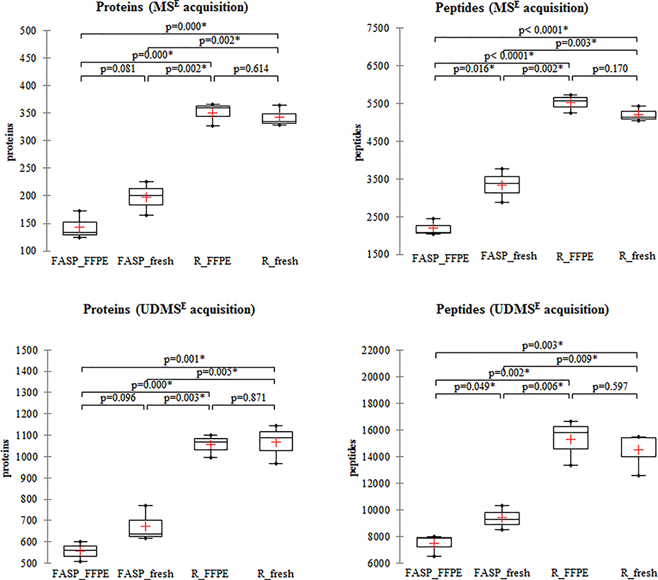
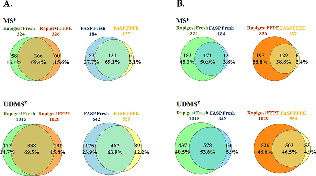
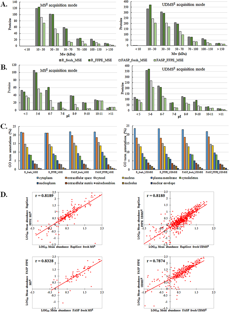
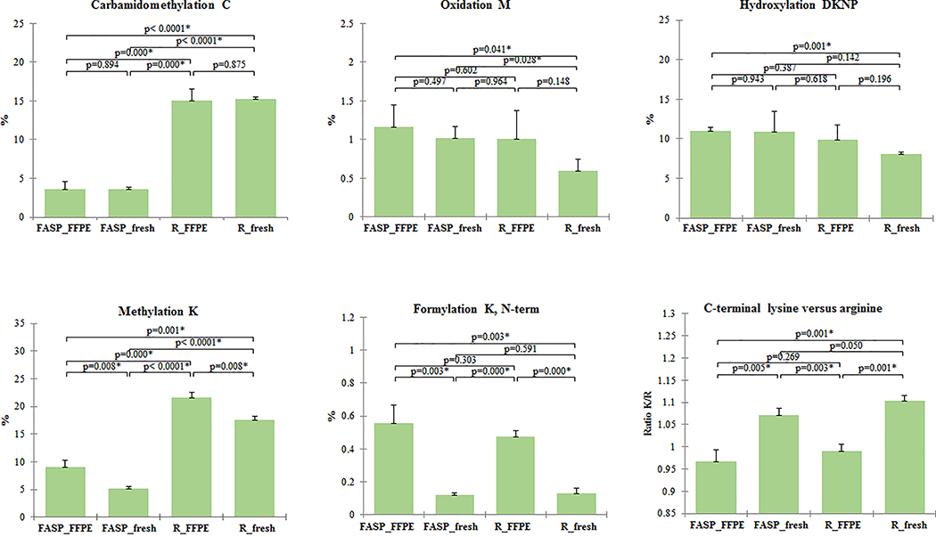
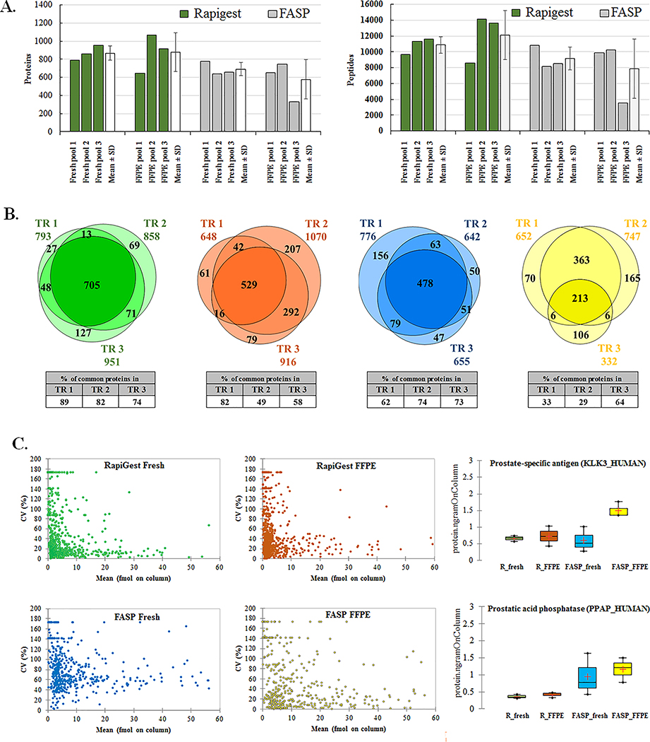
The proteomics of formalin-fixed, paraffin-embedded (FFPE) samples has advanced significantly during the last two decades, but there are many protocols and few studies comparing them directly. There is no consensus on the most effective protocol for shotgun proteomic analysis. We compared the in-solution digestion with RapiGest and Filter Aided Sample Preparation (FASP) of FFPE prostate tissues stored 7 years and mirroring fresh frozen samples, using two label-free data-independent LC-MS/MS acquisitions. RapiGest identified more proteins than FASP, with almost identical numbers of proteins from fresh and FFPE tissues and 69% overlap, good preservation of high-MW proteins, no bias regarding isoelectric point, and greater technical reproducibility. On the other hand, FASP yielded 20% fewer protein identifications in FFPE than in fresh tissue, with 64-69% overlap, depletion of proteins >70 kDa, lower efficiency in acidic and neutral range, and lower technical reproducibility. Both protocols showed highly similar subcellular compartments distribution, highly similar percentages of extracted unique peptides from FFPE and fresh tissues and high positive correlation between the absolute quantitation values of fresh and FFPE proteins. In conclusion, RapiGest extraction of FFPE tissues delivers a proteome that closely resembles the fresh frozen proteome and should be preferred over FASP in biomarker and quantification studies. SIGNIFICANCE: Here we analyzed the performance of two sample preparation methods for shotgun proteomic analysis of FFPE tissues to give a comprehensive overview of the obtained proteomes and the resemblance to its matching fresh frozen counterparts. These findings give us better understanding towards competent proteomics analysis of FFPE tissues. It is hoped that it will encourage further assessments of available protocols before establishing the most effective protocol for shotgun proteomic FFPE tissue analysis.

Keywords: FASP; FFPE; LC-MS/MS; Label-free data-independent acquisition; Protein extraction; RapiGest.


Conflict of interest statement

Declaration of Competing Interest

The authors declare no conflict of interest.

Figures

Figure 1.
Figure 1.. Protein and peptide identifications from fresh frozen and FFPE samples by RapiGest and FASP protocol.
Representative S POOL samples from fresh frozen and FFPE tissues obtained with RapiGest and FASP protocols respectively were run in triplicate in MSE and UDMSE acquisition modes. The number of proteins and peptides obtained by processing with PLGS are plotted for each acquisition mode and technical replicate. In the box plot graphs, median (−), 25th and 75th percentiles and mean (+) are shown. Asterisk (*) indicate significant statistical difference (t-test, p <0.05). (Note that scales differ for MSE and UDMSE acquisition modes.)
Figure 2.
Figure 2.. Comparison of the identified common and unique proteins between tissue types and protocols for sample preparation.
(A) Comparison of the fresh and FFPE tissues extracted with RapiGest and FASP protocols (B) Comparison of protocol efficiency over fresh and FFPE tissues. S POOL samples representative proteomes for each tissue type and protocol were selected based on proteins identified in at least two out of three technical replicates in MSE and UDMSE acquisition modes, respectively. For each comparison, total number of the quantified proteins per tissue type and protocol as well as common and unique proteins have been presented as number and percentage.
Figure 3.
Figure 3.. Qualitative and quantitative analysis of the fresh/frozen and FFPE tissue proteomes extracted with RapiGest and FASP protocol.
S POOL samples representative proteomes were selected based on proteins identified in at least two out of three technical replicates. Analysis was done based on the distribution of proteins according to: (A) molecular weight, (B) protein pI, (C) subcellular localization and (D) correlation between absolute abundance estimation values in the representative proteome from fresh frozen and FFPE samples. The mean protein abundances from the 3 technical replicates were calculated for common identified proteins between fresh/frozen and FFPE samples with two protocols and two modes of LC-MS/MS acquisition. Mean abundance values expressed as a mean fmols on column were Log10 transformed and values from fresh frozen proteins were plotted against corresponding values from FFPE samples. Pearson correlation coefficients are also reported.
Figure 4.
Figure 4.. Assessment of the effects of fixation and subsequent tissue processing on FFPE tissues.
Individually extracted pool samples (250 ng) from fresh frozen and FFPE tissues with RapiGest and FASP were searched for post-translational modifications (carbamidomethyl cysteine (C), oxidized methionine (M), hydroxylation of asparagine, aspartic acid, proline or lysine (DKNP), methylation of lysine (K), and formylation of lysine, N-term) and ratios of C-terminal lysine versus arginine (R) peptides. Mean and SD values for 3 technical replicates per protocol/tissue type are shown. The peptides with post-translational modifications were expressed as percentage of the total number of peptides. Asterisk (*) indicate significant statistical difference (t-test, p <0.05).
Figure 5.
Figure 5.. Technical reproducibility analysis of RapiGest and FASP protocols.
Pool samples prepared from fresh frozen and FFPE samples and independently extracted in triplicate were run in UDMSE acquisition mode. (A) The number of proteins and peptides are plotted for each replicate with average number of the identified proteins/peptides (Mean ± SD) and CV for each triplicate set. (B) Overlap in protein identifications among independently extracted replicates: RapiGest fresh (green), RapiGest FFPE (orange), FASP fresh (blue), FASP FFPE (yellow). (C) Accuracy of quantification among independently extracted replicates. The mean absolute abundance of the common identified proteins in independently extracted technical replicates is plotted against its corresponding CV (%) for each protocol/tissue type combination. The reproducibility of quantitation is demonstrated for two prostate specific proteins: prostate-specific antigen (KLK3_HUMAN) and prostatic acid phosphatase (PPAP_HUMAN) with absolute quantity of the protein in nanograms (ng) plotted for each technical replicate. In the box plot graphs, median (−), 25th and 75th percentiles and mean (+) are shown.

Similar articles

Cited by

References

    1. Belczacka I, Latosinska A, Metzger J, Marx D, Vlahou A, Mischak H,Frantzi M, Proteomics biomarkers for solid tumors: Current status and future prospects, Mass Spectrom Rev 38 (2019) 49–78, 10.1002/mas.21572. - DOI - PubMed
    1. Zhang B, Whiteaker JR, Hoofnagle AN, Baird GS, Rodland KD,Paulovich AG, Clinical potential of mass spectrometry-based proteogenomics, Nat Rev Clin Oncol 16 (2019) 256–268, 10.1038/s41571-018-0135-7 - DOI - PMC - PubMed
    1. Blonder J,Veenstra TD, Clinical proteomic applications of formalin-fixed paraffin-embedded tissues, Clin Lab Med 29 (2009) 101–13, 10.1016/j.cll.2009.01.006 - DOI - PubMed
    1. Reimel BA, Pan S, May DH, Shaffer SA, Goodlett DR, McIntosh MW, Yerian LM, Bronner MP, Chen R,Brentnall TA, Proteomics on Fixed Tissue Specimens - A Review, Curr Proteomics 6 (2009) 63–69, 10.2174/157016409787847420. - DOI - PMC - PubMed
    1. Specht K, Richter T, Muller U, Walch A, Werner M,Hofler H, Quantitative gene expression analysis in microdissected archival formalin-fixed and paraffin-embedded tumor tissue, Am J Pathol 158 (2001) 419–29, https://doi.org/S0002-9440(10)63985-5 [pii] - PMC - PubMed

Publication types