. 2021 Mar:555:44-55.
doi: 10.1016/j.virol.2020.10.009. Epub 2020 Nov 12.

Lassa-VSV chimeric virus targets and destroys human and mouse ovarian cancer by direct oncolytic action and by initiating an anti-tumor response

Affiliations

Lassa-VSV chimeric virus targets and destroys human and mouse ovarian cancer by direct oncolytic action and by initiating an anti-tumor response

A N van den Pol et al. Virology. 2021 Mar.

Abstract

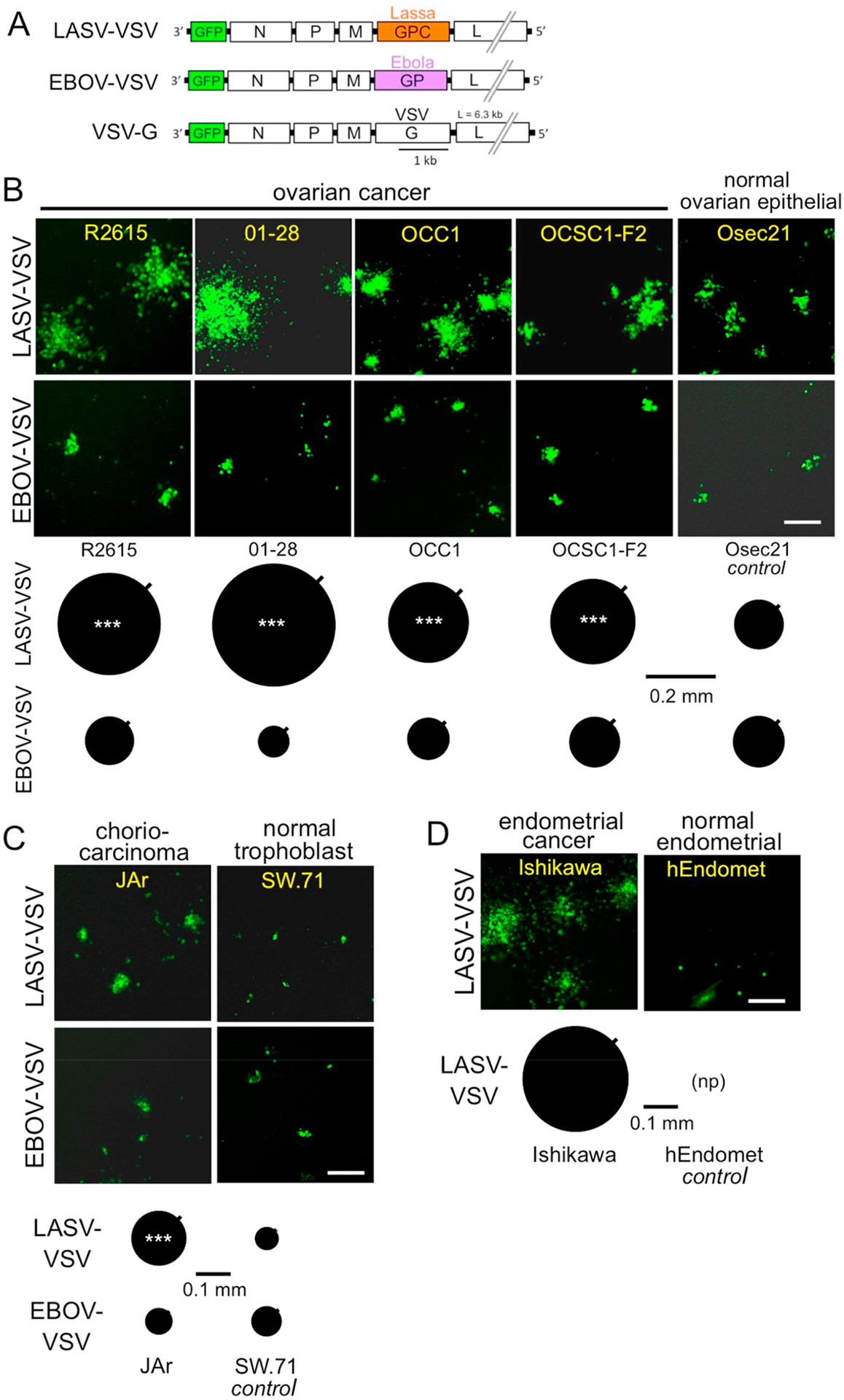
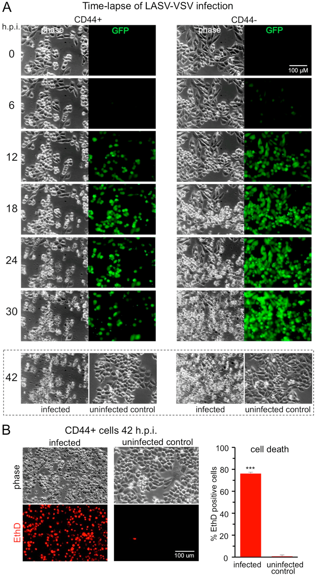
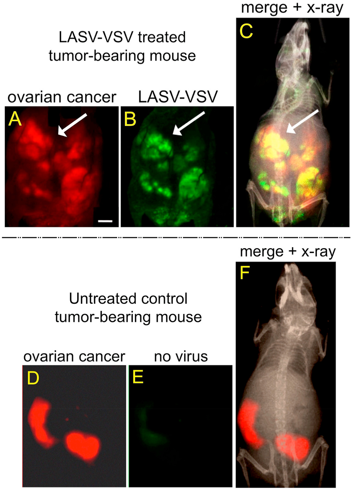
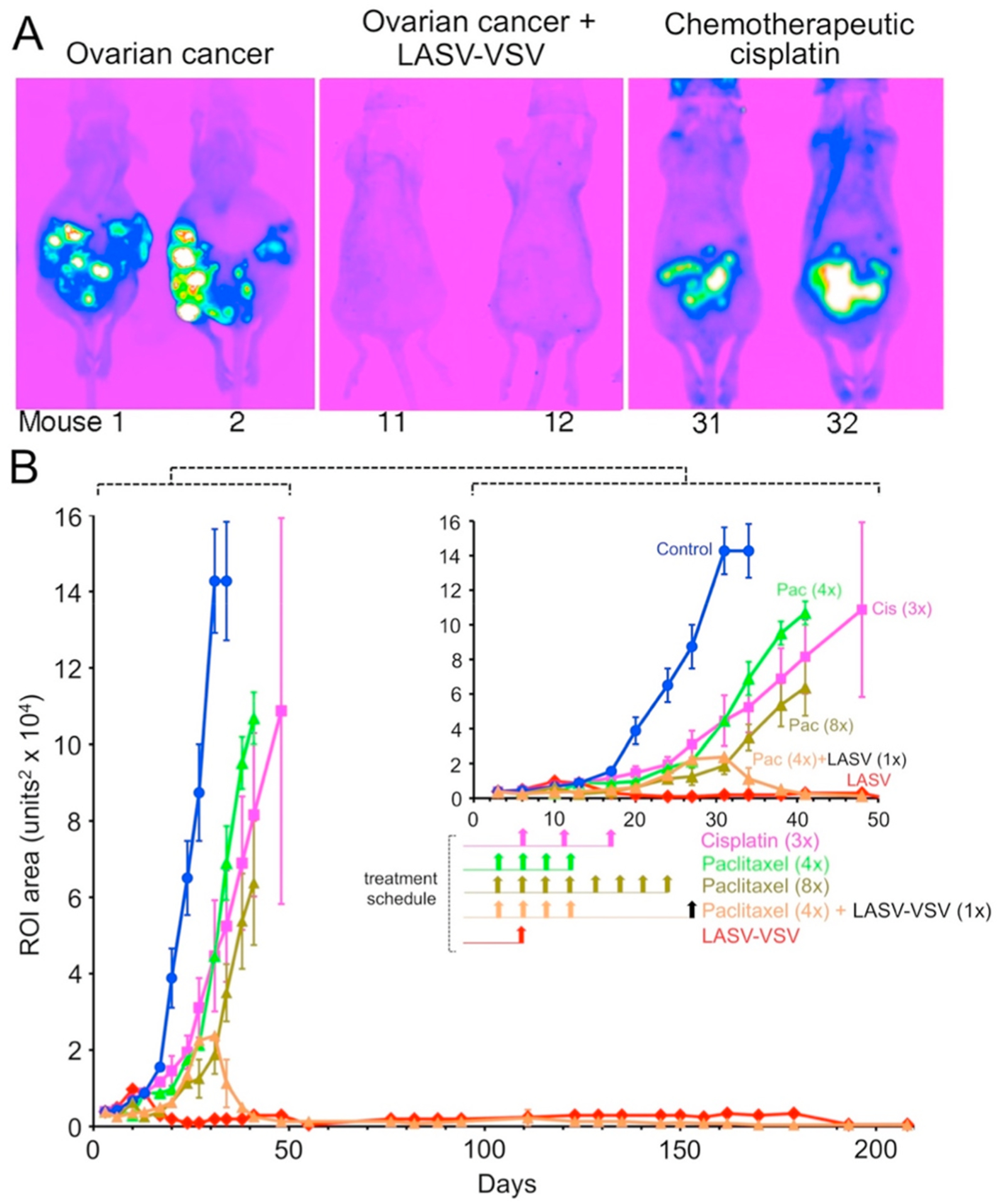
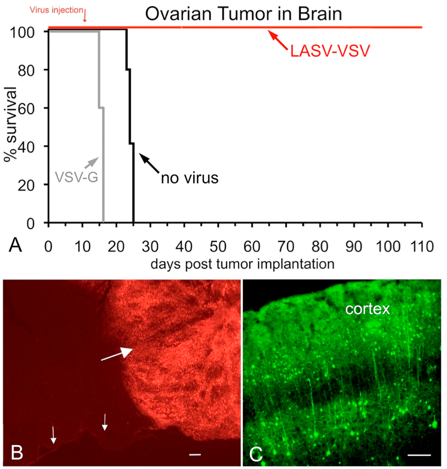
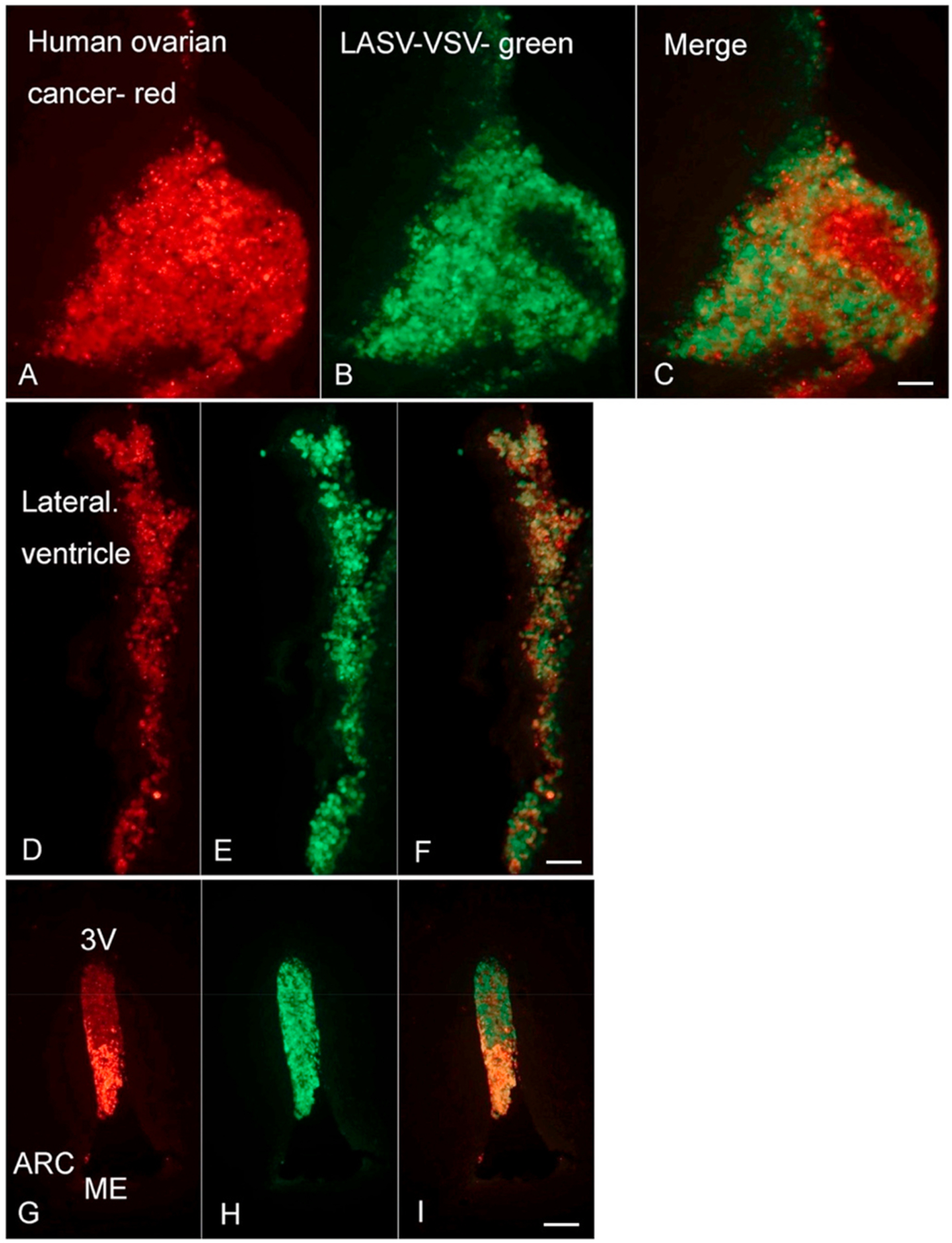
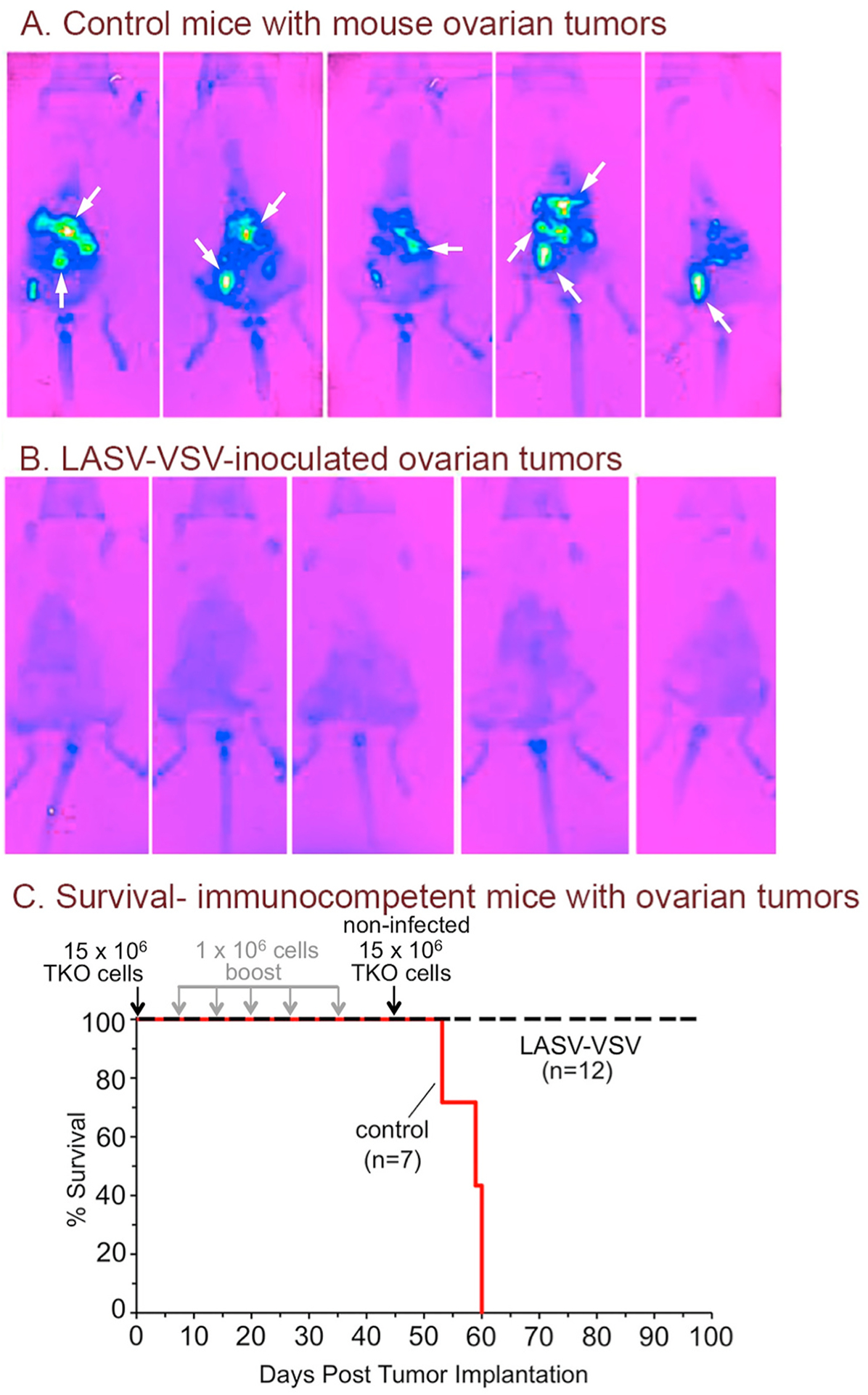
Ovarian cancer is the third most common female cancer, with poor survival in later stages of metastatic spread. We test a chimeric virus consisting of genes from Lassa and vesicular stomatitis viruses, LASV-VSV; the native VSV glycoprotein is replaced by the Lassa glycoprotein, greatly reducing neurotropism. Human ovarian cancer cells in immunocompromised nude mice were lethal in controls. Chemotherapeutic paclitaxel and cisplatin showed modest cancer inhibition and survival extension. In contrast, a single intraperitoneal injection of LASV-VSV selectively infected and killed ovarian cancer cells, generating long-term survival. Mice with human ovarian cancer cells in brain showed rapid deterioration; LASV-VSV microinjection into brain blocked cancer growth, and generated long-term survival. Treatment of immunocompetent mice with infected mouse ovarian cancer cells blocked growth of non-infected ovarian cancer cells peritoneally and in brain. These results suggest LASV-VSV is a viable candidate for further study and may be of use in the treatment of ovarian cancer.


Conflict of interest statement

Declaration of competing interest

Av has a financial interest in Implicyte. Other authors declare no conflict.

Figures

Fig. 1.
Fig. 1.
LASV-VSV produces larger plaques in cultures of human ovarian cancer, choriocarcinoma, and endometrial cancer cells compared to normal cells. (A) Diagram depicting genomic composition of two recombinant chimeric VSVs (top, middle) and one non-chimeric VSV control (bottom) used in the present study. Schematic shows gene order, gene size and replacement of VSV glycoprotein gene (VSV-G; white) with Lassa glycoprotein complex gene (LASV-GPC; orange) or Ebola glycoprotein gene (EBOV-GP; violet). Green fluorescent protein gene (GFP) is indicated in green. (B) Representative images of GFP fluorescent LASV-VSV (first row) and EBOV-VSV (second row) plaques that developed after 24 h on cultures of ovarian cancer cells (R2615, 01–28, OCC1, OCSC1–F2) and normal ovarian surface epithelial cells (Osec21). Quantification of plaque sizes are shown below. Circles indicate mean plaque size (n = 60 per condition) and the small square projecting from each indicates the SE. Stars indicate statistical significance compared with control (***P < 0.001; one-way ANOVA with Tukey-Kramer multiple comparisons test). (C) Representative images (above) of plaques that developed on choriocarcinoma cells (JAr) and control normal trophoblast cells (SW.71) after 24 h; corresponding quantification of plaque sizes also shown (below). (D) Representative images (above) and corresponding plaque sizes (below) of endometrial cancer cells (Ishikawa) and normal endometrial cells (hEndomet). No plaque formation is indicated by (np) with only scattered individual cells being infected. Image scale bars 0.25 mm.
Fig. 2.
Fig. 2.
Time lapse imaging of Lassa-VSV infection of human ovarian cancer cells. (A) H.p.i., hours post infection. By 12 hpi, many of the ovarian cancer cells show infection by expression of the GFP viral reporter gene. By 30 hpi, the majority of the cells show a lytic response to viral infection. CD44+ is correlated with greater metastatic potential and chemotherapeutic resistance. OCSC1 (CD44+) and OCC1 cells (CD44) showed similar cytolytic responses to the virus. In contrast, the uninfected control cells shown at 42 hpi (dashed box) appear normal with no apparent lytic activity and no green fluorescence. (B) OCSC1 (CD44+) cells were infected as in (A) and 42 h.p.i. labeled with the red fluorescent cell death indicator ethidium homodimer (EthD-1). Bar graph (right) shows the quantification of dead EthD-1 positive cells. Bars represent the mean ± SE of triplicate wells for each condition and *** indicates P < 0.001; Student’s t-test.
Fig. 3.
Fig. 3.
Lassa-VSV selectively infects human ovarian cancer cells. LASV-VSV treated tumor-bearing mouse (A–C). (A) The red fluorescent ovarian cancer cells are shown. (B) The same field shows that the GFP reporter in LASV-VSV is generally expressed in the same regions. The green area at the bottom of the image shows the pelvic fat area where ovarian cancer micrometastases are commonly found and where adipocytes promote ovarian cancer growth (Alvero et al., 2017; Nieman et al., 2011). (C) A merged micrograph shows the overlap of green virus in red ovarian cancer tissue; this image also includes an x-ray of the same mouse. Images were taken 9 days after virus inoculation. Untreated control tumor-bearing mouse (D–F). (D) Red fluorescent ovarian cancer tumor. (E) Same field using GFP imaging. (F) Merged image that includes x-ray of the same mouse. Scale bar (A–F), 3 mm.
Fig. 4.
Fig. 4.
Lassa-VSV anti-tumoral effect prevents recurrence in a human ovarian cancer animal model. (A) Fluorescence imaging examples from 3 groups of mice are shown. Control mice (mouse 1,2; left image) received ip injection of human ovarian cancer cells, shown here in pseudocolor; tumors are depicted in light blue, green, and white. LASV-VSV-treated mice (mouse 11,12; center image) received a single injection of LASV-VSV after establishment of ovarian tumors. A single administration of LASV-VSV eliminated detection of ovarian cancer cells. Cisplatin-treated mice (mouse 31,32; right image displayed reduced tumor size but cisplatin did not eliminate tumors. (B) Graph shows relative size of OCSC1–F2 human ovarian tumors in live mice at different stages of tumor development. Untreated tumor-bearing control mice (n = 5; blue) showed rapid tumor expansion. Paclitaxel (n = 5; green, 4 treatments) and (n = 5, 8 treatments) and cisplatin (n = 5; pink, 3 treatments) -treated tumors are attenuated by these chemotherapeutics, but continue to expand. LASV-VSV (n = 5; red) causes a rapid reduction in tumor size and presence. Untreated control, Paclitaxel, and cisplatin-treated mice all showed a lethal response to tumor growth before day 50. Virus-treated mice survived past day 200. Vertical bars indicate SEM. (C) Horizontal bars show survival for each of the six groups. With the exception of the mice treated with the single injection of LASV-VSV, mice from other groups showed a lethal response to tumor expansion by 50 days post-tumor administration. LASV-VSV-treated mice survived past 210 days.
Fig. 5.
Fig. 5.
Intracranial LASV-VSV eliminates human ovarian cancer in mouse brain. (A) Survival curve shows all control mice (n = 5) with ovarian cancer in brain showed a lethal response by 25 days after tumor implant (black line); tumor implanted mice (n = 5) injected with VSV-G all died within 5–6 days after virus injection (gray line). In contrast, complete survival of tumor implanted mice (n = 4) that received intracranial LASV-VSV is shown (red line). (B) Human red fluorescent ovarian cancer cells after injection into the brain and subsequent tumor expansion in a mouse that succumbed to the expanding tumor. Large arrow indicates tumor, small arrows show ventral surface of brain. Bar, 100 μm. (C) Cortical section from a VSV-G infected mouse prepared shortly after death 6 days after virus injection. GFP indicated widespread infection throughout the brain. Bar, 100 μm.
Fig. 6.
Fig. 6.
Virus selectively infects human ovarian cancer metastases within brain. (A–C) Large red tumor in brain selectively infected by green LASV-VSV. Bar, 150 μm. (D–F) Ovarian cancer cells migrated into the lateral ventricle. LASV-VSV (green) selectively infects these red cells. Bar, 150 μm. (G–I) The ovarian cancer cells (red) also migrated into the third ventricle (3 V) medial to the arcuate nucleus (ARC) and dorsal to the median eminence (ME). In the normal brain, the ventricular system is generally cell-free. Cancer cells that have been infected to the point of cytolysis show a reduced red fluorescence due to the loss of the fluorescent molecules with breakdown of the plasma membrane. Virus (green) selectively infected the cancer cells within the ventricles. Bar, 300 μm.
Fig. 7.
Fig. 7.
LASV-VSV enhances survival of mice with mouse ovarian tumors. (A) Examples of fluorescent imaging of live mice 9 ± 2 days after implant of 15 million TKO ovarian cancer cells. Light blue color indicates expanding tumors and arrows indicate large tumor masses. (B) Mice previously treated with mouse ovarian cancer cells inoculated with LASV-VSV 9 ± 2 days, after implant of 15 million non-infected ovarian cancer cells. No tumor growth is detected here or later. (C) Mouse ovarian cancer cells (15 × 106) were administered i.p. to controls (red line), or infected with LASV-VSV (MOI = 1.0) and injected into experimental animals (dashed black line). Both experimental and control animals were boosted at weekly intervals using 1 × 10⁶ cells infected (MOI = 1.0) and uninfected with LASV-VSV, respectively. On day 45, experimental animals received a second bolus of 15 × 106 non-infected TKO cells. All tumor bearing mice not treated with the virus showed a lethal response to the tumor by day 60. All mice treated with infected ovarian cancer cells prior to injection of uninfected cancer cells showed complete survival.
Fig. 8.
Fig. 8.
LASV-VSV immunized mice do not grow mouse ovarian brain tumors. (A) Mouse ovarian cancer cells were implanted into brain in control mice and mice that had received LASV-VSV inoculated cancer cells. All controls showed a lethal response to the expanding tumor (B,C), whereas all immunized mice showed long-term survival with no detectable tumor growth. (B) In non-immunized mice, a large red fluorescent mouse ovarian tumor is seen in the middle of the brain (large arrow). A substantial number of ovarian cancer cells are migrating away (small arrows) from the large primary tumor body. Bar, 15 μm. (C) In another non-immunized mouse, again ovarian cancer cells (arrows) are found migrating to the superficial edge of the cerebral cortex. Dotted line indicates edge of cortical section. Bar, 17 μm. (D) Phase-contrast image of a similar cortical section from an immunized mouse 65 days after ovarian cancer cell implantation. (E) Red fluorescent illumination of the same cortical section showing an absence of any surviving cancer cells. Dotted line indicates edge of section. Bar, 25 μm.

Similar articles

Cited by

References

    1. Akeson M, Zetterqvist BM, Dahllof K, Brannstrom M, Horvath G, 2008. Effect of adjuvant paclitaxel and carboplatin for advanced stage epithelial ovarian cancer: a population-based cohort study of all patients in western Sweden with long-term follow-up. Acta Obstet. Gynecol. Scand 87, 1343–1352 - PubMed
    1. Alvero AB, Fishman DA, Qumsiyeh MB, Garg M, Kacinski BM, Sapi E, 2004. Telomerase prolongs the lifespan of normal human ovarian surface epithelial cells without inducing neoplastic phenotype. J. Soc. Gynecol. Invest 11, 553–561. - PubMed
    1. Alvero AB, Chen R, Fu HH, Montagna M, Schwartz PE, Rutherford T, Silasi DA, Steffensen KD, Waldstrom M, Visintin I, Mor G, 2009. Molecular phenotyping of human ovarian cancer stem cells unravels the mechanisms for repair and chemoresistance. Cell Cycle 8, 158–166. - PMC - PubMed
    1. Alvero AB, Heaton A, Lima E, Pitruzzello M, Sumi N, Yang-Hartwich Y, Cardenas C, Steinmacher S, Silasi DA, Brown D, Mor G, 2016. TRX-E-002–1 induces c-Jun-dependent apoptosis in ovarian cancer stem cells and prevents recurrence in vivo. Mol. Canc. Therapeut 15, 1279–1290. - PubMed
    1. Alvero AB, Kim D, Lima E, Sumi NJ, Lee JS, Cardenas C, Pitruzzello M, Silasi DA, Buza N, Fahmy T, Mor G, 2017. Novel approach for the detection of intraperitoneal micrometastasis using an ovarian cancer mouse model. Sci. Rep 7, 40989. - PMC - PubMed

Publication types