. 2021 Mar:270:106531.
doi: 10.1016/j.bpc.2020.106531. Epub 2020 Dec 24.

A lowly populated, transient β-sheet structure in monomeric Aβ1-42 identified by multinuclear NMR of chemical denaturation

Affiliations

A lowly populated, transient β-sheet structure in monomeric Aβ1-42 identified by multinuclear NMR of chemical denaturation

Tayeb Kakeshpour et al. Biophys Chem. 2021 Mar.

Abstract

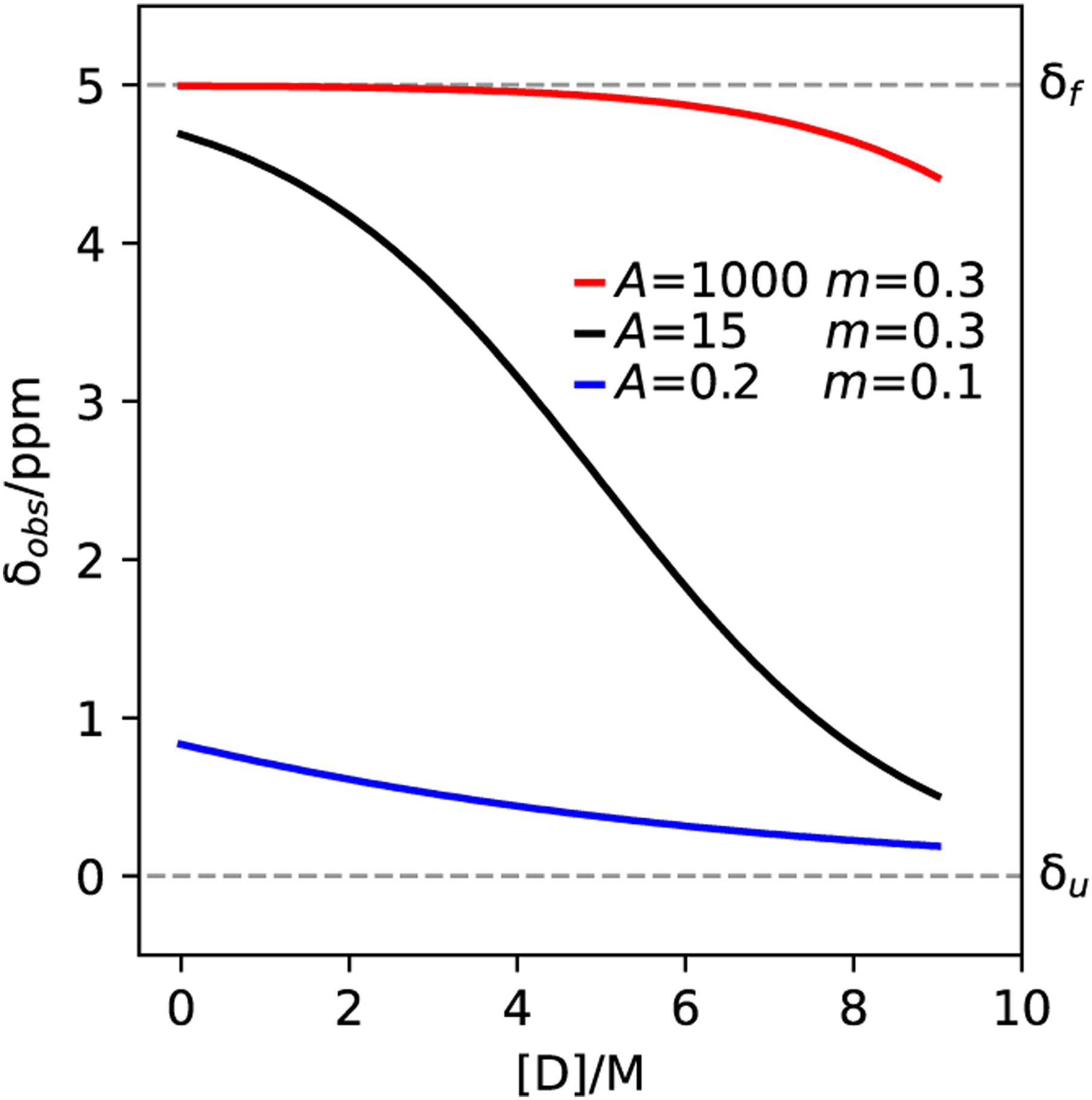
Chemical denaturation is a well-established approach for probing the equilibrium between folded and unfolded states of proteins. We demonstrate applicability of this method to the detection of a small population of a transiently folded structural element in a system that is often considered to be intrinsically fully disordered. The 1HN, 15N, 13Cα, and 13C' chemical shifts of Aβ1-40 and Aβ1-42 peptides and their M35-oxidized variants were monitored as a function of urea concentration and compared to analogous urea titrations of synthetic pentapeptides of homologous sequence. Fitting of the chemical shift titrations yields a 10 ± 1% population for a structured element at the C-terminus of Aβ1-42 that folds with a cooperativity of m = 0.06 kcal/mol·M. The fit also yields the chemical shifts of the folded state and, using a database search, for Aβ1-42 these shifts identified an antiparallel intramolecular β-sheet for residues I32-A42, linked by a type I' β-turn at G37 and G38. The structure is destabilized by oxidation of M35. Paramagnetic relaxation rates and two previously reported weak, medium-range NOE interactions are consistent with this transient β-sheet. Introduction of the requisite A42C mutation and tagging with MTSL resulted in a small stabilization of this β-sheet. Chemical shift analysis suggests a C-terminal β-sheet may be present in Aβ1-40 too, but the turn type at G37 is not type I'. The approach to derive Transient Structure from chemical Denaturation by NMR (TSD-NMR), demonstrated here for Aβ peptides, provides a sensitive tool for identifying the presence of lowly populated, transiently ordered elements in proteins that are considered to be intrinsically disordered, and permits extraction of structural data for such elements.

Keywords: Chemical shift perturbation; Intrinsically disordered protein; Paramagnetic relaxation enhancement; Protein folding; Triple resonance NMR; Urea denaturation.


Figures

Figure 1.
Figure 1.
Three simulated chemical denaturation curves for a typical folded protein (black), a protein that is highly resistant to chemical denaturation (red) and a transiently ordered IDP (blue). For this example, the chemical shifts of the folded and unfolded states are chosen to be 5 and 0 ppm, respectively, and are marked by dashed lines. Plots show the simulated chemical shifts under the condition of rapid exchange as a function of denaturant concentration, [D].
Figure 2.
Figure 2.
Overlay of selected regions of projections of 34 700-MHz 3D HNCO spectra, recorded for four uniformly 15N/13C-enriched Aβ peptides at increasing urea concentrations, marked by decreasing intensity of color. Panels A and B correspond to the projections on the 15N-1HN plane; panels C and D are projections on the 13C’−1HN plane; panel E and F are projections on the 15N-13C’ plane for residues G37 and G38 (with the projection for panel E restricted to the 8.9–8.6 ppm region in the 1H dimension). The black/grey dots mark the corresponding chemical shifts in the reference pentapeptide, with grey corresponding to the highest urea concentration. Urea concentrations (M) used for the titrations were Aβ1–42: 0.00, 0.76, 1.63, 2.67, 4.04, 5.65, 7.15, 8.51; Aβ1–42-Ox: 0.00, 0.87, 1.72, 2.62, 3.57, 4.86, 6.01, 6.98, 7.95; Aβ1–40: 0.00, 0.81, 1.61, 2.70, 4.20, 5.78, 7.36, 8.85; Aβ1–40-Ox: 0.00, 0.76, 1.58, 2.58, 3.45, 4.84, 5.95, 6.91, 7.78. Ref. peptide for G37 (QVGGQ): 0.00, 2.76, 5.38, 7.78; Ref. peptide for G38 (QGGVQ): 0.00, 2.72, 5.22, 7.82.
Figure 3.
Figure 3.
Urea titrations of the backbone chemical shifts of the five Aβ peptides. Curves for other residues are shown in SI Figure S4. Dashed lines correspond to fits to Eq. 6, using global m and A values across residues I32-I41 (I32-V39 for Aβ1–40). Legend colors match those in Figure 2.
Figure 4.
Figure 4.
Contour plot of χ22min, obtained for Aβ1–42 from a 100 × 100 grid search of m and A, followed by best-fitting of the chemical shift parameters. The ‘+’ marker at A = 0.11 and m = 0.06 kcal/mol·M corresponds to the global minimum, χ2 = 5.8 × 10⁻³. Analogous plots for the other four peptides are shown in SI Figure S5.
Figure 5.
Figure 5.
Overlay of nine protein segments with secondary chemical shifts and residue types that best match the folded chemical shift (δf) values and residue types of I32-I41 obtained for Aβ1–42 (cf Eq. 8). These segments resulted from a search over the SPARTA+ database that contains experimental chemical shifts for 580 proteins [50], with the best-matching segments listed in Table 3. Colors match those of the keys in Figure 8.
Figure 6.
Figure 6.
Overlay of the 800-MHz 1H-15N HSQC spectra of 75 μM Aβ1–42-A42C-MTSL in 44 mM Tris-HCl, 10 mM NaCl, 2 mM EDTA, pH 7.4, 278 K, recorded before (black) and after (red) reduction of the MTSL spin label by addition of a 50-fold molar excess of sodium ascorbate, pH 7.4. Both HSQC spectra were processed with 15 Hz exponential line broadening in the 1H dimension. Smaller panels correspond to expanded regions of the full spectrum.
Figure 7.
Figure 7.
Peak intensity ratios in 1H-15N HSQC spectra of the paramagnetic and diamagnetic forms of Aβ1–42-A42C-MTSL. (A) Spectra recorded in the absence of urea (Figure 6). (B) Ratios obtained from spectra recorded under identical conditions except for the addition of 8 M urea. Red bars represent the upper limit for the attenuation ratios, based on the absence of detectable intensity in the paramagnetic state.
Figure 8.
Figure 8.
Plot of the inverse sixth power of the distance between amide protons and the Cβ atom of the residue that matches C42 in Aβ1–42-A42C-MTSL. The intramolecular distances are shown for the ten best matching protein fragments listed in Table 3, nine of which contain an antiparallel β-sheet. For 2E7P, which contains a Gly residue at the position that matched C42, a pseudo-Cβ atom was defined by extending the length of the Cα-Hα3 bond.

References

    1. Meisl G, Yang X, Hellstrand E, Frohm B, Kirkegaard JB, Cohen SIA, Dobson CM, Linse S, and Knowles TPJ, Differences in nucleation behavior underlie the contrasting aggregation kinetics of the A beta 40 and A beta 42 peptides. Proc. Natl. Acad. Sci. U. S. A, 2014. 111(26): p. 9384–9389. - PMC - PubMed
    1. Roher AE, Lowenson JD, Clarke S, Woods AS, Cotter RJ, Gowing E, and Ball MJ, Beta-amyloid-(1–42) is a major component of cerebrovascular amyloid deposits - Implications for pathology of Alzheimer-disease. Proc. Natl. Acad. Sci. U. S. A, 1993. 90(22): p. 10836–10840. - PMC - PubMed
    1. van der Lee R, Buljan M, Lang B, Weatheritt RJ, Daughdrill GW, Dunker AK, Fuxreiter M, Gough J, Gsponer J, Jones DT, Kim PM, Kriwacki RW, Oldfield CJ, Pappu RV, Tompa P, Uversky VN, Wright PE, and Babu MM, Classification of Intrinsically Disordered Regions and Proteins. Chem. Rev, 2014. 114(13): p. 6589–6631. - PMC - PubMed
    1. Honjo K, Black SE, and Verhoeff N, Alzheimer’s Disease, Cerebrovascular Disease, and the beta-amyloid Cascade. Canadian Journal of Neurological Sciences, 2012. 39(6): p. 712–728. - PubMed
    1. Walsh DM and Selkoe DJ, Oligomers in the brain: The emerging role of soluble protein aggregates in neurodegeneration. Protein Pept. Lett, 2004. 11(3): p. 213–228. - PubMed

Publication types

LinkOut - more resources