. 2020 Dec 18;295(51):17398-17410.
doi: 10.1074/jbc.RA120.014243.

Hydrogen/deuterium exchange memory NMR reveals structural epitopes involved in IgE cross-reactivity of allergenic lipid transfer proteins

Affiliations

Hydrogen/deuterium exchange memory NMR reveals structural epitopes involved in IgE cross-reactivity of allergenic lipid transfer proteins

Martina Di Muzio et al. J Biol Chem. .

Abstract

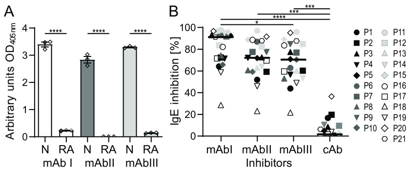
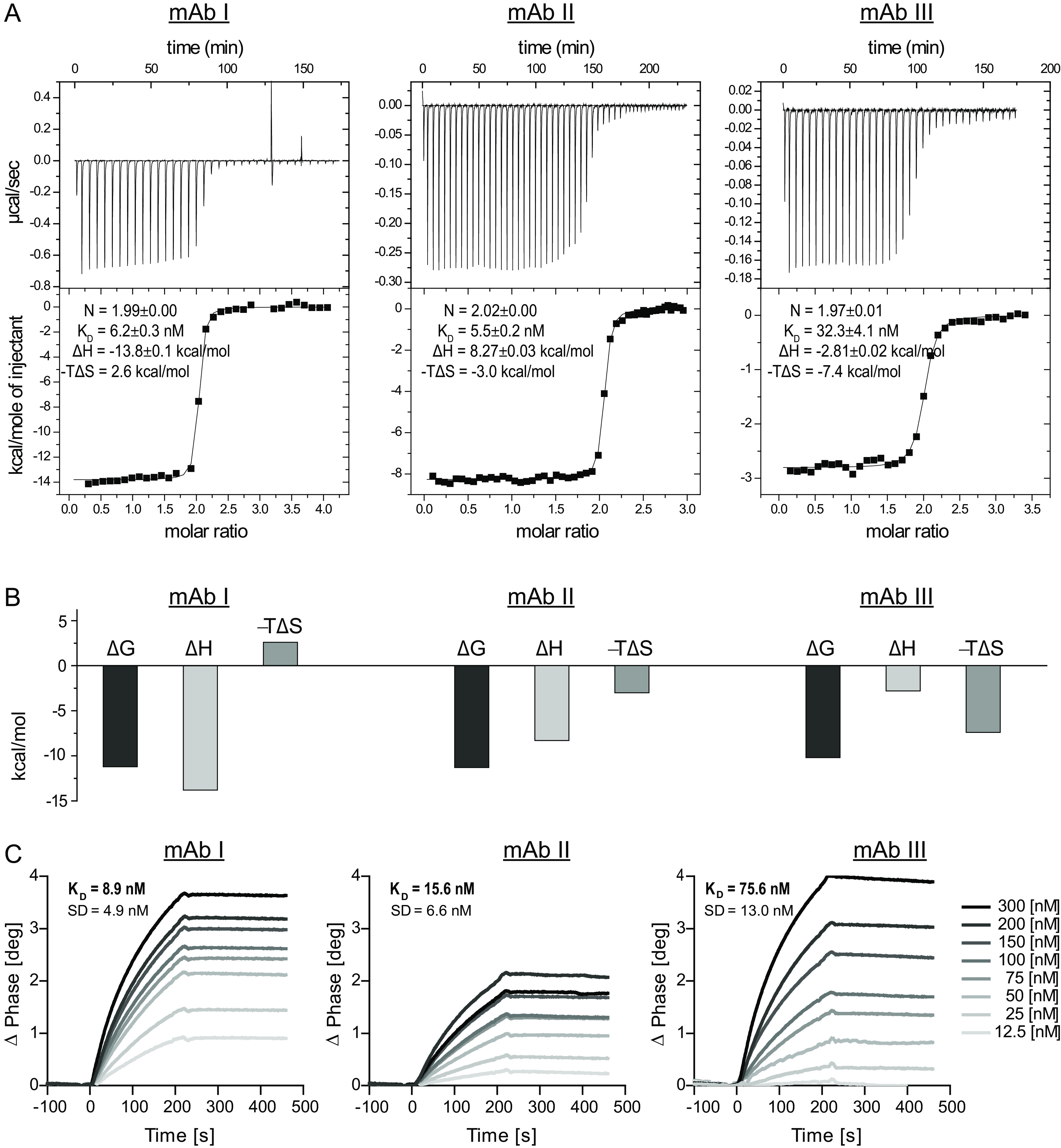
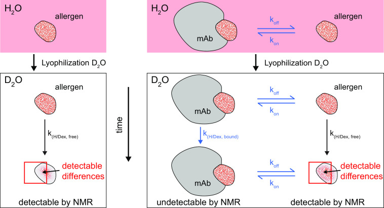
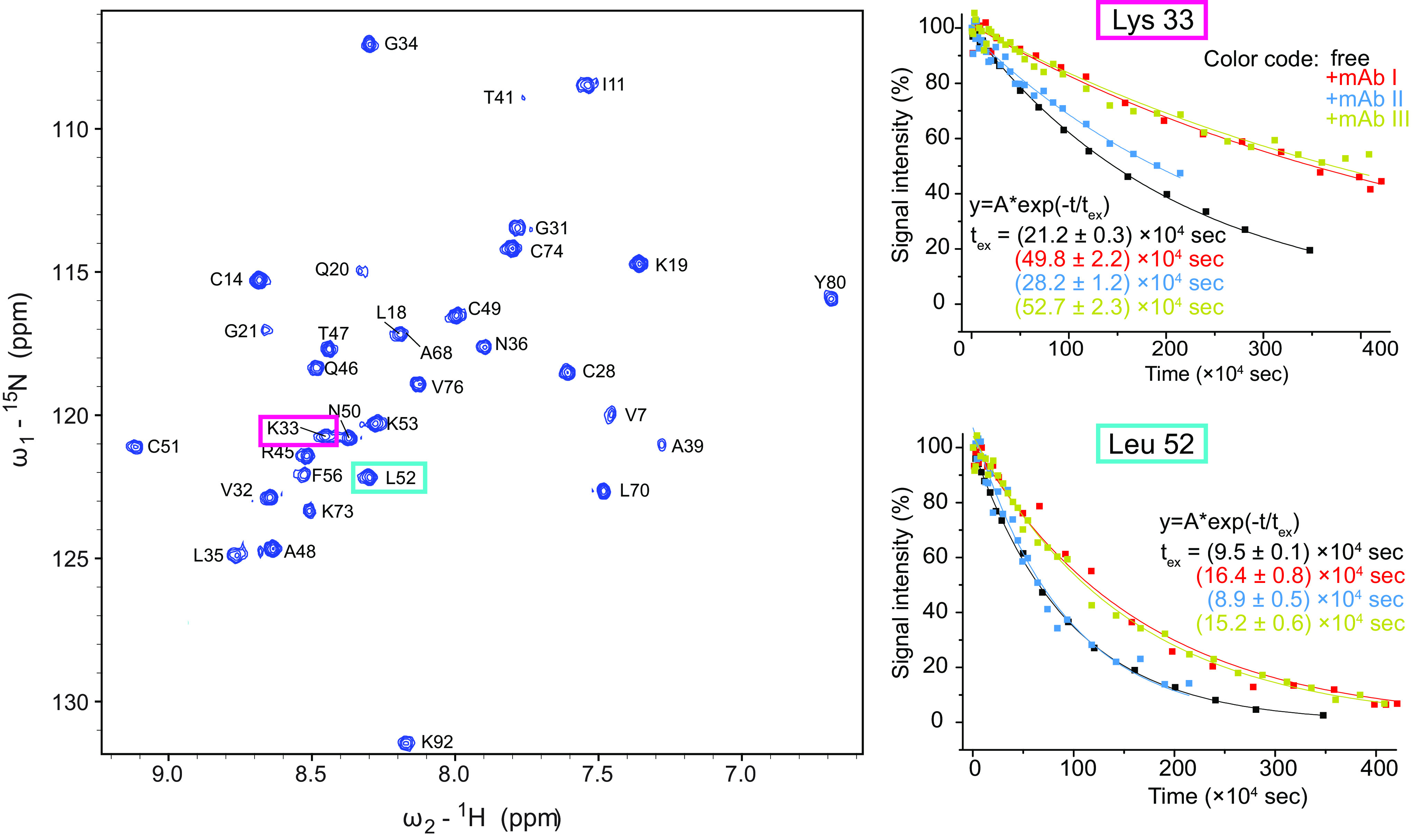
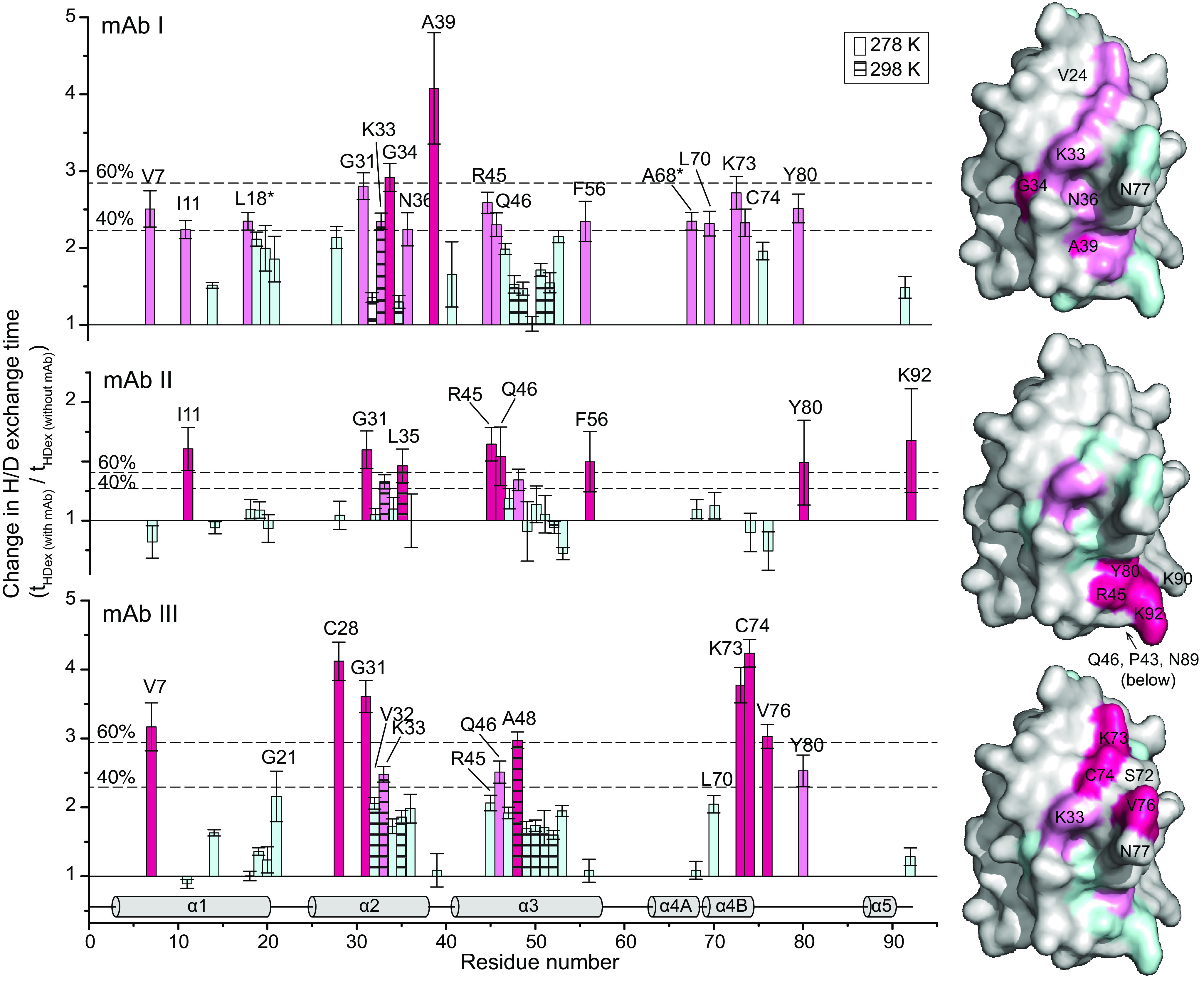
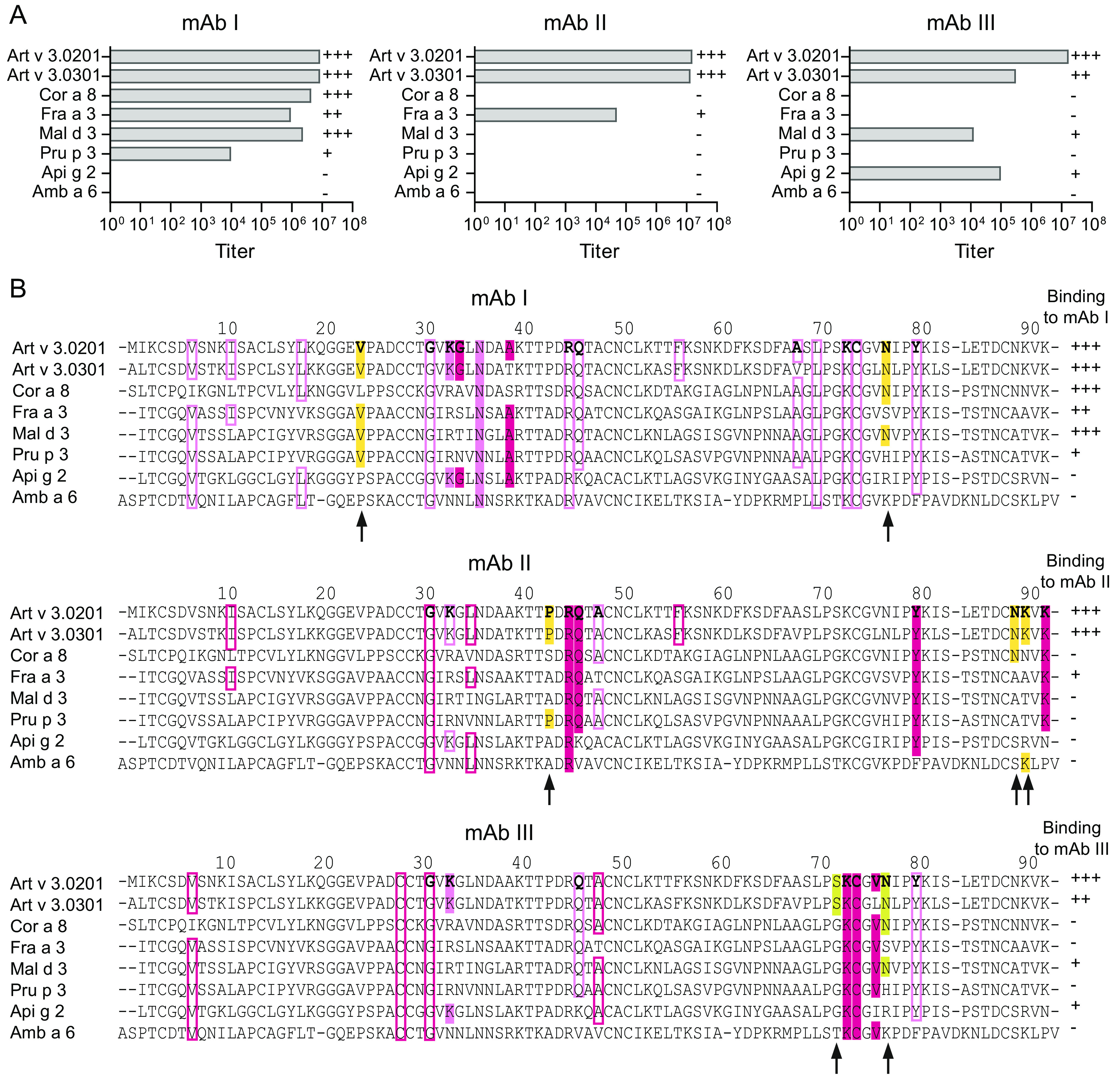
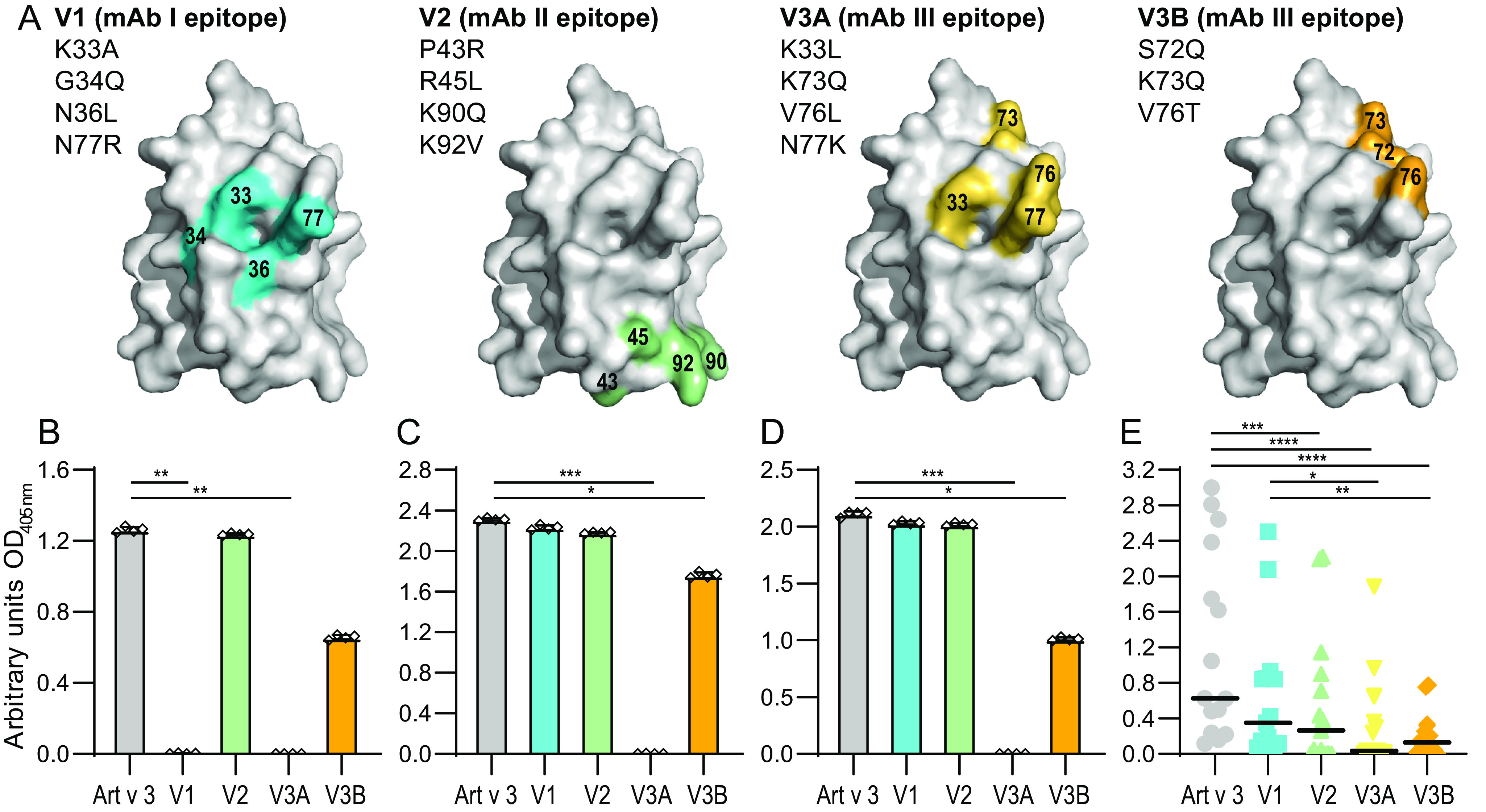
Identification of antibody-binding epitopes is crucial to understand immunological mechanisms. It is of particular interest for allergenic proteins with high cross-reactivity as observed in the lipid transfer protein (LTP) syndrome, which is characterized by severe allergic reactions. Art v 3, a pollen LTP from mugwort, is frequently involved in this cross-reactivity, but no antibody-binding epitopes have been determined so far. To reveal human IgE-binding regions of Art v 3, we produced three murine high-affinity mAbs, which showed 70-90% coverage of the allergenic epitopes from mugwort pollen-allergic patients. As reliable methods to determine structural epitopes with tightly interacting intact antibodies under native conditions are lacking, we developed a straightforward NMR approach termed hydrogen/deuterium exchange memory (HDXMEM). It relies on the slow exchange between the invisible antigen-mAb complex and the free 15N-labeled antigen whose 1H-15N correlations are detected. Due to a memory effect, changes of NH protection during antibody binding are measured. Differences in H/D exchange rates and analyses of mAb reactivity to homologous LTPs revealed three structural epitopes: two partially cross-reactive regions around α-helices 2 and 4 as well as a novel Art v 3-specific epitope at the C terminus. Protein variants with exchanged epitope residues confirmed the antibody-binding sites and revealed strongly reduced IgE reactivity. Using the novel HDXMEM for NMR epitope mapping allowed identification of the first structural epitopes of an allergenic pollen LTP. This knowledge enables improved cross-reactivity prediction for patients suffering from LTP allergy and facilitates design of therapeutics.

Keywords: Art v 3; NMR spectroscopy; allergen; allergen epitope mapping; epitope mapping; hydrogen/deuterium exchange; immunoglobulin E (IgE); immunoglobulin G (IgG); lipid transfer protein; monoclonal antibody; mugwort pollen; nuclear magnetic resonance (NMR).


Conflict of interest statement

Conflict of interest—R. v. R. reports consultancies from HAL Allergy BV, Citeq BV, and Amgany Inc. and speaker's fees from HAL Allergy BV, and Thermo Fisher Scientific. F. F. is a member of the Scientific Advisory Boards of HAL Allergy BV, AllergenOnline, and SIAF. G. G. is member of the Scientific Advisory Board of Bencard.

R. v. R. reports consultancies from HAL Allergy BV, Citeq BV, and Amgany Inc. and speaker's fees from HAL Allergy BV, and Thermo Fisher Scientific. F. F. is a member of the Scientific Advisory Boards of HAL Allergy BV, AllergenOnline, and SIAF. G. G. is member of the Scientific Advisory Board of Bencard

Figures

Figure 1.
Figure 1.
Characterization and IgE inhibition capacities of Art v 3–specific mAbs. A, binding of mAbs I, II, and III to native Art v 3 (N) or reduced and alkylated Art v 3 (RA) was determined by ELISA. Measurements were performed in triplicates; means and S.E. (error bars) are given. B, IgE binding of allergic patients (P1–P21) to immobilized Art v 3 inhibited by the respective monoclonal antibodies was determined by ELISA. An unrelated anti-Amb a 1 antibody was used as control Ab (cAb). Black lines, medians. ****, p ≤ 0.0001; ***, p ≤ 0.001; *, p ≤ 0.05.
Figure 2.
Figure 2.
Isothermal titration calorimetry and surface acoustic wave data of the interaction between Art v 3 and mAb I, mAb II and mAb III. A, raw data and integrated heats as a function of the molar ratio with the best fit. B, illustration of the thermodynamic components ΔG, ΔH, and −TΔS for each of the interactions. C, representative binding curves between the respective mAb and Art v 3 monitored by surface acoustic wave technology. Average KD values and S.D. of 12 channels are given.
Figure 3.
Figure 3.
Schematic presentation of the HDXMEM NMR methodology. The 15N-labeled allergen is dissolved in H2O and contains nearly 100% amide protons schematically indicated by red dots. After lyophilization and dissolving in D2O, the amide protons start slowly exchanging with deuterium (white dots) and become invisible in the 1H-detected NMR spectra. For each observable proton, the exchange rates are measured. A similar experiment is done in the presence of a mAb at a ratio at which the allergen is present as a mixture of free and bound states (1:1). Only the amide signals of the free allergen are detectable, but because there is an equilibrium between free and bound allergen, a mixture of H/D exchange rates is observed containing contributions of the free and bound state. Regions that are bound to the mAb are expected to be protected from H/D exchange and therefore show a lower exchange rate compared with the free allergen.
Figure 4.
Figure 4.
HDXMEM data of Art v 3 in the presence and absence of mAbs. Left, 15N-1H HSQC of Art v 3 in the absence of a mAb 10 min after dissolving the sample in D2O. The signals of the two amino acids Lys-33 and Leu-52 are highlighted. Right, the intensities of the two example residues (Lys-33 and Leu-52) are plotted over time in the absence and presence of mAb I, II, and III measured at 298 K with an exponential fit and the extracted exchange times as fitting parameter.
Figure 5.
Figure 5.
Effects of mAb binding on the H/D exchange times with residue resolution. Shown are the factors at which the H/D exchange times for each Art v 3 residue changed in the presence of 0.25 equivalents mAb compared with free Art v 3. Hatched bars, data recorded at 298 K; all other bars, data obtained at 278 K. Thresholds at 40 and 60% of the largest change are indicated as dotted lines. The secondary structure is shown at the bottom, and the surface presentations on the right are based on the crystal structure (PDB entry 6FRR, chain A). Changes in the H/D exchange rates are color-coded in hot pink (>60%), moderate changes in pink (>40%), and changes below the lowest threshold in pale cyan. Residues without a detectable NH group are depicted in gray. Error bars, S.D.
Figure 6.
Figure 6.
Recognition pattern of mAbs to Art v 3 and homologous LTPs. A, titers of mAb I, mAb II, and mAb III for Art v 3 and six other LTPs were measured by ELISA. Binding strengths of mAbs are depicted as follows: titers >106 (+++), titers between 105 and 106 (++), titers between 104 and 105 (+), and titers <104 (−). B, sequence alignment of Art v 3 and homologs to refine binding patterns of individual mAbs. Color-coded hotspots observed during the interaction with mAbs are used in accordance with Fig. 4 (filled boxes, amino acids involved in the epitope; rimmed boxes, amino acids that have been identified by HDXMEM NMR and are thus influenced by binding but not part of the epitope). Yellow boxes, amino acids invisible by HDXMEM NMR that potentially contribute to the epitope. Surface-exposed residues involved in mAb binding of Art v 3.0201 are shown in boldface type. Binding strengths of different homologs to the mAbs are indicated on the right. Residues that correlate with the mAb-binding pattern are highlighted with an arrow.
Figure 7.
Figure 7.
Art v 3 epitope variants and recognition by mAbs and patients' serum IgE. A, surface representation of residues exchanged for epitope variants V1, V2, V3A, and V3B based on the crystal structure (PDB entry 6FRR, chain A). Shown is ELISA antibody reactivity to Art v 3 and epitope variants using mAb I (B), mAb II (C), and mAb III (D). Bars, mean of four technical replicates; whiskers, S.D. E, IgE reactivity of allergic patients' sera (n = 15) to Art v 3 and epitope variants. Black lines, medians. ****, p ≤ 0.0001; ***, p ≤ 0.001; **, p ≤ 0.01; *, p ≤ 0.05.

References

    1. Abbott W. M., Damschroder M. M., and Lowe D. C. (2014) Current approaches to fine mapping of antigen-antibody interactions. Immunology 142, 526–535 10.1111/imm.12284 - DOI - PMC - PubMed
    1. Mirza O., Henriksen A., Ipsen H., Larsen J. N., Wissenbach M., Spangfort M. D., and Gajhede M. (2000) Dominant epitopes and allergic cross-reactivity: complex formation between a Fab fragment of a monoclonal murine IgG antibody and the major allergen from birch pollen Bet v 1. J. Immunol. 165, 331–338 10.4049/jimmunol.165.1.331 - DOI - PubMed
    1. Padavattan S., Flicker S., Schirmer T., Madritsch C., Randow S., Reese G., Vieths S., Lupinek C., Ebner C., Valenta R., and Markovic-Housley Z. (2009) High-affinity IgE recognition of a conformational epitope of the major respiratory allergen Phl p 2 as revealed by X-ray crystallography. J. Immunol. 182, 2141–2151 10.4049/jimmunol.0803018 - DOI - PubMed
    1. Willison L. N., Zhang Q., Su M., Teuber S. S., Sathe S. K., and Roux K. H. (2013) Conformational epitope mapping of Pru du 6, a major allergen from almond nut. Mol. Immunol. 55, 253–263 10.1016/j.molimm.2013.02.004 - DOI - PubMed
    1. Zhang Q., Yang J., Bautista J., Badithe A., Olson W., and Liu Y. (2018) Epitope mapping by HDX-MS elucidates the surface coverage of antigens associated with high blocking efficiency of antibodies to birch pollen allergen. Anal. Chem. 90, 11315–11323 10.1021/acs.analchem.8b01864 - DOI - PubMed

Publication types