. 2020 Dec 18;295(51):17413-17424.
doi: 10.1074/jbc.RA120.015595.

N-acetylglucosamine drives myelination by triggering oligodendrocyte precursor cell differentiation

Affiliations

N-acetylglucosamine drives myelination by triggering oligodendrocyte precursor cell differentiation

Michael Sy et al. J Biol Chem. .

Abstract

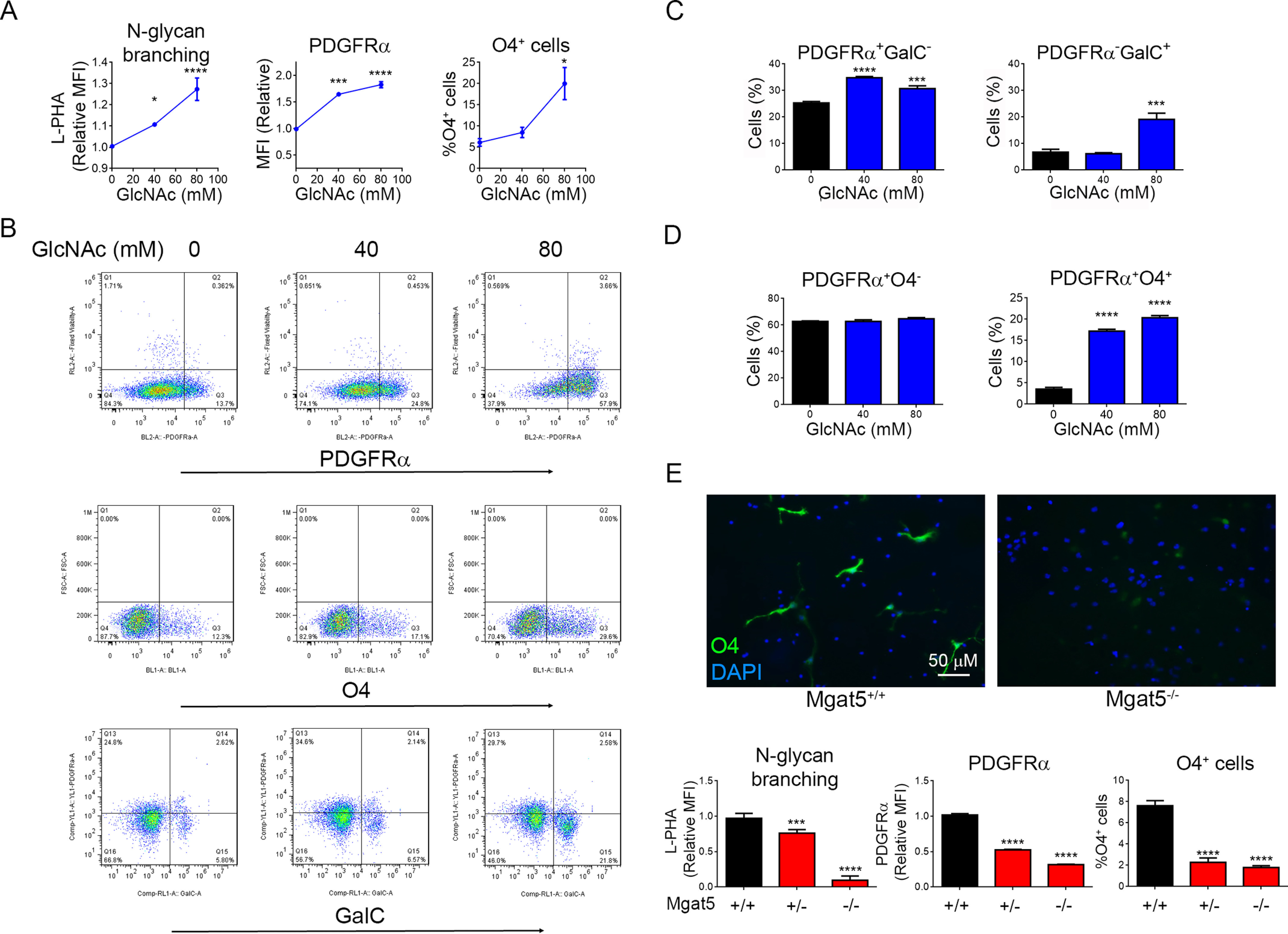
Myelination plays an important role in cognitive development and in demyelinating diseases like multiple sclerosis (MS), where failure of remyelination promotes permanent neuro-axonal damage. Modification of cell surface receptors with branched N-glycans coordinates cell growth and differentiation by controlling glycoprotein clustering, signaling, and endocytosis. GlcNAc is a rate-limiting metabolite for N-glycan branching. Here we report that GlcNAc and N-glycan branching trigger oligodendrogenesis from precursor cells by inhibiting platelet-derived growth factor receptor-α cell endocytosis. Supplying oral GlcNAc to lactating mice drives primary myelination in newborn pups via secretion in breast milk, whereas genetically blocking N-glycan branching markedly inhibits primary myelination. In adult mice with toxin (cuprizone)-induced demyelination, oral GlcNAc prevents neuro-axonal damage by driving myelin repair. In MS patients, endogenous serum GlcNAc levels inversely correlated with imaging measures of demyelination and microstructural damage. Our data identify N-glycan branching and GlcNAc as critical regulators of primary myelination and myelin repair and suggest that oral GlcNAc may be neuroprotective in demyelinating diseases like MS.

Keywords: N-acetylglucosamine; N-glycan branching; N-linked glycosylation; metabolism; multiple sclerosis; myelin; myelin repair; myelination; oligodendrocyte; oligodendrocyte precursor cell; oligodendrocytes.


Conflict of interest statement

Conflict of interest—A. U. B., F. P., J. D., and M. D. are named as inventors on a patent that describes GlcNAc as a biomarker for multiple sclerosis. J. D. and M. D. are named as inventors on a patent for use of GlcNAc in multiple sclerosis and were cofounders of Glixis Therapeutics, a company developing analogs of GlcNAc for multiple sclerosis and other autoimmune diseases.

A. U. B., F. P., J. D., and M. D. are named as inventors on a patent that describes GlcNAc as a biomarker for multiple sclerosis. J. D. and M. D. are named as inventors on a patent for use of GlcNAc in multiple sclerosis and were cofounders of Glixis Therapeutics, a company developing analogs of GlcNAc for multiple sclerosis and other autoimmune diseases

Figures

Figure 1.
Figure 1.
GlcNAc and N-glycan branching promotes oligodendrogenesis. AD, flow cytometry of E12.5 NSCs from CD1 (B and C) or C57BL/6 (D) mice cultured in growth media (FGF + EGF) ± GlcNAc for 48 h. Cell surface binding levels of L-PHA and PDGFRα are measured as mean fluorescence intensity (MFI). E, flow cytometry and immunofluorescence microscopy of E12.5 NSCs in differentiation media (FGF + PDGF-AA) from Mgat5+/+, Mgat5+/−, and Mgat5−/− C57BL/6 mice. Data are three technical replicates per group (AE), representative of 3 (AC) or two (D and E) experiments. p-values are by one-way ANOVA with Sidak's multiple comparison test. All error bars are standard error. *p < 0.05, **p < 0.01, ***p < 0.001, ****p < 0.0001.
Figure 2.
Figure 2.
GlcNAc and N-glycan branching promote primary myelination. AC, newborn PL/J mice were given exogenous GlcNAc or [U13C]GlcNAc by providing their nursing mothers with GlcNAc or [U13C]GlcNAc at 1 mg/ml in drinking water from P3–P8. The pups and mothers were sacrificed at P8 and brains were analyzed by LC–MS/MS for UDP-[U13C]HexNAc (A, n = 3, 3 adult, n = 4,4 P8 pups), flow cytometry (B, n = 3, 3) or immunofluorescence microscopy (C, n = 5,5) for the indicated markers. L-PHA staining in (B) is gated on PDGFRα+ cells. Data in (C) is the average of fluorescence intensity of the area depicted in red of three brain slices per mouse. One-sided t test. D and E, the indicated adult mice (10 weeks old) were treated with tamoxifen at weeks 0 and 4 and sacrificed at week 8, and brains were analyzed by immunofluorescence microscopy for MBP, myelin (FluoroMyelin), Olig2+, and CC1+ cells (n = 5 (2 male, 3 female), 8 (6 male, 2 female) (D), and n = 5 (2 male, 3 female), 4 (2 male, 2 female) (E); one-sided t test). Each data point in the graphs represents average fluorescence or cell counts of the highlighted area from three (D) or two (E) different brain slices per mouse.
Figure 3.
Figure 3.
Oral GlcNAc promotes remyelination and limits axonal injury. A, the ability of GlcNAc to promote myelin repair in vivo was assessed using the cuprizone model, with oral GlcNAc treatment (1 mg/ml in drinking water) during the last 3 weeks of a 6-week cuprizone exposure (I, active phase treatment) or after 5 weeks of cuprizone treatment for 1, 2, or 4 weeks (II, III, and IV, recovery phase treatment). WT (I and III) or Mgat5 heterozygous (II and IV) C57BL/6 mice were used. B, area of the medial corpus callosum (CC) analyzed. CF, shown is the latency to fall in an accelerated rotarod test and immunofluorescence staining of the corpus callosum for MBP, degraded MBP (dMBP), APP, myelin (FluoroMyelin), and/or CC1/Olig2 from WT mice with active phase GlcNAc treatment (C, n = 5,5, all male), Mgat5 heterozygous mice with recovery phase GlcNAc treatment for 1 week (D, n = 8 (5 male, 3 female), 7 (4 male, 3 female)), WT mice with recovery phase GlcNAc treatment for 2 weeks (E, n = 11,14 for rotarod, n = 5,7 for immunofluorescence, all male), or Mgat5 heterozygous mice with active phase GlcNAc for 4 weeks (F, n = 6,6 with 4 male and 2 female per group). Data points represents average fluorescence from 3–4 different brain slices per mouse. Rotarod p-values by 2-way ANOVA with Sidak's multiple comparisons post-test. Immunofluorescence p-values by one-tailed t test. Scale bar = 50 µm. G, the CC of mice from the 4-week recovery phase treatment group in panel (F) were analyzed by EM (n = 3,3 with 2 male and 1 female per group). Representative electron micrographs in control and GlcNAc treatment groups are shown, scale bar = 1 µm. Filled and empty arrowheads indicate examples of myelinated and unmyelinated dystrophic axons, respectively. Plot of g-ratio versus axon diameter (n = 214 and 222 axons) was counted blindly from two fields (105µm2) per mouse (p-value comparing best fit curves from nonlinear regression, R2 is the goodness of fit for each group). Numbers of total axons, myelinated axons, and dystrophic axons (axon diameter > 0.7 µm) were counted blindly in six fields (105µm2) per mouse in each treatment group (n = 18, 18, p-value by one-sided t test). All error bars are standard error.
Figure 4.
Figure 4.
Serum HexNAc correlates with markers of myelin-axon microstructural damage in MS patients. AC, association of serum HexNAc levels with MRI measures of myelin-axon microstructural damage in a cohort of n = 180 MS patients. T2w lesion volume (A) and T1w/T2w ratio in normal-appearing white matter (NAWM, B) and gray matter (GM, C) is shown. Coefficient B, standard error (S.E.), and R2 are from linear regression models correcting for age and sex (in B and C). Black lines in regression models represent coefficients from noncorrected models, gray areas show the 95% confidence interval. Value in nM are serum HexNAc levels.

Similar articles

Cited by

References

    1. Reich D. S., Lucchinetti C. F., and Calabresi P. A. (2018) Multiple sclerosis. N. Engl. J. Med. 378, 169–180 10.1056/NEJMra1401483 - DOI - PMC - PubMed
    1. Fields R. D. (2008) White matter in learning, cognition and psychiatric disorders. Trends Neurosci. 31, 361–370 10.1016/j.tins.2008.04.001 - DOI - PMC - PubMed
    1. Lee Y., Morrison B. M., Li Y., Lengacher S., Farah M. H., Hoffman P. N., Liu Y., Tsingalia A., Jin L., Zhang P. W., Pellerin L., Magistretti P. J., and Rothstein J. D. (2012) Oligodendroglia metabolically support axons and contribute to neurodegeneration. Nature 487, 443–448 10.1038/nature11314 - DOI - PMC - PubMed
    1. Fünfschilling U., Supplie L. M., Mahad D., Boretius S., Saab A. S., Edgar J., Brinkmann B. G., Kassmann C. M., Tzvetanova I. D., Möbius W., Diaz F., Meijer D., Suter U., Hamprecht B., Sereda M. W., et al. (2012) Glycolytic oligodendrocytes maintain myelin and long-term axonal integrity. Nature 485, 517–521 10.1038/nature11007 - DOI - PMC - PubMed
    1. Mei F., Lehmann-Horn K., Shen Y.-A. A., Rankin K. A., Stebbins K. J., Lorrain D. S., Pekarek K., A Sagan S., Xiao L., Teuscher C., von Büdingen H.-C., Wess J., Lawrence J. J., Green A. J., Fancy S. P., et al. (2016) Accelerated remyelination during inflammatory demyelination prevents axonal loss and improves functional recovery. eLife 5, 10.7554/eLife.18246 - DOI - PMC - PubMed

Publication types

MeSH terms

Substances