. 2020 Dec 18;295(51):17514-17534.
doi: 10.1074/jbc.RA120.014924.

Evolving the naturally compromised chorismate mutase from Mycobacterium tuberculosis to top performance

Affiliations

Evolving the naturally compromised chorismate mutase from Mycobacterium tuberculosis to top performance

Jūrate Fahrig-Kamarauskait et al. J Biol Chem. .

Abstract

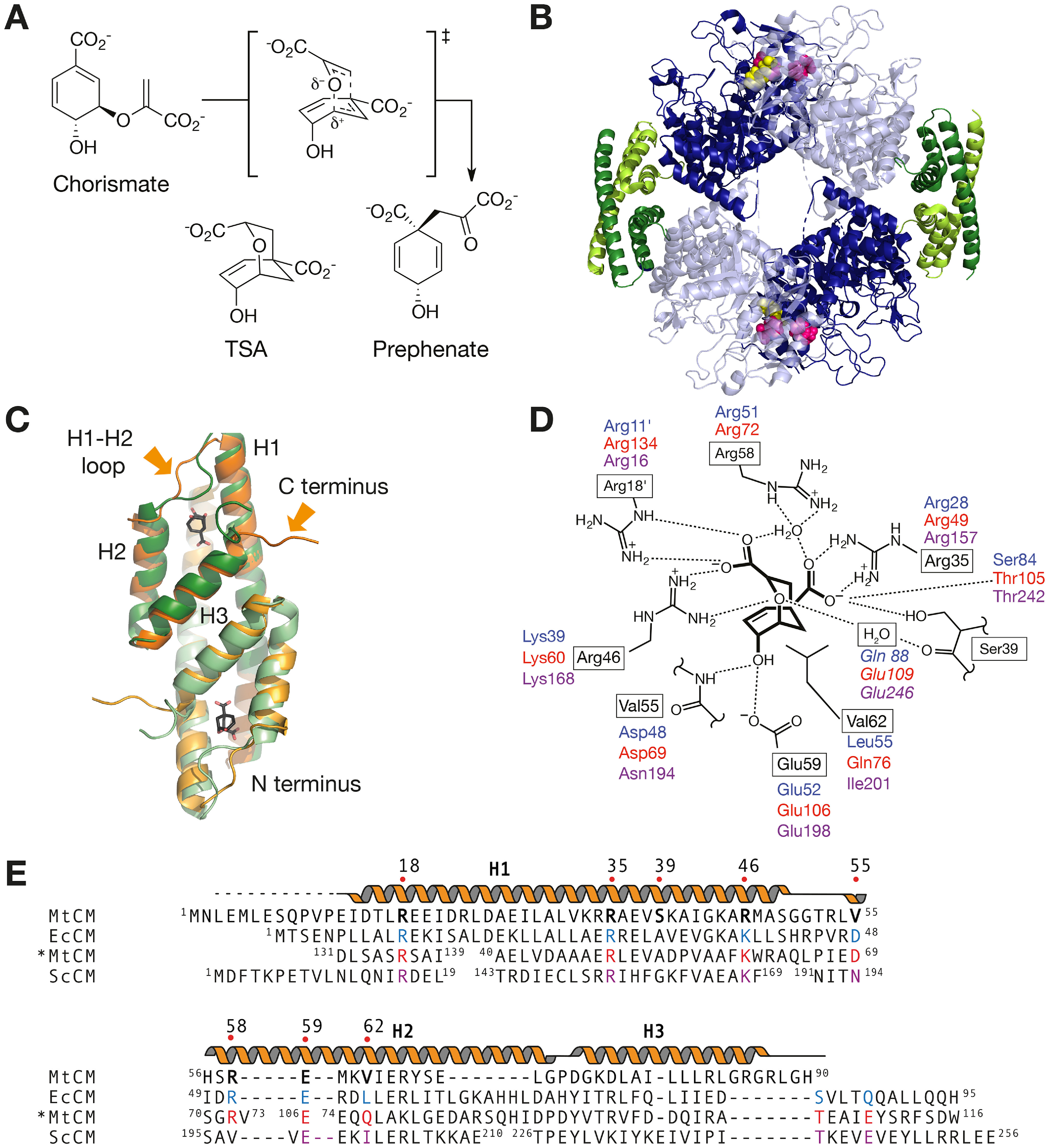
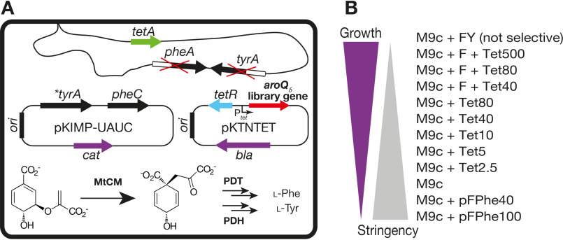
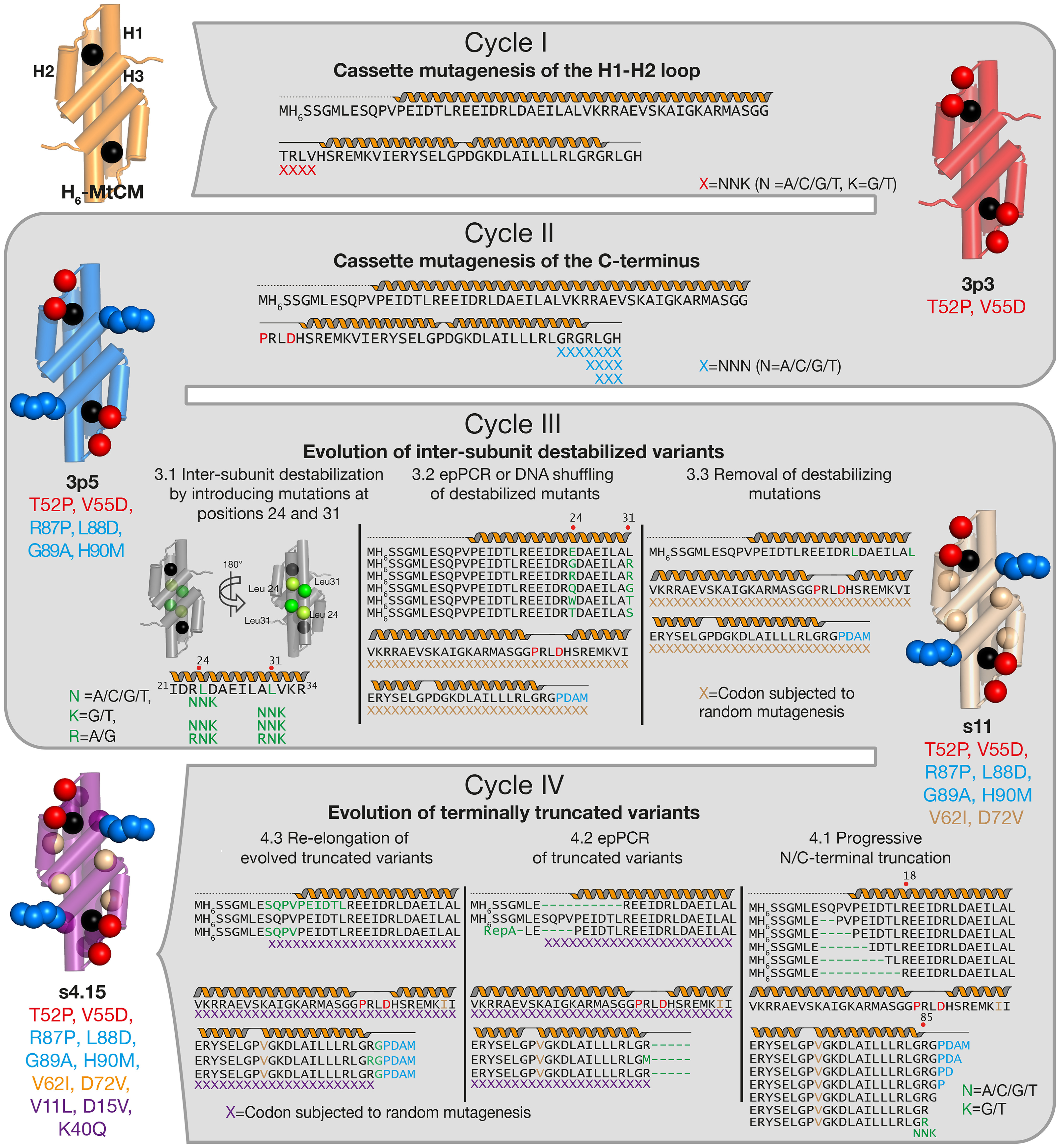
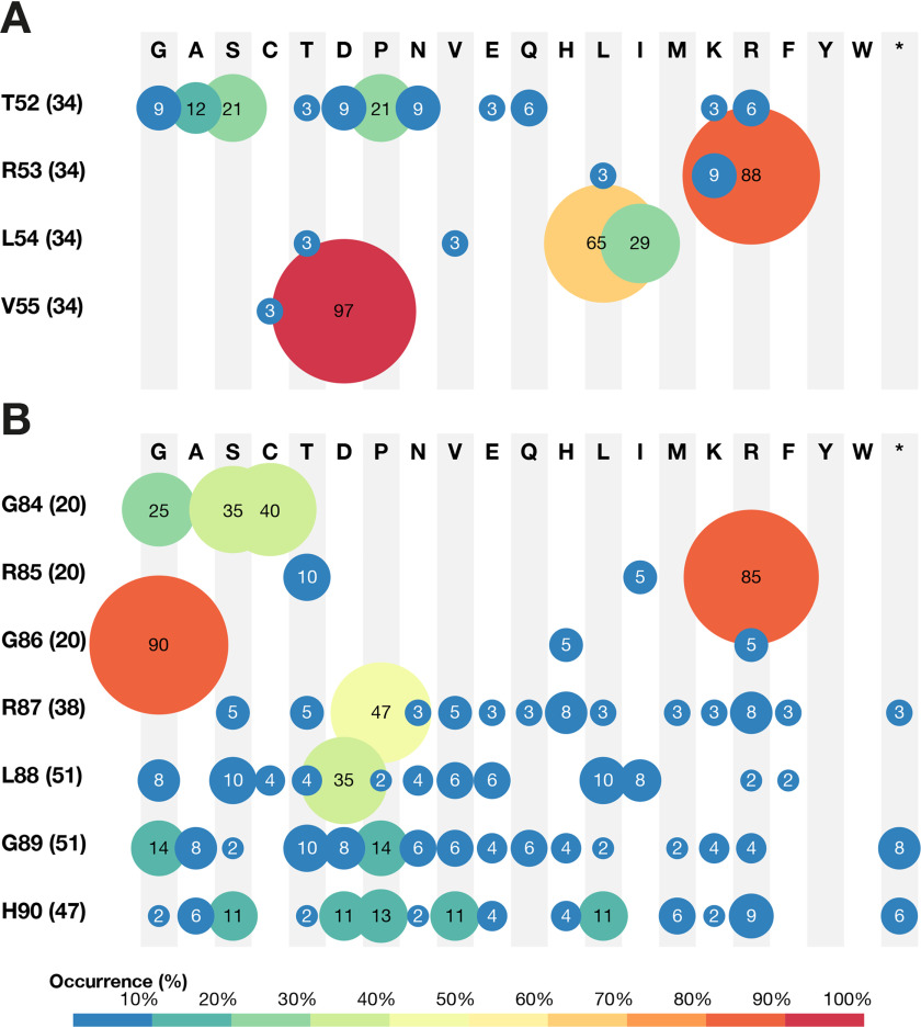
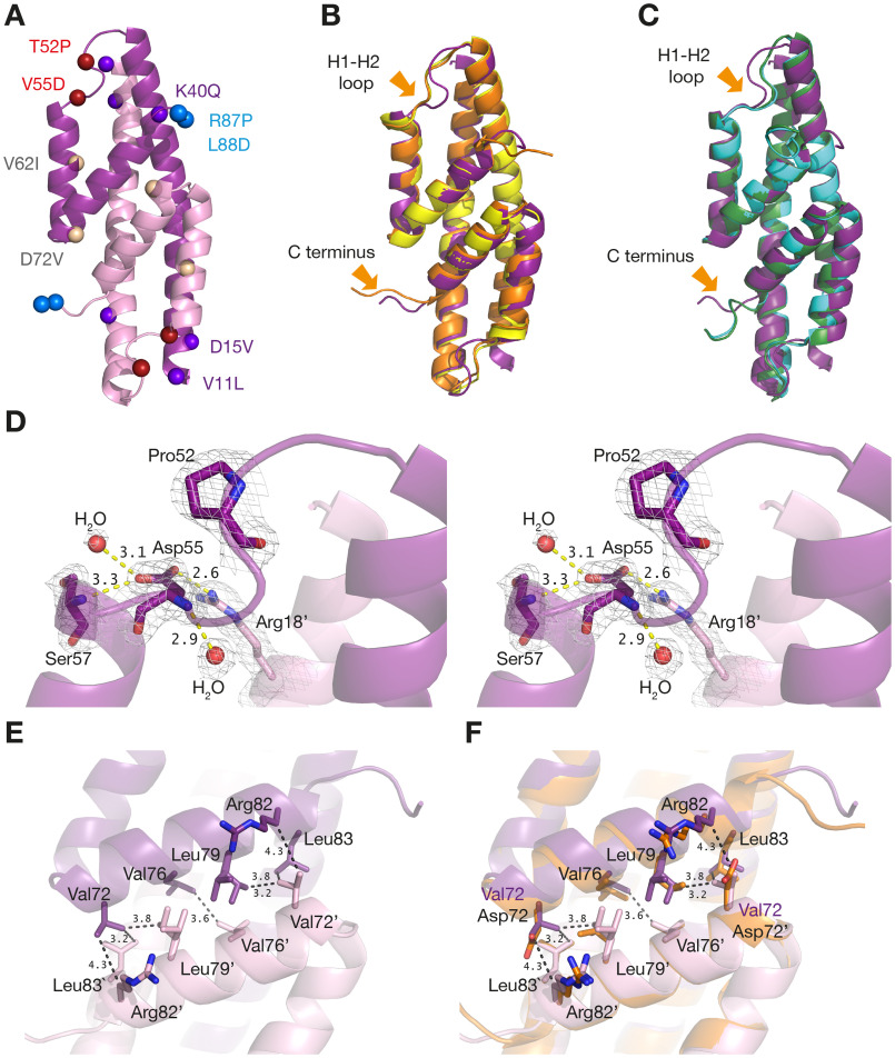
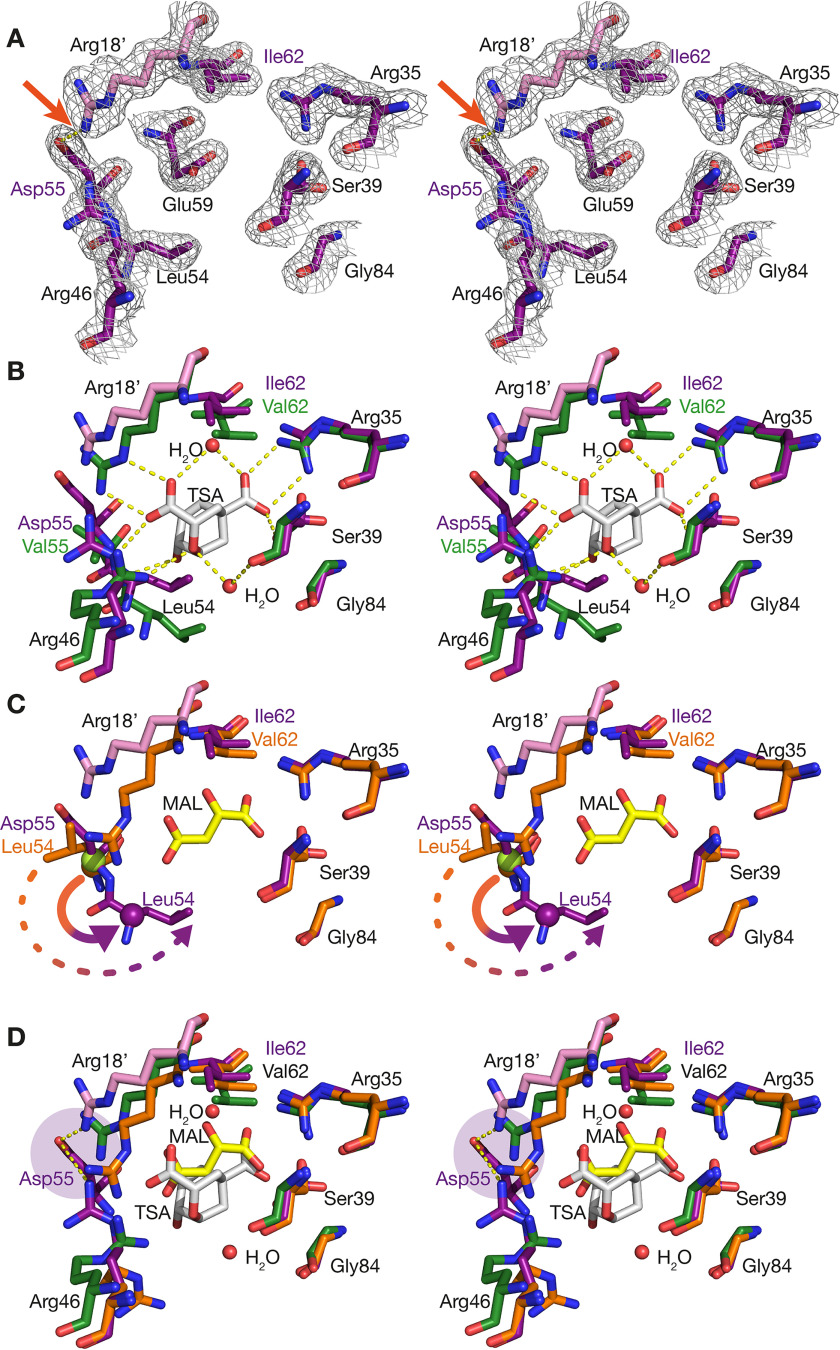
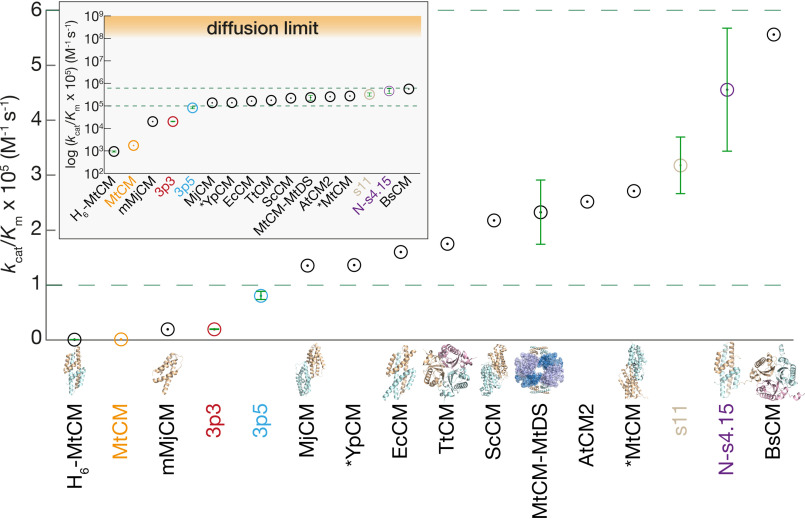
Chorismate mutase (CM), an essential enzyme at the branch-point of the shikimate pathway, is required for the biosynthesis of phenylalanine and tyrosine in bacteria, archaea, plants, and fungi. MtCM, the CM from Mycobacterium tuberculosis, has less than 1% of the catalytic efficiency of a typical natural CM and requires complex formation with 3-deoxy-d-arabino-heptulosonate 7-phosphate synthase for high activity. To explore the full potential of MtCM for catalyzing its native reaction, we applied diverse iterative cycles of mutagenesis and selection, thereby raising kcat/Km 270-fold to 5 × 10⁵m-1s-1, which is even higher than for the complex. Moreover, the evolutionarily optimized autonomous MtCM, which had 11 of its 90 amino acids exchanged, was stabilized compared with its progenitor, as indicated by a 9 °C increase in melting temperature. The 1.5 Å crystal structure of the top-evolved MtCM variant reveals the molecular underpinnings of this activity boost. Some acquired residues (e.g. Pro52 and Asp55) are conserved in naturally efficient CMs, but most of them lie beyond the active site. Our evolutionary trajectories reached a plateau at the level of the best natural enzymes, suggesting that we have exhausted the potential of MtCM. Taken together, these findings show that the scaffold of MtCM, which naturally evolved for mediocrity to enable inter-enzyme allosteric regulation of the shikimate pathway, is inherently capable of high activity.

Keywords: X-ray crystallography; catalytic efficiency; conformational change; crystal structure; directed evolution; enzyme catalysis; enzyme mutation; molecular evolution; protein structure; structure-activity relationship.


Conflict of interest statement

Conflict of interest—The authors declare that they have no conflicts of interest with the contents of this article.

The authors declare that they have no conflicts of interest with the contents of this article

Figures

Figure 1.
Figure 1.
Biosynthetic reaction, structure, active site, and sequences of AroQ chorismate mutases. A, CM accelerates the Claisen rearrangement of chorismate to prephenate. The reaction proceeds via a transition state with chair-like geometry. Also shown is Bartlett's endo-oxa-bicyclic transition state analog (TSA, (74)), which is a good inhibitor of CMs. B, crystal structure of the MtCM-MtDS complex with the bound feedback inhibitors Phe (pink spheres) and Tyr (yellow spheres) (PDB ID: 5CKX). Two dimers of MtCM (subunits shown in two shades of green) bind to an MtDS homotetramer (shown in two shades of blue). C, structural superimposition of the poorly active malate-bound MtCM (two shades of orange, malate not shown; PDB ID: 2VKL) and MtDS-bound activated MtCM (two shades of green, TSA in black; PDB ID: 2W1A). Helices H1, H2, and H3 of one subunit are labeled; regions showing different conformations (H1-H2 loop and C terminus) are indicated by arrows. D, scheme of active site residues (boxed, black) of MtCM bound to TSA in its naturally activated state in complex with MtDS (PDB ID: 2W1A). Analogous prominent catalytic site residues in EcCM (blue; labels in first line), *MtCM (red; second line), and ScCM (magenta; third line) are shown together with their polar interactions (dotted lines). Italicized residues have no correspondence in MtCM. Residues projecting from a different protomer are labeled with a prime (′). E, sequence alignment of MtCM with the CM subclass prototypes AroQα (EcCM), AroQβ (ScCM), and AroQγ (*MtCM) with an emphasis on matching analogous active site residues as in (D). Helical, straight, and dotted lines above the MtCM sequence indicate α-helices, loops, and unresolved parts of the MtCM structure in the MtCM-MtDS complex (PDB ID: 2W1A), respectively.
Figure 2.
Figure 2.
In vivo selection system for directed evolution. A, CM selection system based on E. coli KA12/pKIMP-UAUC. The strain KA12 has a deletion of the CM-encoding bifunctional pheA and tyrA genes. This defect is only partially complemented by monofunctional versions of prephenate dehydratase (PDT, encoded by pheC) and prephenate dehydrogenase (PDH, encoded by *tyrA) on plasmid pKIMP-UAUC with the replication origin orip15A. Consequently, survival on minimal medium lacking Phe (F) and Tyr (Y) requires introduction of a functional CM library gene (aroQδ) on the compatible pKTNTET-based plasmid (oripUC). The genes bla, cat, pheA, tyrA, tetA, and tetR encode β-lactamase, chloramphenicol acetyltransferase, CM-PDT, CM-PDH, a tetracycline efflux pump, and the repressor of the tetA promoter (Ptet), respectively. B, selection stringencies used for MtCM evolution. The minimal medium M9c was provided with Phe, Tyr, tetracycline (Tet, aroQδ inducer; concentration in ng/ml), and dl-para-fluoro-phenylalanine (pFPhe; concentration in μm) as indicated above. pFPhe causes cell death if incorporated to a significant extent into cellular proteins instead of Phe (75). This is because of the inability of the cell's phenylalanyl-tRNA synthetase to distinguish between the natural amino acid and its analog pFPhe, resulting in accumulation of faulty proteins (76, 77). By adding pFPhe to the medium, we exploit and adapt this observation for a new selection strategy favoring highly efficient CM variants, which provide sufficient endogenously produced Phe to outcompete the toxic pFPhe.
Figure 3.
Figure 3.
Directed evolution strategy. Two cycles of cassette mutagenesis (cycle I, H1-H2 loop; cycle II, C terminus) were followed by two cycles of perturbation-compensation (green letters) involving epPCR and DNA shuffling (cycle III, inter-subunit destabilization; cycle IV, progressive terminal truncations). The four evolutionary cycles and mutated residues are shown in different colors (I, red; II, light blue; III, orange; IV, purple). Representative variant names are listed. The mutated positions are indicated as spheres mapped onto CM cartoons; the black sphere pinpoints the location of the active site.
Figure 4.
Figure 4.
Selected MtCM sequences from evolutionary cycles I and II. A, results from cycle I of directed evolution. Shown is the amino acid distribution at the randomized positions as derived from sequencing of 34 TRLV library members growing on selective M9c plates. B, cycle II sequencing data. Included are 52 variants selected on M9c+pFPhe from libraries CT7, CT-LGH, and CT-RLGH. Circle size and color correlate with the frequency of individual encoded residues with the color code shown below the panels. Amino acids are listed with their one-letter abbreviation. The percentage of a particular residue at each randomized position is given within the circle, and the absolute number of codons considered in the compilations is indicated in parentheses next to the WT residue listed on the left for each sampled position.
Figure 5.
Figure 5.
Structure of top-evolved variant N-s4.15 and comparison with native MtCM. A, overall structure of N-s4.15. The two subunits are colored in different shades of purple. Colored spheres locate the mutations identified during the course of directed evolution. B, superimposition of N-s4.15 (purple) with malate-bound (orange; PDB ID: 2VKL) and apo (yellow; PDB ID: 2QBV) MtCM WT structures. C, superimposition of N-s4.15 (purple) with MtDS-bound activated MtCM with (green; PDB ID: 2W1A) or without (cyan; PDB ID: 2W19) TSA (ligand not shown). The largest structural differences are pointed out with arrows. Note that there are crystal contacts in both of these regions (see Fig. S5). D, close-up stereo view of the H1-H2 loop of N-s4.15. The electron density is contoured at 1σ. Pro52 induces a kink in the H1-H2 loop and Asp55 N-caps helix H2. Polar interactions of Asp55 (distance in Å) are indicated with yellow dashed lines. E, position of Val72 at the N-s4.15 dimer interface. Prominent van der Waals interactions are indicated by black dashed lines, and relevant residues from the other protomer are denoted by a prime (′). F, N-s4.15 dimer interface as in E superimposed with malate-bound (unactivated) MtCM (orange, with the WT Asp72; PDB ID: 2VKL). The structure of N-s4.15 is represented by PDB ID: 5MPV (this work) in all panels.
Figure 6.
Figure 6.
Stereo images of the active site of MtCM. The structure of the top-evolved variant N-s4.15 (PDB ID: 5MPV, this work) is shown in purple/pink. Some residues (e.g. Glu59) were omitted for clarity in individual panels. A, N-s4.15 active site with corresponding 2mFo-DFc electron density (gray mesh), contoured at 1σ. The hydrogen bond between residues Asp55 and Arg18 (prime) of the other subunit is highlighted by an arrow. B, superimposition of N-s4.15 and MtDS-activated WT MtCM (green; PDB ID: 2W1A) in complex with TSA (white sticks), including relevant water molecules and H-bonds. C, superimposition of N-s4.15 and WT MtCM (orange; PDB ID: 2VKL) bound to malate (yellow). A register shift in the corresponding H1-H2 loops is apparent from the identical location of the Cα atoms of Leu54 of WT MtCM (orange sphere) and Asp55 of N-s4.15 (green sphere on top). It is also visualized by the relocation of the orange Leu54 Cα sphere to the corresponding purple Cα in N-s4.15 (solid arrow) with a concomitant shift of the Leu54 side chain by ∼8 Å (tip to tip; dashed arrow). D, superimposition of N-s4.15 with TSA-bound MtDS-activated (green; PDB ID: 2W1A) and malate-bound WT MtCM (orange; PDB ID: 2VKL) with a focus on the position of the guanidinium group of the catalytic residue Arg46. For the evolved variant N-s4.15, relevant H-bonds with Asp55 are shown (purple disk).
Figure 7.
Figure 7.
Catalytic efficiencies of evolved MtCM variants compared with natural CMs. The range of kcat/Km values typically measured for natural CMs is delineated by two green dashed lines both in a linear and logarithmic (inset) representation. The corresponding kinetic parameters and their references are provided in Table S9. Key variants evolved in this work are color-coded according to the evolutionary scheme of Fig. 3 and provided with experimental error bars. Where available, the protein structures are displayed for illustration (not drawn to scale), including MtCM (PDB ID: 2VKL, (11)), mMjCM (an engineered monomeric Methanocaldococcus jannaschii CM; PDB ID: 2GTV, (78)), *YpCM (secreted Yersinia pestis CM; PDB ID: 2GBB, (41)), EcCM (PDB ID: 1ECM, (13)), TtCM (Thermus thermophilus CM; PDB ID: 1UI9), ScCM (PDB ID: 1CSM, (15)), MtCM-MtDS (PDB ID: 2W19, (11)), *MtCM (secreted M. tuberculosis CM; PDB ID: 2FP1, (20)), N-s4.15 (PDB ID: 5MPV, this work), and BsCM (Bacillus subtilis CM; PDB ID: 1DBF, (79)).

Similar articles

Cited by

References

    1. Munack S., Roderer K., Ökvist M., Kamarauskaite˙ J., Sasso S., van Eerde A., Kast P., and Krengel U. (2016) Remote control by inter-enzyme allostery: a novel paradigm for regulation of the shikimate pathway. J. Mol. Biol. 428, 1237–1255 10.1016/j.jmb.2016.01.001 - DOI - PubMed
    1. Burschowsky D., Thorbjørnsrud H. V., Heim J. B., Fahrig-Kamarauskaitė J., Würth-Roderer K., Kast P., and Krengel U. (2018) Inter-enzyme allosteric regulation of chorismate mutase in Corynebacterium glutamicum: structural basis of feedback activation by Trp. Biochemistry 57, 557–573 10.1021/acs.biochem.7b01018 - DOI - PubMed
    1. Haslam E. (1993) Shikimic Acid: Metabolism and Metabolites, John Wiley, New York
    1. Herrmann K. M., and Weaver L. M. (1999) The shikimate pathway. Annu. Rev. Plant Physiol. Plant Mol. Biol. 50, 473–503 10.1146/annurev.arplant.50.1.473 - DOI - PubMed
    1. Roberts F., Roberts C. W., Johnson J. J., Kyle D. E., Krell T., Coggins J. R., Coombs G. H., Milhous W. K., Tzipori S., Ferguson D. J., Chakrabarti D., and McLeod R. (1998) Evidence for the shikimate pathway in apicomplexan parasites. Nature 393, 801–805 10.1038/31723 - DOI - PubMed

Publication types

MeSH terms

LinkOut - more resources