Review
. 2020 Dec 18;295(51):17602-17623.
doi: 10.1074/jbc.REV120.007746.

Bacterial iron detoxification at the molecular level

Affiliations
Review

Bacterial iron detoxification at the molecular level

Justin M Bradley et al. J Biol Chem. .

Abstract

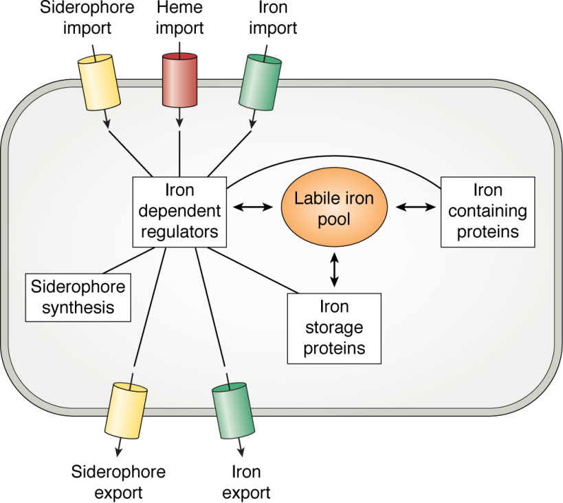
Iron is an essential micronutrient, and, in the case of bacteria, its availability is commonly a growth-limiting factor. However, correct functioning of cells requires that the labile pool of chelatable "free" iron be tightly regulated. Correct metalation of proteins requiring iron as a cofactor demands that such a readily accessible source of iron exist, but overaccumulation results in an oxidative burden that, if unchecked, would lead to cell death. The toxicity of iron stems from its potential to catalyze formation of reactive oxygen species that, in addition to causing damage to biological molecules, can also lead to the formation of reactive nitrogen species. To avoid iron-mediated oxidative stress, bacteria utilize iron-dependent global regulators to sense the iron status of the cell and regulate the expression of proteins involved in the acquisition, storage, and efflux of iron accordingly. Here, we survey the current understanding of the structure and mechanism of the important members of each of these classes of protein. Diversity in the details of iron homeostasis mechanisms reflect the differing nutritional stresses resulting from the wide variety of ecological niches that bacteria inhabit. However, in this review, we seek to highlight the similarities of iron homeostasis between different bacteria, while acknowledging important variations. In this way, we hope to illustrate how bacteria have evolved common approaches to overcome the dual problems of the insolubility and potential toxicity of iron.

Keywords: Bfr; Dps; DtxR; Ftn; Fur; Irr; RirA; bacterioferritin; encapsulin; encapsulins; ferritin; gene regulation; iron; iron metabolism; iron regulation; iron storage; iron toxicity; reactive oxygen species (ROS).


Conflict of interest statement

Conflict of interest—The authors declare that they have no conflicts of interest with the contents of this article.

The authors declare that they have no conflicts of interest with the contents of this article

Figures

Figure 1.
Figure 1.
Routes of iron trafficking in bacterial cells. Heavy arrows depict intracellular movement of iron, light arrows show the movement of iron or iron-bearing compounds across the cell membrane, and lines connect the transcriptional regulators to systems under their control. When the concentration of the labile iron pool increases, iron, or an iron-containing group, binds to the transcriptional regulator. This leads to down-regulation of processes such as siderophore synthesis, export of apo-siderophores, import of Fe3+-siderophores, heme import, and Fe2+ uptake systems. Simultaneously, expression of iron-containing and iron storage proteins is up-regulated together, occasionally, with iron efflux pumps. Reduction in the labile iron pool leads to dissociation of iron/iron-containing groups from the regulators, resulting in the opposite transcriptional responses.
Scheme 1.
Scheme 1.
Figure 2.
Figure 2.
Domain movements induced by the binding of divalent metals to Fur. Binding of divalent metal ions to the regulatory site of Fur induces a rotation of the DNA-binding domain relative to the dimerization domain, bringing the DNA recognition helices into more favorable alignment for binding to the Fur box. Residues Lys-15, Tyr-56, and Arg-57, which form favorable interactions with the nucleotide, are highlighted in red. Reproduced from PDB depositions 4RAY and 4RB1 (27).
Figure 3.
Figure 3.
Binding of DtxR to a 21-base pair model oligonucleotide. Identical DtxR dimers bind to opposite faces of the nucleotide, but only one of the four SH3-like domains is resolved crystallographically. The inset shows the N-terminal region of the protein with residues 3–6 highlighted in red. Upon binding of the regulatory metal ion, the highlighted region undergoes a helix-to-coil transition that relieves what would otherwise be an unfavorable steric interaction between protein and DNA. Also highlighted in red are residues Arg-27, Ala-28, Arg-29, Thr-40, Ser-42, Arg-47, Arg-50, and Arg-60, which form favorable interactions with the nucleotide. Reproduced using PDB deposition 1C0W (52).
Figure 4.
Figure 4.
Structures of representative proteins involved in bacterial iron acquisition. A, HasR, a β-barrel porin involved in transport of heme across the periplasmic membrane in complex with HasA. The importers of siderophores exhibit very similar topology. Also shown are chaperone proteins FhuD (B) and HmuT (C), which shuttle siderophores and heme, respectively, across the periplasmic space as well as the ABC transporter HmuUV (D), which transports heme across the cytoplasmic membrane. ABC transporters involved in siderophore transport exhibit similar topology. Reproduced from PDB depositions 3CSL (101), 1EFD (223), 3NU1 (224), and 4G1U (225).
Figure 5.
Figure 5.
The bacterial ferritins. A, the dodecameric assembly of L. innocua Dps (a mini-ferritin) viewed along one of the ferritin-like 3-fold channels. B, single iron ion observed bound to the L. innocua Dps ferroxidase center. C, the 24-meric assembly adopted by both Ftn and Bfr viewed along the channel formed at the 3-fold symmetry axis. D, the ligands to iron bound at the ferroxidase center of a typical bacterial Ftn together with the associated site C (left) compared with the more symmetrical iron-binding environment in E. coli Bfr and the distinct coordination environment of the iron ion located on the inner surface of the protein (right). In Ftn, the higher-affinity site A has a higher coordination number than site B. E, expanded view of the ferritin B-channel showing Fe2+ bound to Asp-132 of one monomer with the potential ligands Asp-30 and Asn-63 of the two other monomers forming the channel also highlighted. F, side view of the ferritin 3-fold channel showing the conserved Cys (top), Glu (middle), and Asp (bottom) residues thought to guide the Fe2+ substrate toward the interior of the protein. G, schematic representation of the displacement mechanism that operates in some ferritins. Two equivalents of Fe2+ bind to the apo-ferroxidase center. Oxygen (or peroxide) binds and is reduced to peroxide (or water) by the simultaneous oxidation of both Fe2+ ions to Fe3+. Hydrolysis of the transient diferric peroxo intermediate liberates peroxide and forms a ferric-oxo precursor of the mineral core. This is displaced from the catalytic site, completing the cycle by regenerating the apo-ferroxidase center. Images produced using PDB depositions 1QGH (184) (Dps), 4ZTT (226) (Ftn), and 3E1P (161) (Bfr).
Figure 6.
Figure 6.
Encapsulated ferritins. A, the ferritin fold is made up of two homologous pairs of anti-parallel α-helices (136), here colored green and cyan. In the true, cage-forming ferritins, these are connected via a loop joining helices B and C. Short helices running perpendicular to the long axis of the bundle help to template cage formation in the mini-ferritins (top) or 24-meric examples (middle). Members of the superfamily that do not form cages, such as EncFtn (bottom), are associated with further extended secondary structure elements, such as the membrane-spanning helices of MbfA or the large additional helices of EncFtn, which prevent assembly into cages. B, the annular pentamer of dimers adopted by the majority of encapsulated ferritins. C, the ferroxidase center of a typical encapsulated ferritin highlighting the noncrystallographic 2-fold symmetry of the iron environment. For clarity, only the ligands provided by the lower of the two protomers have been labeled. Images produced using PDB deposition 5N5E (206) (EncFtn).
Figure 7.
Figure 7.
Schematic overview of the major components of iron sensing and detoxification found in bacterial cells. Note that not all of these components are present in a single bacterial cell. Regulatory proteins are shown here as repressors but, in some cases, can also act as activators. Encapsulins are large protein compartments that house EncFtn ferritin-like proteins. The fate of iron stored in encapsulins and in Dps proteins is not clear, although it is likely that at some point, it becomes bioavailable again. Ftn, Bfr, and Dps do not appear to be distributed according to phyla. Fur is the transcriptional regulator in most bacteria but is replaced by DtxR/IdeR in some actinobacteria. In the α-proteobacteria, Fur plays a diminished role in iron homeostasis, with the majority of these functions being performed by Irr. In some rhizobiales, this is achieved in conjunction with a second global regulator, RirA. Import of siderophores and heme across the cytoplasmic membrane (IM) is performed by ABC transporters in all known cases, and Feo is the major importer of Fe2+. In Gram-negative bacteria, heme and siderophores are imported to the periplasm by outer-membrane (OM) porins, whereas a network of heme-binding proteins transports this cofactor across the cell wall of the Gram-positive bacteria. Characterized Fe2+ export systems are rare, but P-type ATPases are the most widely distributed. IceT of S. typhimurium is the only example of the MFS characterized to date, whereas the CDF proteins are limited to γ-proteobacteria and the MbfA proteins to α-proteobacteria. YiiP from E. coli is the only Fe2+ efflux pump for which the structure has been solved (227).

References

    1. Beinert H., Holm R. H., and Münck E. (1997) Iron-sulfur clusters: nature's modular, multipurpose structures. Science 277, 653–659 10.1126/science.277.5326.653 - DOI - PubMed
    1. Gamba I., Codolà Z., Lloret-Fillol J., and Costas M. (2017) Making and breaking of the O-O bond at iron complexes. Coord. Chem. Rev. 334, 2–24 10.1016/j.ccr.2016.11.007 - DOI
    1. Berks B. C., Ferguson S. J., Moir J. W. B., and Richardson D. J. (1995) Enzymes and associated electron transport systems that catalyse the respiratory reduction of nitrogen oxides and oxyanions. Biochim. Biophys. Acta 1232, 97–173 10.1016/0005-2728(95)00092-5 - DOI - PubMed
    1. Kappler U., and Maher M. J. (2013) The bacterial SoxAX cytochromes. Cell. Mol. Life Sci. 70, 977–992 10.1007/s00018-012-1098-y - DOI - PMC - PubMed
    1. Poulos T. L. (2014) Heme enzyme structure and function. Chem. Rev. 114, 3919–3962 10.1021/cr400415k - DOI - PMC - PubMed

Publication types

MeSH terms