. 2020 Dec 18;295(51):17646-17658.
doi: 10.1074/jbc.RA120.015492.

The HRDC domain oppositely modulates the unwinding activity of E. coli RecQ helicase on duplex DNA and G-quadruplex

Affiliations

The HRDC domain oppositely modulates the unwinding activity of E. coli RecQ helicase on duplex DNA and G-quadruplex

Fang-Yuan Teng et al. J Biol Chem. .

Abstract

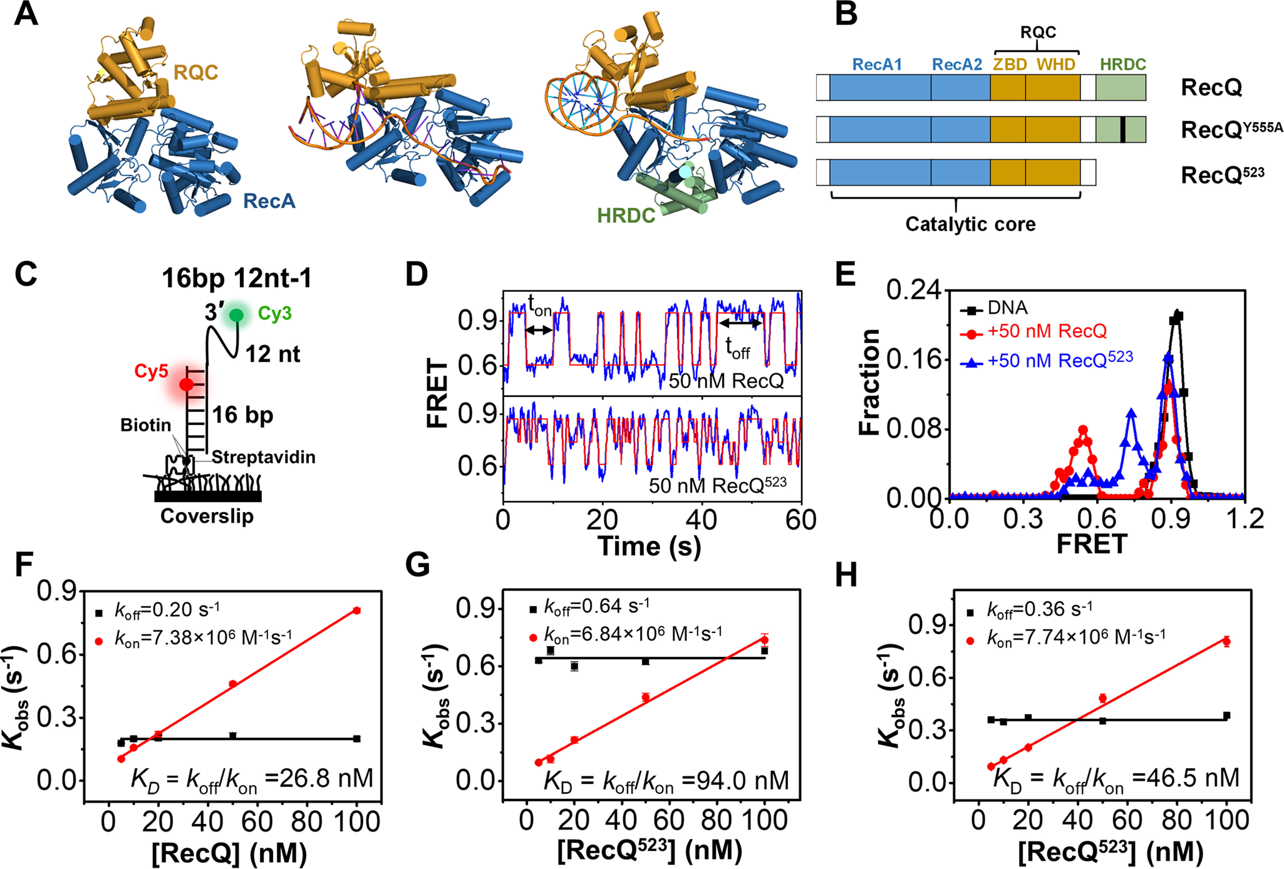
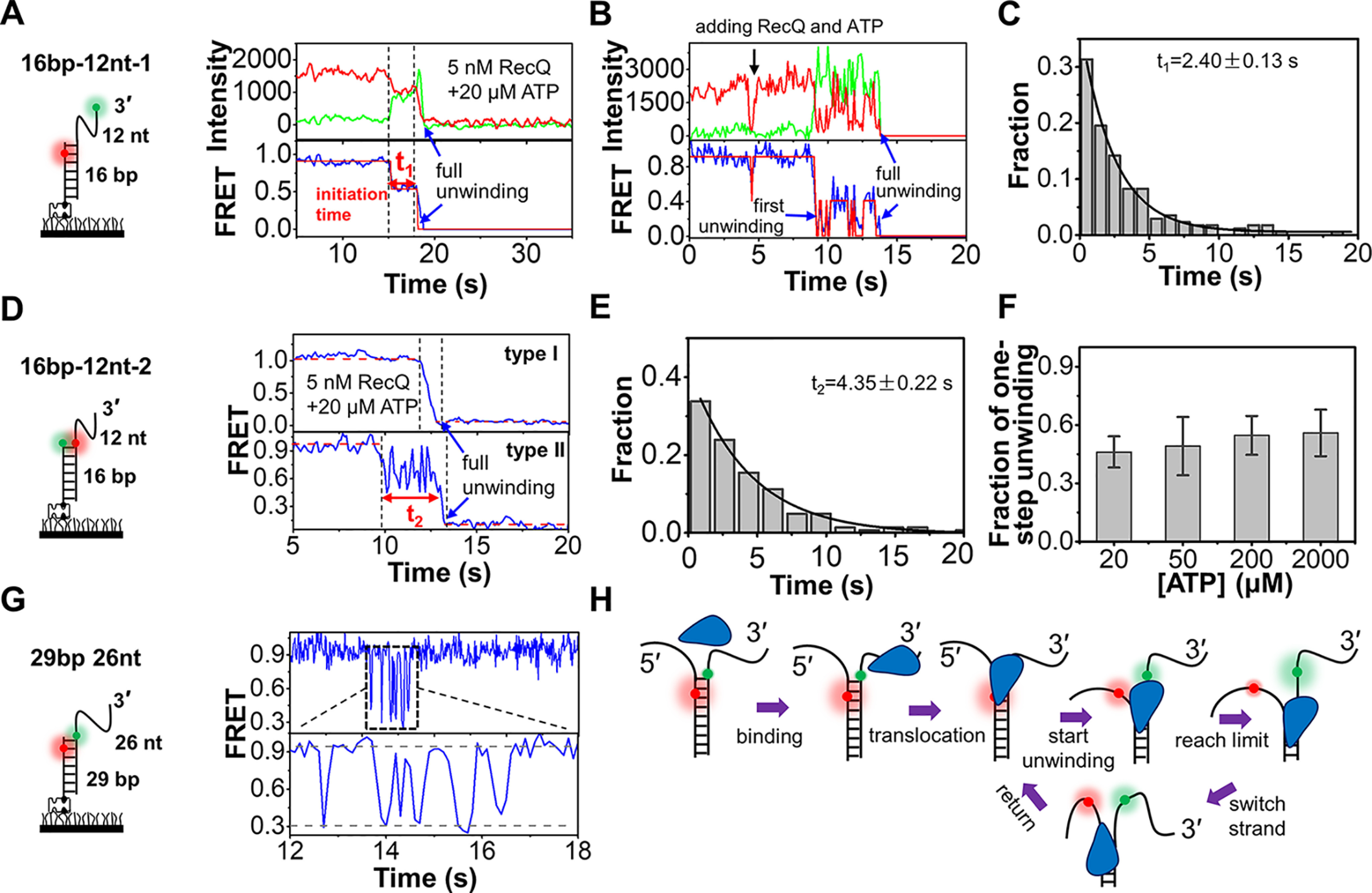
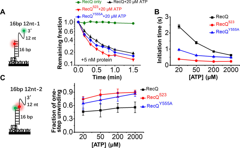
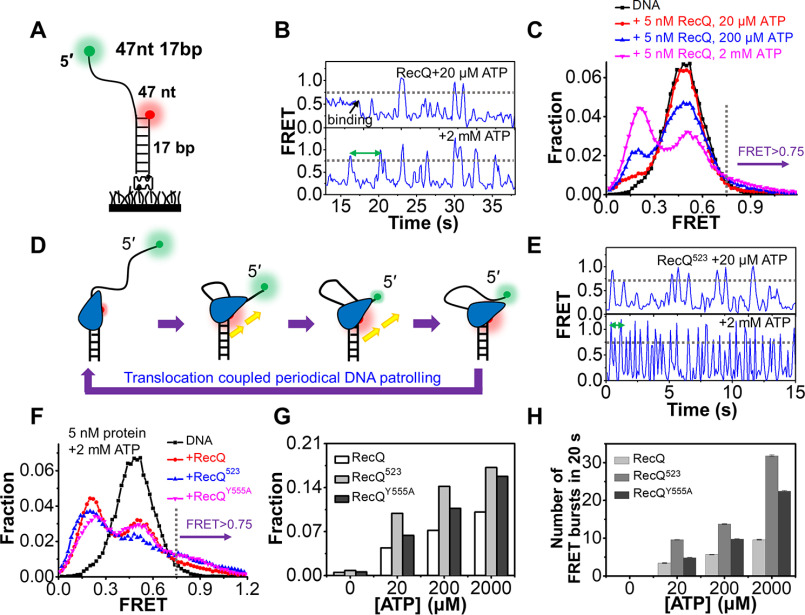
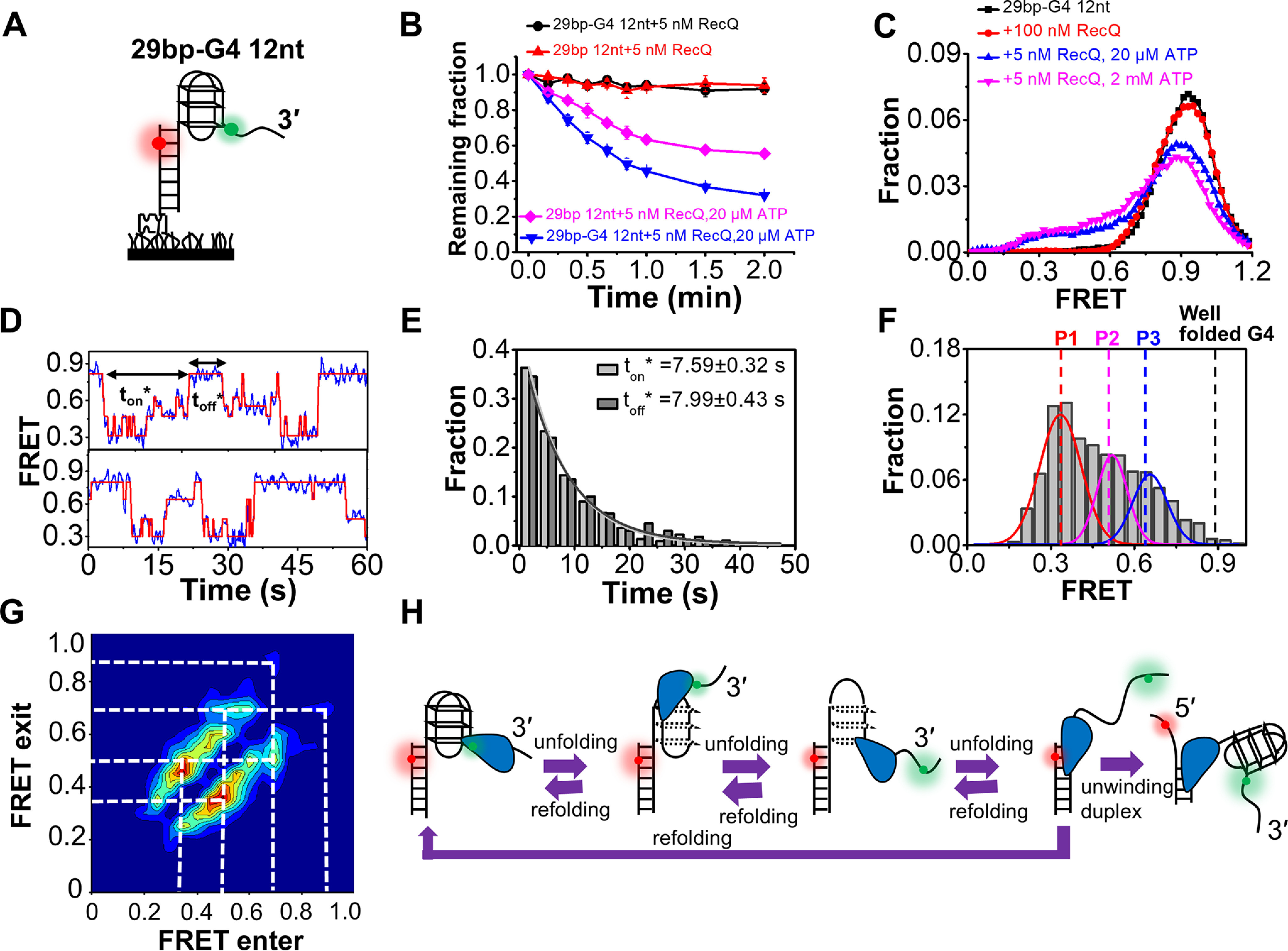
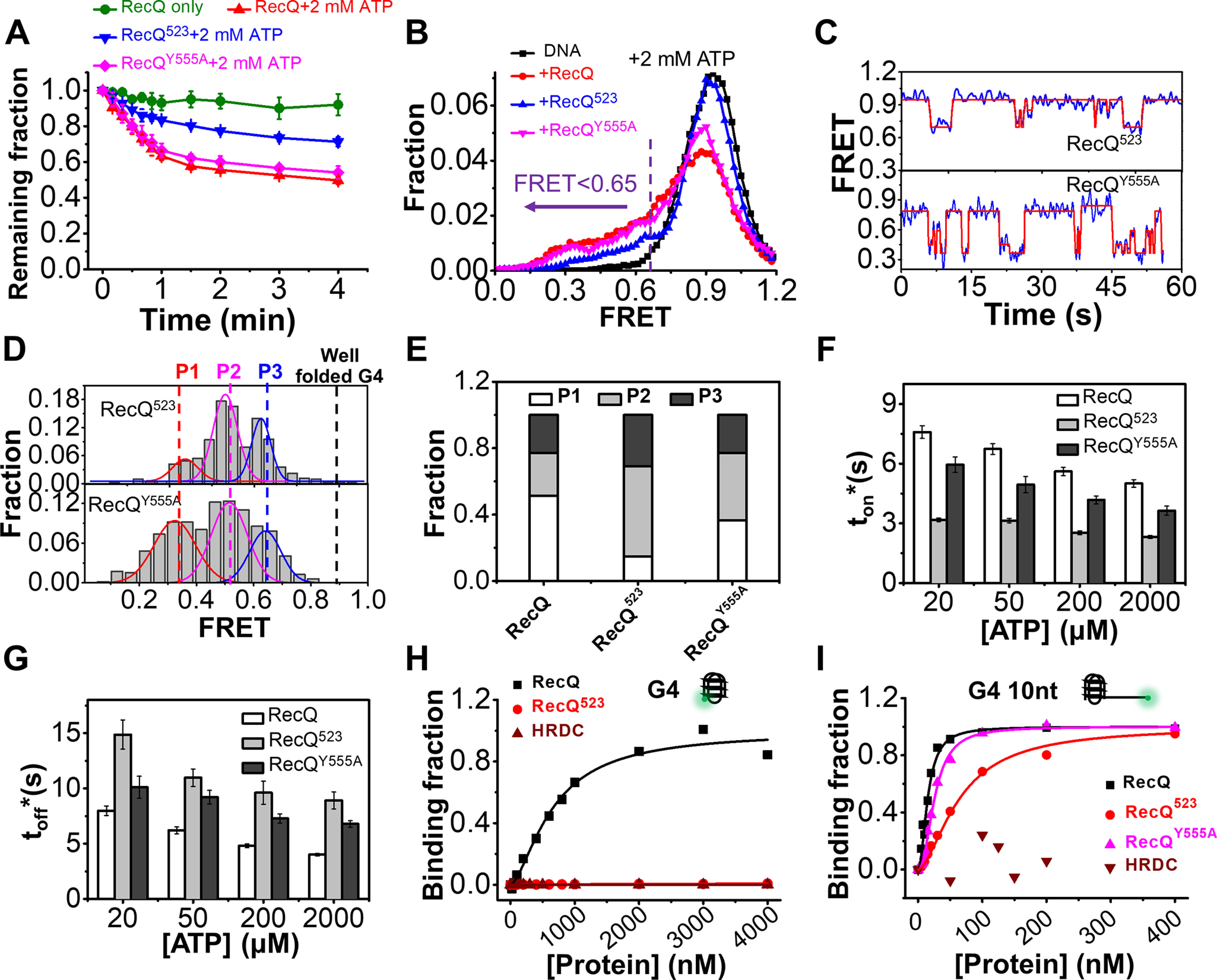
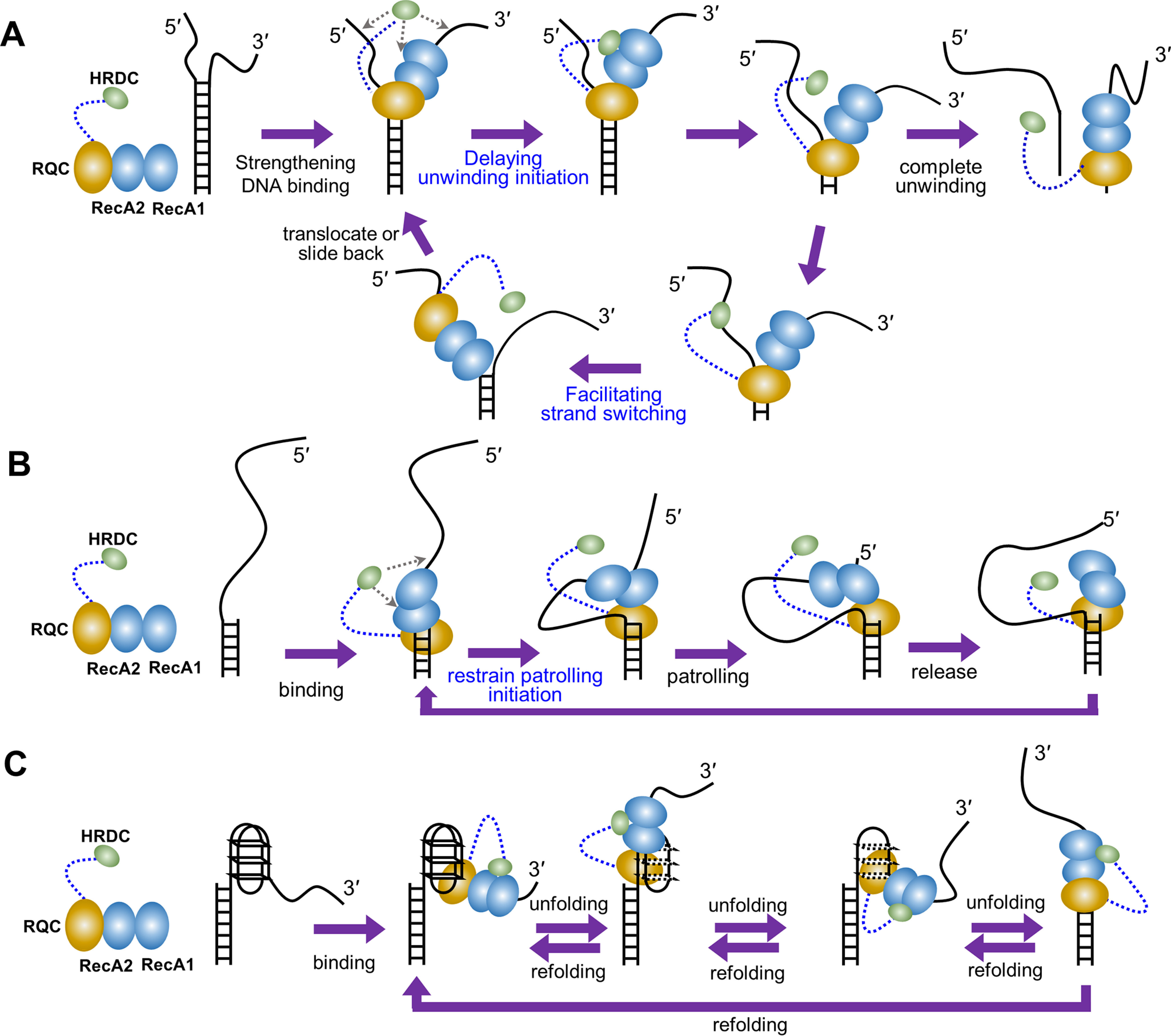
RecQ family helicases are highly conserved from bacteria to humans and have essential roles in maintaining genome stability. Mutations in three human RecQ helicases cause severe diseases with the main features of premature aging and cancer predisposition. Most RecQ helicases shared a conserved domain arrangement which comprises a helicase core, an RecQ C-terminal domain, and an auxiliary element helicase and RNaseD C-terminal (HRDC) domain, the functions of which are poorly understood. In this study, we systematically characterized the roles of the HRDC domain in E. coli RecQ in various DNA transactions by single-molecule FRET. We found that RecQ repetitively unwinds the 3'-partial duplex and fork DNA with a moderate processivity and periodically patrols on the ssDNA in the 5'-partial duplex by translocation. The HRDC domain significantly suppresses RecQ activities in the above transactions. In sharp contrast, the HRDC domain is essential for the deep and long-time unfolding of the G4 DNA structure by RecQ. Based on the observations that the HRDC domain dynamically switches between RecA core- and ssDNA-binding modes after RecQ association with DNA, we proposed a model to explain the modulation mechanism of the HRDC domain. Our findings not only provide new insights into the activities of RecQ on different substrates but also highlight the novel functions of the HRDC domain in DNA metabolisms.

Keywords: DNA repair; E. coli; G-quadruplex; RecQ; helicase; single-molecule; unwinding.


Conflict of interest statement

Conflict of interest—The authors declare that they have no conflicts of interest with the contents of this article.

The authors declare that they have no conflicts of interest with the contents of this article

Figures

Figure 1.
Figure 1.
RecQ helicase repetitively associates with and dissociates from the 3′-partial duplex DNA. A, crystal structures of an HRDC-deleted E. coli RecQ (PDB: 1OYW), HRDC-deleted C. sakazakii RecQ in complex with DNA (PDB: 4TMU), and HRDC-containing human BLM in complex with DNA (4O3M). B, domain map of E. coli RecQ constructs used in this study. C, schematic set-up of the smFRET experiment. D, the typical smFRET traces of 16 bp 12 nt-1 in 50 nm RecQ and RecQ523. An automated step-finding algorithm was used to identify the different FRET states (red line). E, FRET histograms of 16 bp 12 nt-1 in 50 nm RecQ or RecQ523 based on the fitted FRET traces. In all the following figures, the FRET histograms were collected from more than 200 traces. FH, the dissociation rate (koff =1/ton′, red) and the binding rate (kon=1/toff′, black) as a function of protein concentrations. As expected for a binary reaction, the dissociation rate is independent of protein concentration whereas the binding rate has a linear dependence on it. The dissociation constant is thus determined as KD = koff/kon. Error bars denote the standard deviations.
Figure 2.
Figure 2.
Unwinding of duplex DNA by RecQ. A and B, the typical fluorescence and FRET traces of 16 bp 12 nt-1 in 5 nm RecQ and 20 µm ATP. More reaction traces were exhibited in Fig. S4A. t1 represented the initiation time before duplex unwinding. C, distribution of t1 obtained from single-exponential fitting. In all the following figures, the time histograms were collected from more than 200 traces. D, two typical FRET traces of 16 bp 12 nt-2. Type I represents one-step unwinding, and type II represents repetitive unwinding with total unwinding time t2. E, distribution of t2 obtained from single-exponential fitting. F, the fractions of type I increase with the increase in ATP concentrations. G, repetitive unwinding can be observed in the substrate 29 bp 26 nt. H, a proposed model for RecQ-catalyzed duplex unwinding.
Figure 3.
Figure 3.
HRDC suppresses the duplex unwinding activity of RecQ. A, fractions of remaining 16 bp 12 nt-1 molecules on coverslip versus time after addition of 5 nm RecQ and 20 µm ATP. Lines are the simple connections of the individual data points by Origin 8.0. B, the initiation time of 16 bp 12 nt-1 in 5 nm RecQ, RecQ523, and RecQY555A in different concentrations of ATP. C, the fractions of 16 bp 12 nt-2 traces showing one-step unwinding in 5 nm RecQ, RecQ523, and RecQY555A in different concentrations of ATP.
Figure 4.
Figure 4.
RecQ periodically patrols on the 5′-ssDNA. A, the 5′-partial duplex 47 nt 17 bp contains a 17 bp duplex and a 47 nt 5′-ssDNA. B, the typical FRET traces of 47 nt 17 bp in 5 nm RecQ and indicated ATP concentrations. C, FRET distributions of 47 nt 17 bp only and in 5 nm RecQ and different concentrations of ATP. D, a proposed action of RecQ on the 5′-partial duplex. E, the typical FRET traces of 47 nt 17 bp in 5 nm RecQ523 and indicated ATP concentrations. F, FRET distributions of 47 nt 17 bp in 5 nm RecQ, RecQ523, and RecQY555A with 2 mm ATP. G, the fractions of DNA at looping states (FRET > 0.75) in different reaction conditions. H, the number of FRET bursts within the 20-s time window in 5 nm RecQ, RecQ523, and RecQY555A with indicated ATP concentration.
Figure 5.
Figure 5.
RecQ can unfold the G4 DNA structure. A, the G4-containing DNA substrate 29 bp-G4 12 nt. B, fractions of remaining DNA molecules on coverslip versus time after the addition of RecQ and ATP. C, FRET distributions of G4 substrate in different concentrations of RecQ and ATP. D, in 5 nm RecQ and 20 µm ATP, the FRET values of 29 bp-G4 12 nt fluctuate between different levels. The automated step-finding algorithm was used to identify the individual steps (red line) during G4 unfolding and refolding. The time for the continuous FRET oscillations was defined as ton*, and the time interval between two lasting oscillations was defined as toff*. E, distributions of ton* and toff*. F, distributions of the FRET oscillation regions of 29 bp-G4 12 nt in 5 nm RecQ and 20 µm ATP. G, transition density plot for oscillation regions of 29 bp-G4 12 nt from ∼200 FRET oscillation regions. H, schematic diagram of our model to explain how RecQ unfolds the G4 structure repetitively.
Figure 6.
Figure 6.
The HRDC domain of RecQ is necessary for the efficient unfolding of the G4 structure. A, fractions of remaining 29 bp-G4 12 nt molecules on coverslip versus time after the addition of 5 nm protein and 2 mm ATP. B, FRET distributions of 29 bp-G4 12 nt in 2 mm ATP and different RecQ constructs. According to the FRET peaks in Fig. 5F, a criterion at E0.65 was set artificially, below which the G4 structure was recognized as being disrupted. C, in 5 nm RecQ523 or RecQY555A and 20 µm ATP, the FRET values of 29 bp-G4 12 nt fluctuate between different levels. D, distributions of the FRET oscillation regions of the G4 substrate from ∼100 traces. E, the fractions of P1, P2, and P3 in the FRET histograms of G4 substrate in different types of RecQ. F and G, histograms of ton* and toff* in RecQ, RecQ523, and RecQY555A. H and I, RecQ constructs binding to G4 (H) or G4 10 nt (I) measured by equilibrium DNA binding assay. The dissociation constant (KD) of RecQ bound to G4 is 658.2 ± 79 nm; KD of RecQ523 and HRDC bound to G4 were both not available. KD of RecQ, RecQ523, and RecQY555A bound to G4 10 nt were 15.1 ± 1.1 nm, 63.7 ± 4.1 nm, and 25.6 ± 1 nm, respectively; KD of HRDC bound to G4 was not available.
Figure 7.
Figure 7.
The proposed modulation mechanism of the HRDC domain. A, in duplex DNA unwinding, HRDC delays the unwinding initiation of RecQ possibly by inhibiting ATP hydrolysis, ATP binding, and ADP and/or Pi release due to the interaction with the RecA core. Moreover, HRDC promotes the strand-switch of RecQ by dynamically binding to the 5′-ssDNA, thereby inhibiting the unidirectional unwinding. B, in the periodically patrolling process, HRDC restrains the patrolling initiation of RecQ possibly by inhibiting ATP hydrolysis, ATP binding, and ADP and/or Pi release due to the interaction with the RecA core. C, in G4 unfolding, HRDC reinforces the association of RecQ on the DNA by interacting with the RecA core, leading to the complete and long-time G4 unfolding.

Similar articles

Cited by

References

    1. Brosh R. M. (2013) DNA helicases involved in DNA repair and their roles in cancer. Nat. Rev. Cancer 13, 542–558 10.1038/nrc3560 - DOI - PMC - PubMed
    1. Vindigni A., Marino F., and Gileadi O. (2010) Probing the structural basis of RecQ helicase function. Biophys. Chem. 149, 67–77 10.1016/j.bpc.2010.03.012 - DOI - PubMed
    1. Croteau D. L., Popuri V., Opresko P. L., and Bohr V. A. (2014) Human RecQ helicases in DNA repair, recombination, and replication. Ann. Rev. Biochem. 83 83, 519–552 10.1146/annurev-biochem-060713-035428 - DOI - PMC - PubMed
    1. Larsen N. B., and Hickson I. D. (2013) RecQ helicases: Conserved guardians of genomic integrity. Adv. Exp. Med. Biol. 973, 161–184 10.1007/978-1-4614-5037-5_8 - DOI - PubMed
    1. Mohaghegh P., Karow J. K., Brosh R. M., Bohr V. A., and Hickson I. D. (2001) The Bloom's and Werner's syndrome proteins are DNA structure-specific helicases. Nucleic Acids Res. 29, 2843–2849 10.1093/nar/29.13.2843 - DOI - PMC - PubMed

Publication types

LinkOut - more resources