. 2020 Dec 18;295(51):17684-17697.
doi: 10.1074/jbc.RA120.014077.

Mycobacterium tuberculosis infection up-regulates MFN2 expression to promote NLRP3 inflammasome formation

Affiliations

Mycobacterium tuberculosis infection up-regulates MFN2 expression to promote NLRP3 inflammasome formation

Fang Xu et al. J Biol Chem. .

Abstract

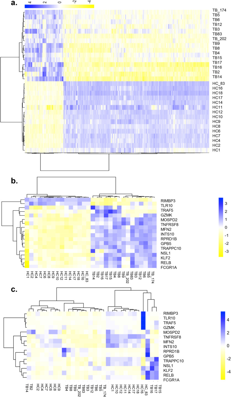
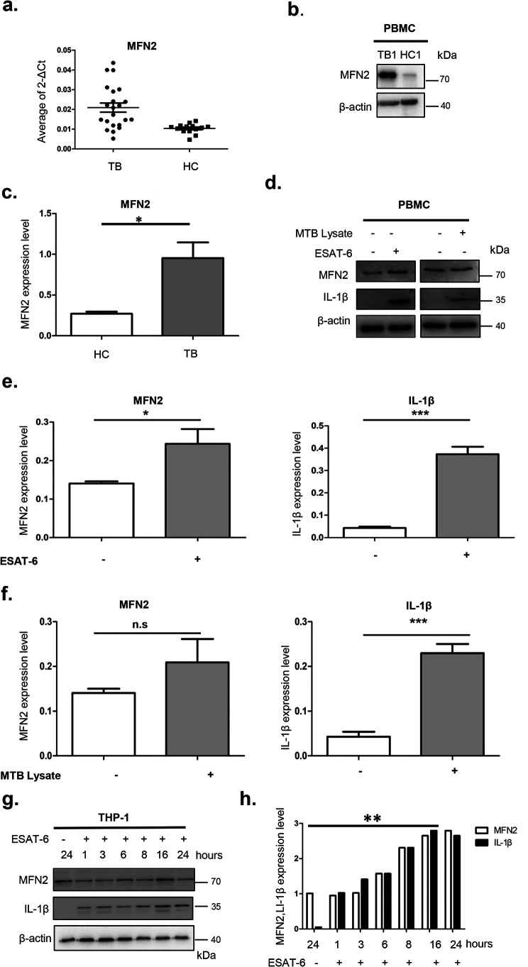
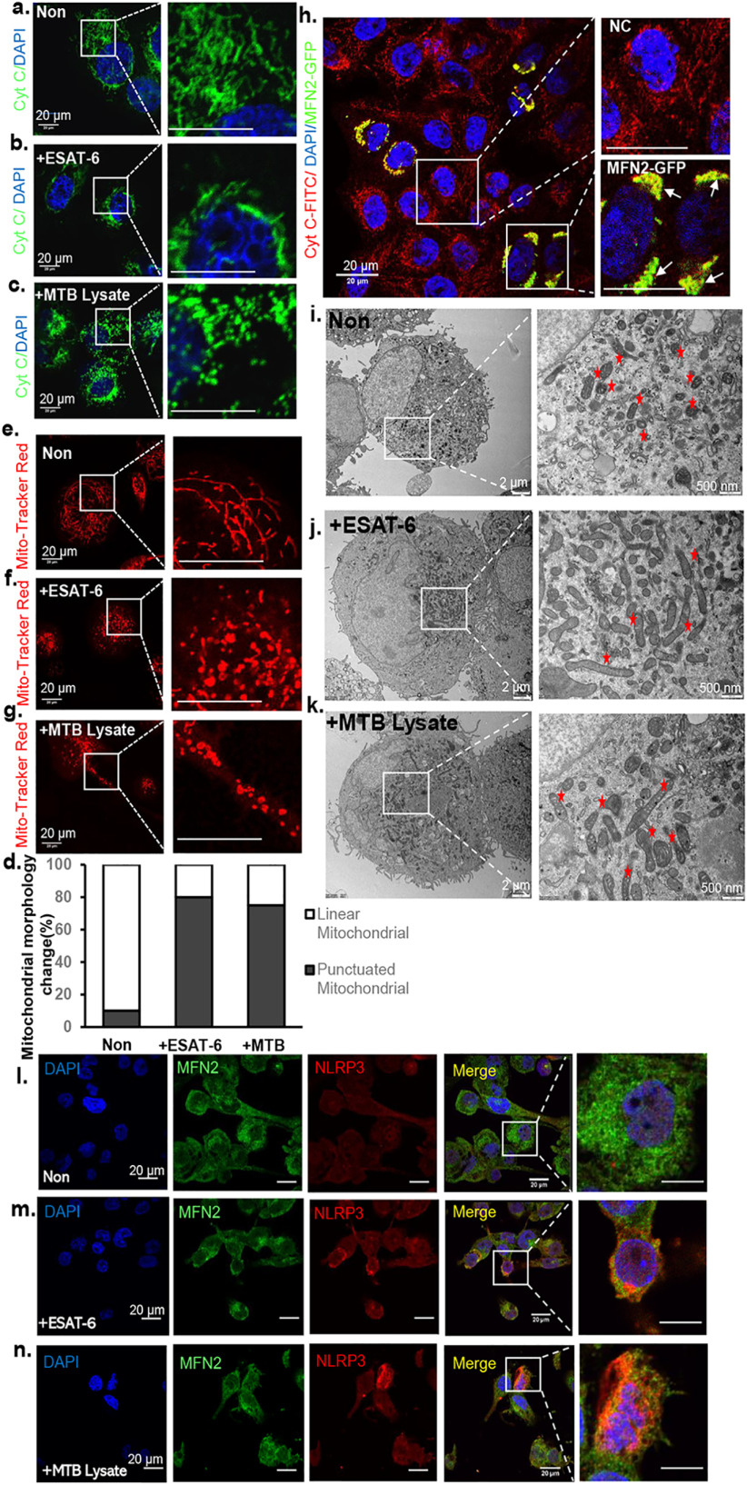
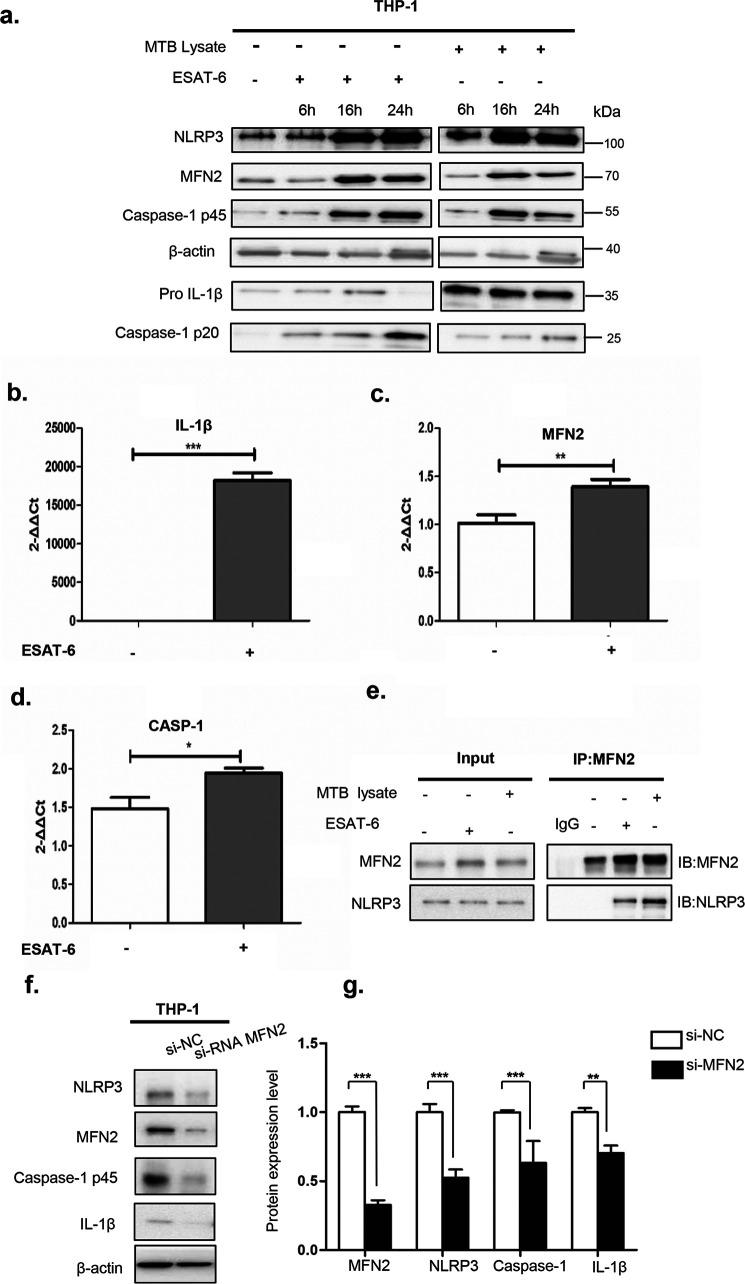
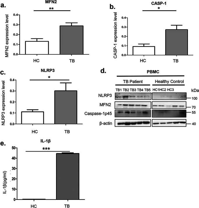
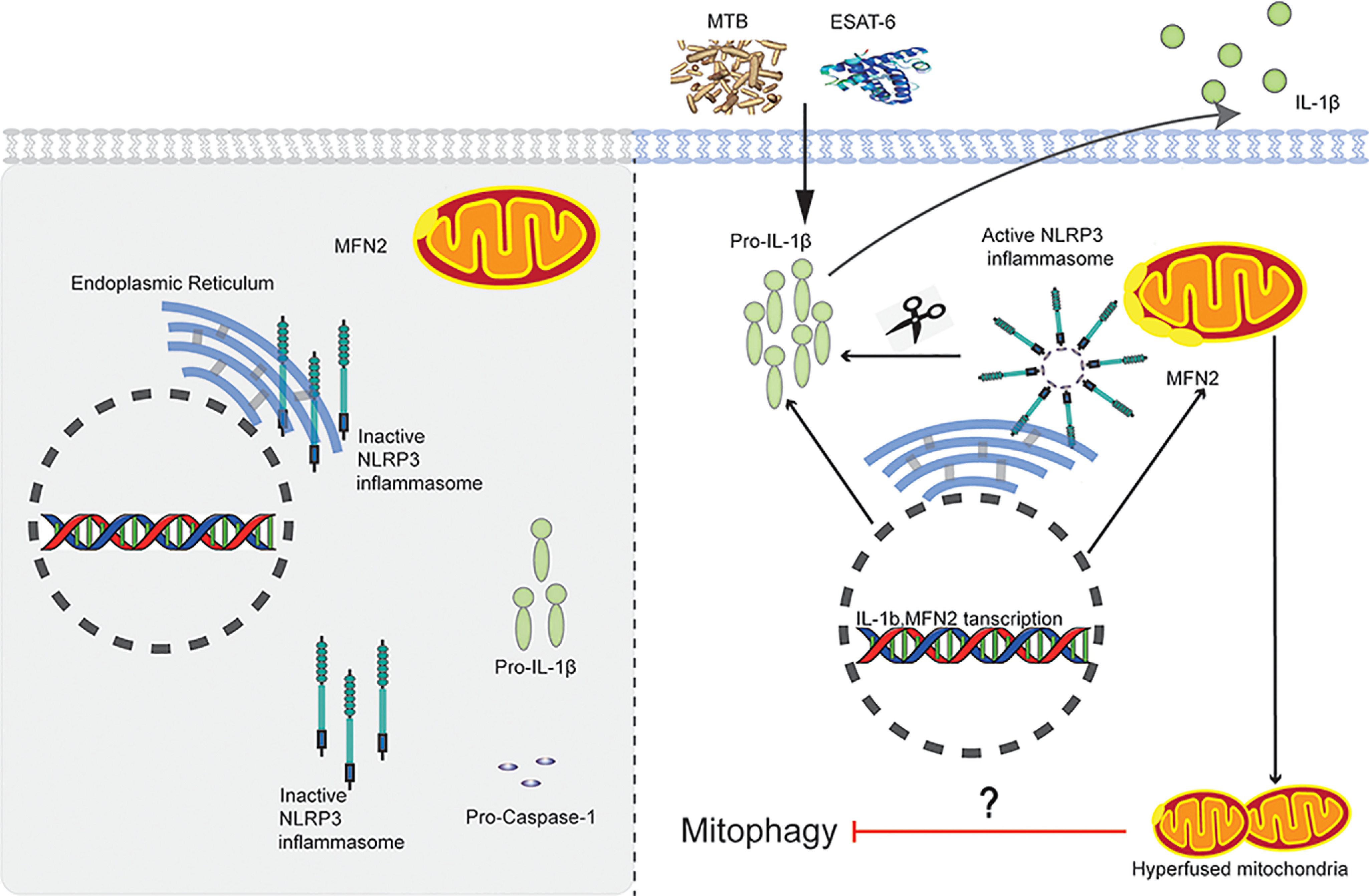
Tuberculosis (TB), caused by the infection of Mycobacterium tuberculosis (MTB), is one of the leading causes of death worldwide, especially in children. However, the mechanisms by which MTB infects its cellular host, activates an immune response, and triggers inflammation remain unknown. Mitochondria play important roles in the initiation and activation of the nucleotide-binding oligomerization domain-like receptor with a pyrin domain 3 (NLRP3) inflammasome, where mitochondria-associated endoplasmic reticulum membranes (MAMs) may serve as the platform for inflammasome assembly and activation. Additionally, mitofusin 2 (MFN2) is implicated in the formation of MAMs, but, the roles of mitochondria and MFN2 in MTB infection have not been elucidated. Using mircroarry profiling of TB patients and in vitro MTB stimulation of macrophages, we observed an up-regulation of MFN2 in the peripheral blood mononuclear cells of active TB patients. Furthermore, we found that MTB stimulation by MTB-specific antigen ESAT-6 or lysate of MTB promoted MFN2 interaction with NLRP3 inflammasomes, resulting in the assembly and activation of the inflammasome and, subsequently, IL-1β secretion. These findings suggest that MFN2 and mitochondria play important role in the pathogen-host interaction during MTB infection.

Keywords: Mycobacterium tuberculosis; NLRP3 inflammasome; cytokine; cytokine inflammasome; microarray; microarray mitochondria; mitochondria; mitofusin 2.


Conflict of interest statement

Conflict of interest—The authors declare no competing interests for this work.

The authors declare no competing interests for this work

Figures

Figure 1.
Figure 1.
Genome-wide mRNA profiling of PBMCs from TB patients (TB) and HC. Cluster analysis of differentially expressed mRNAs. a, genome-wide mRNA profiles of PBMCs from TBs and HCs. The heatmap represents the results of a two-way hierarchical clustering of mRNA and samples. Each row represents an mRNA, and each column represents the sample tested. Cluster analysis of differentially expressed mRNAs is shown. The color key indicates the expression level of the mRNAs, red represents mRNAs with an expression level above the mean, and green represents mRNAs with an expression level below the mean. b, heatmap of different expression of 15 candidate genes (microarray). Each row represents an mRNA, and each column represents the sample tested. Cluster analysis of differentially expressed mRNAs is shown. The color key indicates the expression level of the mRNAs, red represents mRNAs with an expression level above the mean, and green represents mRNAs with an expression level below the mean. c, heatmap of different expression of 15 candidate genes (RT-qPCR). Each row represents an mRNA, and each column represents the sample tested. Cluster analysis of differentially expressed mRNAs is shown. The color key indicates the expression level of the mRNAs, red represents mRNAs with an expression level above the mean, and green represents mRNAs with an expression level below the mean.
Figure 2.
Figure 2.
MFN2 is up-regulated after MTB infection or stimulation. a, RT-qPCR measurement of MFN2 mRNA isolated from PBMCs of active TB patients and HC. ** represents a significant difference between HC and TB, p < 0.01, as calculated by comparing the average fold-changes in gene expression of MFN2 in each TB patient (n = 20) sample with normalized average of the gene expression of each healthy control (n = 20). b and c, immunoblotting measurement of MFN2 expression of the PBMCs isolated from 5 TB patients and 3 healthy controls (image shows representative TB1 and HC1). β-Actin is a loading control. A star sign represents a significant difference between HC and TB, p < 0.05. d–f, immunoblotting measurement of MFN2 and IL-1β protein levels of the PBMCs isolated from 2 healthy controls with or without stimulation by heat-killed inactivated MTB lysates or ESAT-6 for 24 h. β-Actin is a loading control. g and h, immunoblotting measurement of MFN2 and IL-1β protein levels protein isolated from THP-1-marchophages stimulated by ESAT-6. MFN2 reached the highest level at 16 h after stimulation. Differences in the protein levels are compared with the previous time points. Images are representative of 3 independent experiments. Data are presented as mean ± S.D. (n = 3). *, p < 0.05; **, p < 0.01; ***, p < 0.001.
Figure 3.
Figure 3.
MTB stimulation alters both mitochondrial membrane potential and MFN2 localization in THP-1 macrophages. a–c, confocal immunofluorescent images of unstimulated THP-1 macrophages (Non), ESAT-6–stimulated macrophages (+ESAT-6), and MTB lysate-stimulated macrophages (+MTB). All these cells were fixed before staining with cytochrome c (green) antibody to label mitochondria, and DAPI to stain nuclei (blue). Note that cytochrome c-labeled mitochondria aggregate in perinuclear region. Scale bar, 20 μm. d, the percentage of the mitochondrial morphology change (from string mitochondria to punctuated mitochondria) with anti-cytochrome c. The number of punctuated mitochondria was counted from ESAT-6 or MTB-stimulated cells in at least 10 randomly chosen fields of view. 100 mitochondria from 10 macrophages in each group were used. The results are representative of 3 independent experiments. e–g, confocal immunofluorescent images of unstimulated THP-1 macrophages (Non), ESAT-6–stimulated macrophages (+ESAT-6), and MTB lysate–stimulated macrophages (+MTB lysate). Cells were stained with MitoTracker Red CMXRos (red) to label live-cell mitochondrial membrane potential. Note that most thread-like shapes (e) are changed to round shapes or donut shapes in f and g. Scale bar, 20 μm. h, images of THP-1macrophages transfected with MFN2-GFP. Arrows point to endogenous MFN2 colocalized with MFN2-GFP. Scale bar, 20 μm. i–k, representative TEM images of THP-1 macrophages stimulated with MTB lysate and ESAT-6 for 24 h. As a control, representative images of unstimulated cells are shown. The mitochondrial morphology in stimulated cells and their magnified images are shown (white box region and marked with a red asterisk). Bar indicates 2 (left panel) or 0.5 μm (right panel). l–n, confocal immunofluorescent images of unstimulated THP-1 macrophages, MTB lysate-stimulated macrophages, and ESAT-6–stimulated macrophages. All these cells were fixed before staining with MFN2 antibody (green) and NLRP3 antibody (red) to label endogenous MFN2 and NLRP3, respectively. DAPI (blue) was used to label nuclei. Insets show 6× magnified images of the boxed region. Scale bar, 20 μm.
Figure 4.
Figure 4.
Endogenous NLRP3 associates with MFN2 after stimulated with MTB. a, immunoblotting analysis of lysates of THP-1 in cultured macrophages. Cell lysates prepared from macrophages following stimulation with MTB or ESAT-6 for 6, 16, and 24 h were analyzed by immunoblotting using anti-MFN2, anti-Caspase-1, anti-IL-1β, and anti-NLRP3 antibodies. β-Actin is a loading control. b–d, RT-qPCR analyses of THP-1 macrophages. ESAT-6–stimulated cells samples were collected at 16 h in parallel with immunoblotting analyses (a). e, endogenous immunoprecipitation (IP) from cell lysates with MFN2-specific antibody were analyzed by immunoblotting with mouse monoclonal antibodies against NLRP3 or MFN2. MTB- and ESAT-6–stimulated cells were collected in parallel with immunoblotting analysis. Stimulated cell samples were collected in the same way with immunoblotting analysis. Rabbit IgG is a negative control. f and g, samples from macrophages with transient expression of siRNA against MFN2 or EGFP control mRNAs (si-NC) were analyzed by immunoblotting after stimulated by MTB stimulation with antibodies against MFN2, Caspase-1, NLRP3, and IL-1β. β-Actin is a loading control. Images are representative of 3 independent experiments. Data are presented as mean ± S.D. (n = 3). *, p < 0.05; **, p < 0.01; ***, p < 0.001.
Figure 5.
Figure 5.
Expression levels of MFN2 and NLRP3 inflammasome marker genes in vivo. a–d, immunoblotting assays showed the endogenous levels of MFN2 (a), Caspase-1 p45 (b), and NLRP3 (c) isolated from PBMCs of active TB patients and PPD-skin test-negative healthy individuals with no prior exposure to TBs (HCs). β-Actin is a loading control. TB, n = 5; HC, n = 5. The experiments were repeated three times and representative blots are shown in d. e, ELISA measurements of IL-1β secretion of serum from the TBs and HCs. *, p < 0.05; **, p < 0.01; ***, p < 0.001.
Figure 6.
Figure 6.
A Proposed model of MFN2-dependent activation of the NLRP3 inflammasome during MTB infection. After stimulated with MTB or ESAT-6, MFN2 promotes the association of NLRP3 and MFN2 through procapase-1 to form the NLRP3 inflammasome at mitochondrial outer member associated with endoplasmic reticulum membranes (MAMs). The NLRP3 inflammasome cleaves the pro-IL-1β to produce its mature form IL-1β for secretion.

Similar articles

Cited by

References

    1. WHO (2019) Global Tuberculosis Report, World Health Organization
    1. Martinon F., Burns K., and Tschopp J. (2002) The inflammasome: a molecular platform triggering activation of inflammatory caspases and processing of proIL-β. Mol. Cell 10, 417–426 10.1016/S1097-2765(02)00599-3 - DOI - PubMed
    1. Fritz J. H., Ferrero R. L., Philpott D. J., and Girardin S. E. (2006) Nod-like proteins in immunity, inflammation and disease. Nat. Immunol. 7, 1250–1257 10.1038/ni1412 - DOI - PubMed
    1. Schroder K., and Tschopp J. (2010) The inflammasomes. Cell 140, 821–832 10.1016/j.cell.2010.01.040 - DOI - PubMed
    1. Pelegrin P., and Surprenant A. (2006) Pannexin-1 mediates large pore formation and interleukin-1beta release by the ATP-gated P2X7 receptor. EMBO J. 25, 5071–5082 10.1038/sj.emboj.7601378 - DOI - PMC - PubMed

Publication types

MeSH terms