. 2020 Dec 18;295(51):17770-17780.
doi: 10.1074/jbc.RA120.014705.

The C-terminal region of the plasmid partitioning protein TubY is a tetramer that can bind membranes and DNA

Affiliations

The C-terminal region of the plasmid partitioning protein TubY is a tetramer that can bind membranes and DNA

Ikuko Hayashi. J Biol Chem. .

Abstract

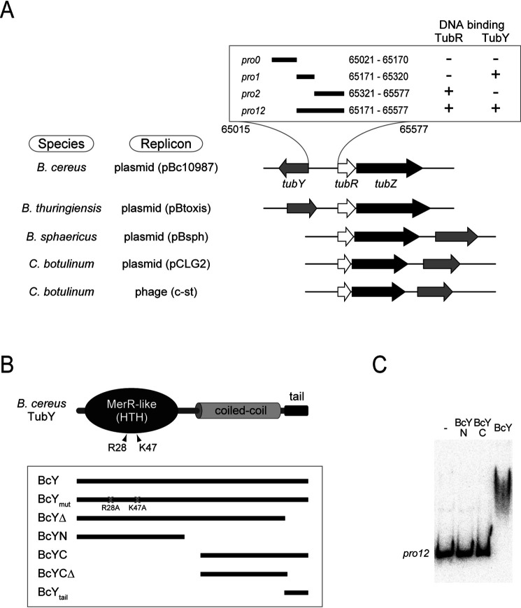
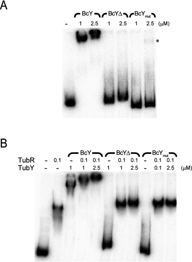
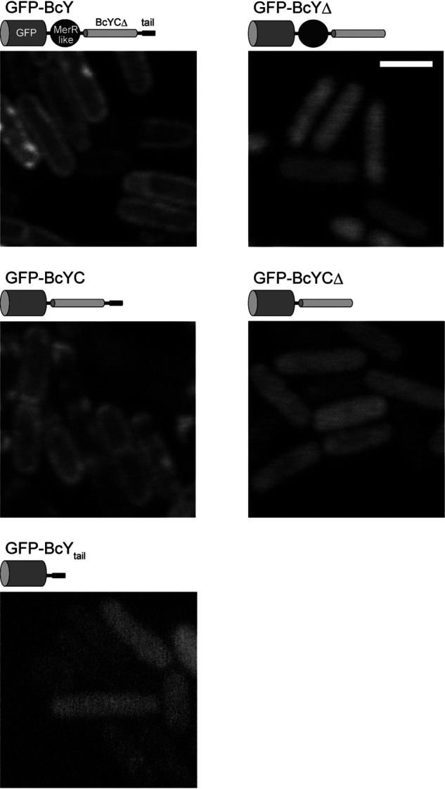
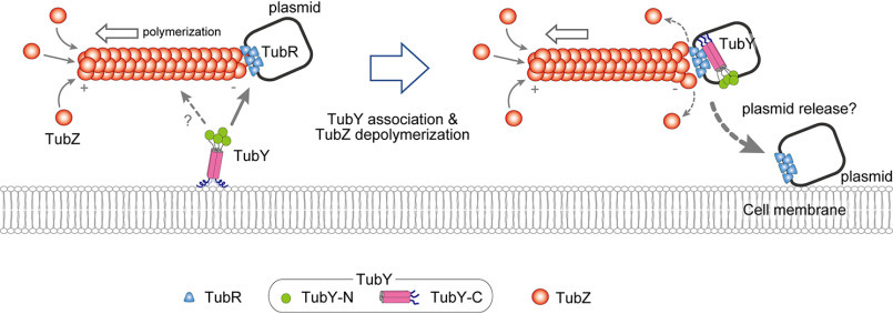
Bacterial low-copy-number plasmids require partition (par) systems to ensure their stable inheritance by daughter cells. In general, these systems consist of three components: a centromeric DNA sequence, a centromere-binding protein and a nucleotide hydrolase that polymerizes and functions as a motor. Type III systems, however, segregate plasmids using three proteins: the FtsZ/tubulin-like GTPase TubZ, the centromere-binding protein TubR and the MerR-like transcriptional regulator TubY. Although the TubZ filament is sufficient to transport the TubR-centromere complex in vitro, TubY is still necessary for the stable maintenance of the plasmid. TubY contains an N-terminal DNA-binding helix-turn-helix motif and a C-terminal coiled-coil followed by a cluster of lysine residues. This study determined the crystal structure of the C-terminal domain of TubY from the Bacillus cereus pXO1-like plasmid and showed that it forms a tetrameric parallel four-helix bundle that differs from the typical MerR family proteins with a dimeric anti-parallel coiled-coil. Biochemical analyses revealed that the C-terminal tail with the conserved lysine cluster helps TubY to stably associate with the TubR-centromere complex as well as to nonspecifically bind DNA. Furthermore, this C-terminal tail forms an amphipathic helix in the presence of lipids but must oligomerize to localize the protein to the membrane in vivo. Taken together, these data suggest that TubY is a component of the nucleoprotein complex within the partitioning machinery, and that lipid membranes act as mediators of type III systems.

Keywords: DNA binding protein; DNA segregation; MerR; TubZ; amphipathic helix; bacteria; crystal structure; cytoskeleton; plasmid; plasmid partioning; plasmid partitioning; segrosome.


Conflict of interest statement

Conflict of interest—The author declares that he has no conflicts of interest with the contents of this article.

The author declares that he has no conflicts of interest with the contents of this article

Figures

Figure 1.
Figure 1.
The tubYRZ regulon. A, Gene organization of the tubYRZ regulon in the plasmids from different Bacillus species (pBc10987 from Bacillus cereus, pBtoxis from Bacillus thuringiensis and pBsph from Bacillus sphaericus), along with the regulons in the pCLG2 plasmid and prophage c-st from Clostridium botulinum. Type III partitioning systems containing tubR, tubZ and tubY are listed. Transcription directions of tubY (gray), tubR (white) and tubZ (black) are shown as arrows. DNA fragments used in this study are shown in the box. Binding of TubR and TubY, determined by the pulldown assay, is indicated. B, Domain organization of Bacillus cereus TubY. Mutations critical for DNA binding are labeled. Constructs used in this study are shown in the box. C, EMSA analysis of BcY and its mutants (BcYN and BcYC) binding to the pro12 region. Protein concentration is 1 μm. Reactions were analyzed by electrophoresis using a 6% polyacrylamide gels.
Figure 2.
Figure 2.
Crystal structure of the tetramerization domain of BcY. A, Sequence alignment of the C-terminal domains in TubY listed in Fig. 1A: BcYC (NCBI Reference Sequence ID: WP_000564666; pBt158 from pBtoxis (WP_086402658); Bsph_p187 from pBsph (WP_069511974); and CLG_A0046 (ACT33709) and CST188 (YP_398618) from pCLG2 and c-st, respectively. The α-helix observed in the crystal structure is highlighted in yellow. The C-terminal amphipathic tail is shown in cyan. Residues involved in the hydrophobic core are indicated by green arrows. Conserved residues in the C-terminal tail are indicated by asterisks. B, Tetramerization domain of BcY. Residues forming the hydrophobic core are shown as sticks and labeled. Each monomer is shown in a different color. The N and C termini are labeled. C, Top view of the tetramerization domain. The orientation in (C) is related to that in (B) by a 90° rotation about a vertical axis. The 2-fold axis is indicated.
Figure 3.
Figure 3.
DNA-binding analysis of BcY and its mutants. A, EMSA analysis of binding of BcY and its mutants BcYΔ and BcYmut to the pro12 region. The amounts of BcY (in µM) are indicated above the lanes. Asterisk indicates very weak binding of BcYmut to pro12. Reactions were analyzed by electrophoresis on 4% polyacrylamide gels. B, EMSA analysis of BcY and TubR with pro12.
Figure 4.
Figure 4.
Centromere binding of TubR and BcY. A, EMSA analysis of TubR and BcY binding to the pro2 region. TubR binds pro2, whereas BcY does not. Addition of both TubR and BcY results in a supershifted pro2 band, indicating that BcY binds to the centromere and TubR complex. Reactions were analyzed by electrophoresis using a 6% polyacrylamide gel. B, Hydroxyl radical footprinting analysis of the tubRZ promoter region. The amounts of TubR and BcY (in nm) are indicated above the lane. The numbers on the right-hand side show the location on the pBc10987 plasmid. Gray bars on the left-hand side indicate the regions containing the TubR-binding sites (21). Hyper-sensitive sites are marked by red asterisks. At higher concentrations of TubR (> 50 nm; red bracket), BcY protects the TubR-binding region (65460–65530 nt).
Figure 5.
Figure 5.
BcY C-terminal tail binds phospholipids. A, Helical wheel representation of the amphipathic helix in BcYtail. Residue numbers are labeled. Hydrophobic residues are highlighted in black. The peptide sequence used in the experiment is given at the bottom: the amphipathic helix region shown in the helical wheel is highlighted in gray (residues 192–199). Hydrophobic residues are shown in a larger font size. B, Far-UV circular dichroic spectra of the BcYtail peptide in the absence (dotted line) or presence (solid line) of phospholipids (pl). C, Co-sedimentation of BcY and BcYΔ with phospholipids. BcY co-sediments with phospholipid vesicles, whereas BcYΔ does not.
Figure 6.
Figure 6.
BcY localization in B. subtilis cells. Fluorescence micrographs of BcY and its mutants (BcYΔ, BcYC, BcYCΔ, BcYtail) fused to GFP in B. subtilis cells. Cartoons of each construct are shown above the micrographs. Scale bar, 2 μm.
Figure 7.
Figure 7.
Model for TubY-mediated plasmid release. Hypothetical model for TubY-mediated plasmid release. In the absence of TubY, the plasmid is transported by the TubZ filament (10). When the segrosome at the minus-end of the filament approaches TubY attached to the cell membrane (left), TubY may dissociate from the membrane to interact with the TubR-centromere complex (right). TubY induces dissociation of the TubR-plasmid complex from the TubZ filament (16), delivering the plasmid to the daughter cell.

Similar articles

Cited by

References

    1. Ebersbach G., and Gerdes K. (2005) Plasmid segregation mechanisms. Annu. Rev. Genet. 39, 453–479 10.1146/annurev.genet.38.072902.091252 - DOI - PubMed
    1. Baxter J. C., and Funnell B. E. (2014) Plasmid Partition Mechanisms. Microbiol. Spectr. 2, - PubMed
    1. Brooks A. C., and Hwang L. C. (2017) Reconstitutions of plasmid partition systems and their mechanisms. Plasmid 91, 37–41 10.1016/j.plasmid.2017.03.004 - DOI - PubMed
    1. Hayes F., and Barillà D. (2006) The bacterial segrosome: a dynamic nucleoprotein machine for DNA trafficking and segregation. Nat Rev Microbiol. 4, 133–143 10.1038/nrmicro1342 - DOI - PubMed
    1. Oliva M. A. (2016) Segrosome Complex Formation during DNA Trafficking in Bacterial Cell Division. Front. Mol. Biosci. 3, 51 10.3389/fmolb.2016.00051 - DOI - PMC - PubMed

Publication types

MeSH terms

LinkOut - more resources