. 2020 Dec 18;295(51):17865-17876.
doi: 10.1074/jbc.RA120.016295.

Antibiotic binding releases autoinhibition of the TipA multidrug-resistance transcriptional regulator

Affiliations

Antibiotic binding releases autoinhibition of the TipA multidrug-resistance transcriptional regulator

Xuguang Jiang et al. J Biol Chem. .

Abstract

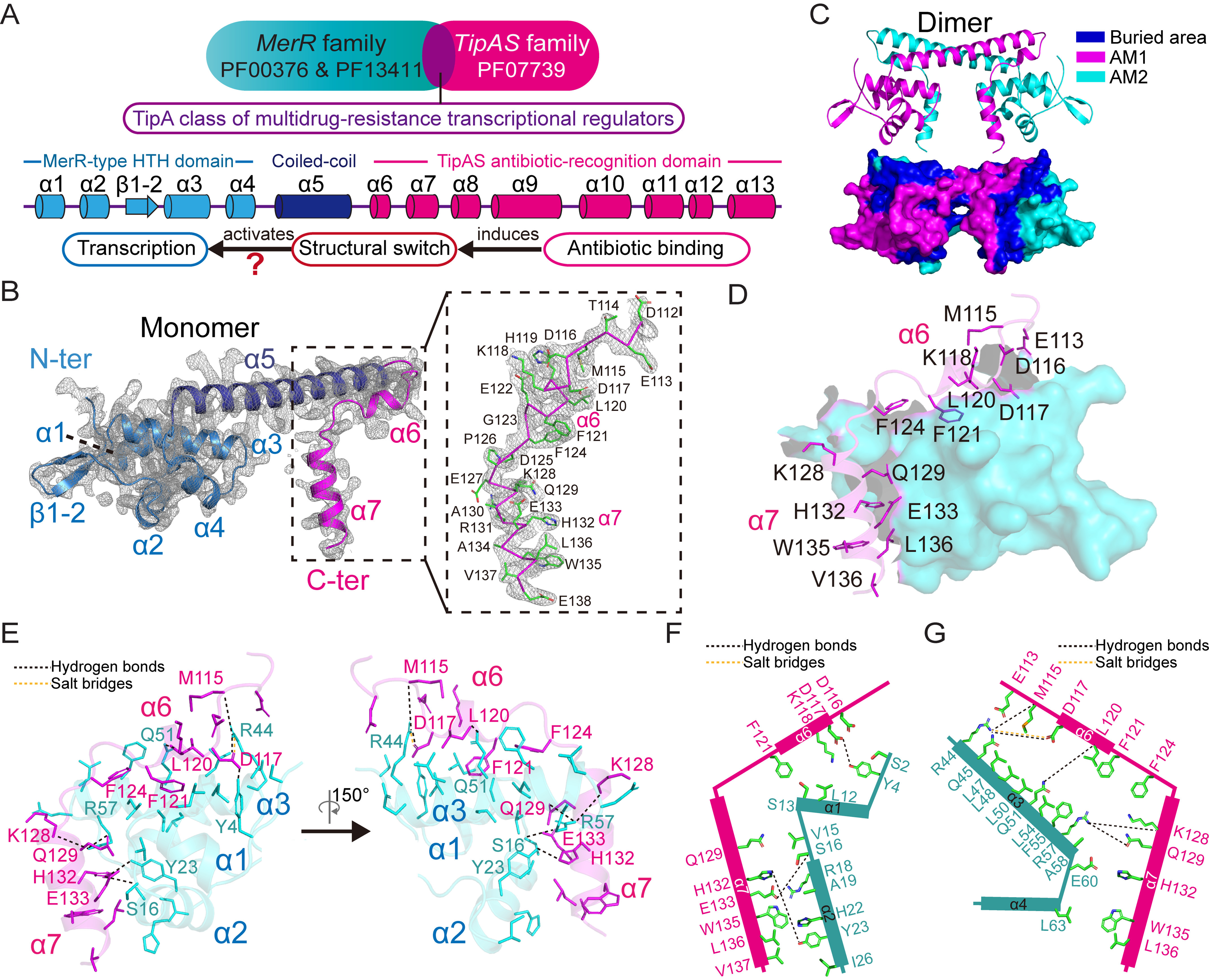
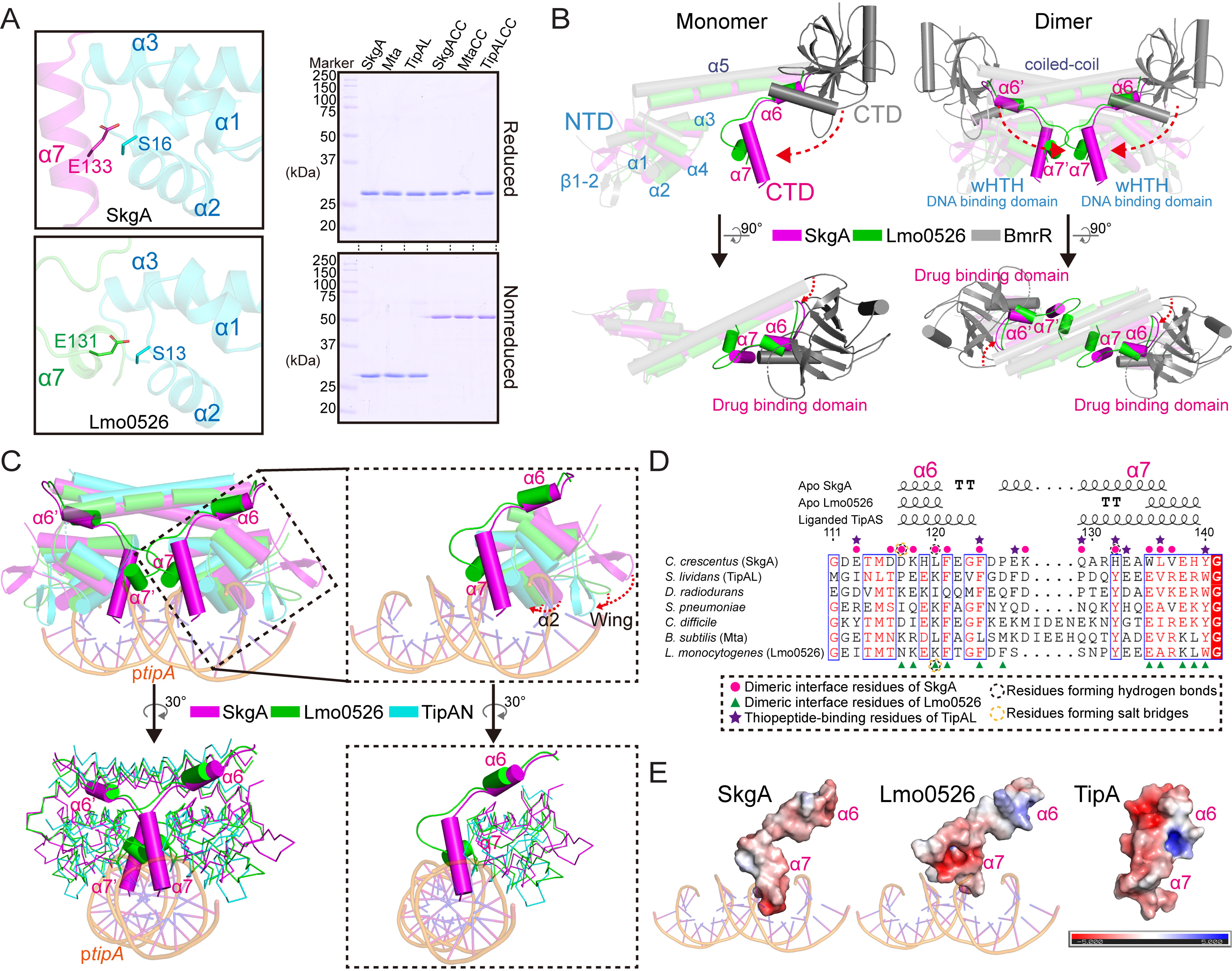
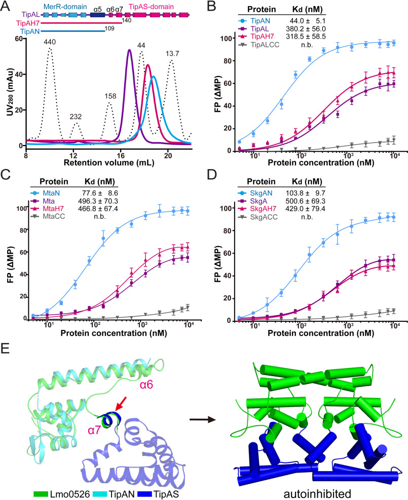
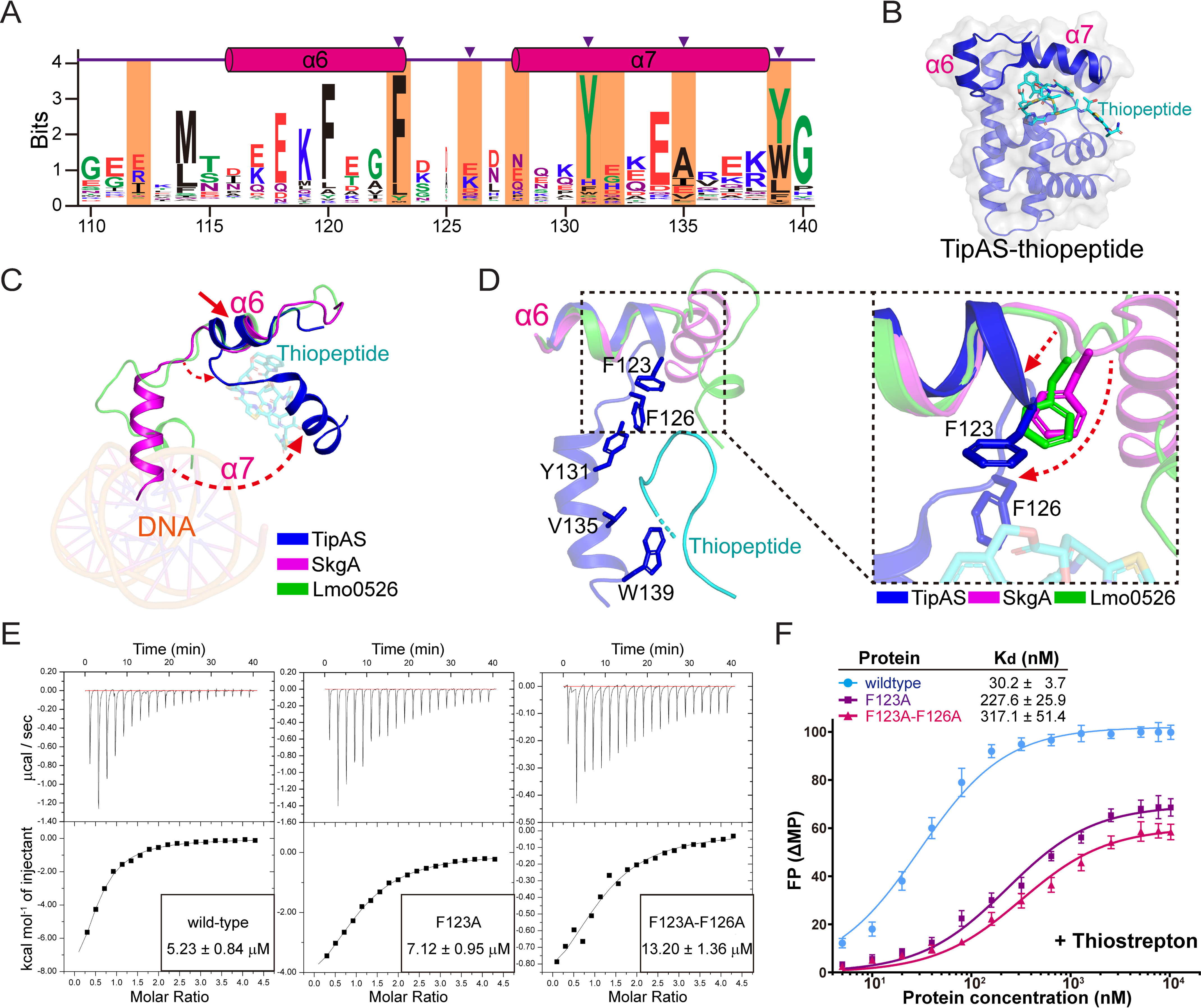
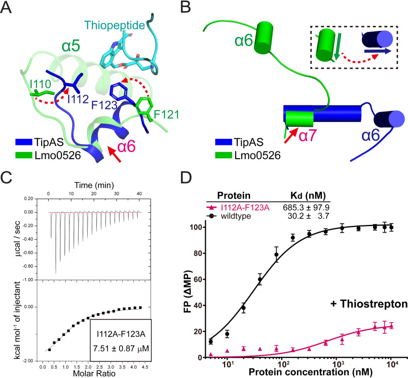
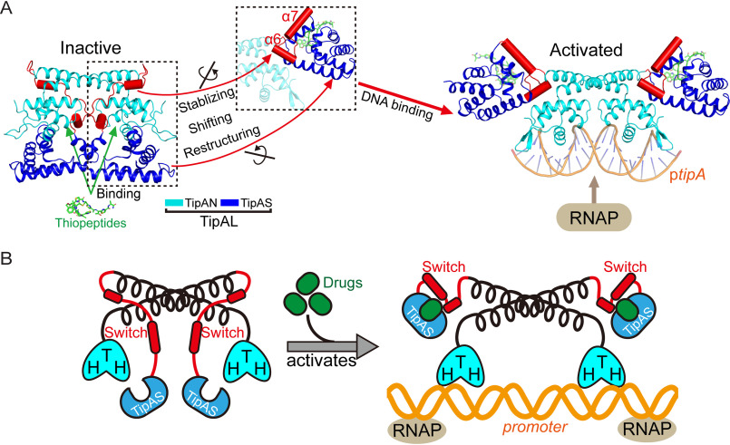
Investigations of bacterial resistance strategies can aid in the development of new antimicrobial drugs as a countermeasure to the increasing worldwide prevalence of bacterial antibiotic resistance. One such strategy involves the TipA class of transcription factors, which constitute minimal autoregulated multidrug resistance (MDR) systems against diverse antibiotics. However, we have insufficient information regarding how antibiotic binding induces transcriptional activation to design molecules that could interfere with this process. To learn more, we determined the crystal structure of SkgA from Caulobacter crescentus as a representative TipA protein. We identified an unexpected spatial orientation and location of the antibiotic-binding TipAS effector domain in the apo state. We observed that the α6-α7 region of the TipAS domain, which is canonically responsible for forming the lid of antibiotic-binding cleft to tightly enclose the bound antibiotic, is involved in the dimeric interface and stabilized via interaction with the DNA-binding domain in the apo state. Further structural and biochemical analyses demonstrated that the unliganded TipAS domain sterically hinders promoter DNA binding but undergoes a remarkable conformational shift upon antibiotic binding to release this autoinhibition via a switch of its α6-α7 region. Hence, the promoters for MDR genes including tipA and RNA polymerases become available for transcription, enabling efficient antibiotic resistance. These insights into the molecular mechanism of activation of TipA proteins advance our understanding of TipA proteins, as well as bacterial MDR systems, and may provide important clues to block bacterial resistance.

Keywords: DNA-binding protein; TipA; activation mechanism; antibiotic resistance; autoinhibition; crystal structure; drug resistance; multidrug resistance (MDR); structure biology; transcription promoter; transcription regulation; transcriptional regulator.


Conflict of interest statement

Conflict of interest—The authors declare no conflicts of interest with the contents of this article.

The authors declare no conflicts of interest with the contents of this article

Figures

Figure 1.
Figure 1.
Crystal structure of C. crescentus TipA-class protein SkgA. A, schematic diagrams showing the domain composition of the TipA-class regulators. The structure of TipA proteins is constituted mostly by α-helices with initial four helices within the DNA-binding domain, an intermediate coiled-coil α5 helix responsible for the dimerization, and the rest of the eight helices composing the TipAS domain. B, crystal structure of SkgA with one molecule in the asymmetric unit. The 2FoFc density map is contoured at 1σ. The α6–α7 region is highlighted in the right panel with ribbon representation for main chain and stick representation for side chain. N-ter, N terminus; C-ter, C terminus. C, dimeric structure of SkgA that is constructed by molecules from two adjacent asymmetric units. The protein surface is shown in the lower panel with buried area colored in blue. AM, asymmetric molecule. D, the α6–α7 region is involved in the dimeric interface. The α6–α7 region of one protomer interacted with the N-terminal DNA-binding domain of the other protomer. Amino acid residues of the α6–α7 region involved in the interaction are labeled with side chains shown as sticks. E, interactions between the α6–α7 region and N-terminal DNA-binding domain. Hydrogen bonds and salt bridges formed between residues are indicated by black and yellow dotted lines, respectively. The other labeled residues are involved in hydrophobic interactions. F, schematic diagram showing the interactions between the α6–α7 region and α1–α2 region in the SkgA dimer. G, schematic diagram showing the interactions between the α6–α7 region and α3–α4 region in the SkgA dimer.
Figure 2.
Figure 2.
Structural analysis revealed a potential inhibition of the TipA–DNA interaction by the conserved α6–α7 region. A, in-solution validation of the specific conformation of the α6–α7 region shown in crystal structure. Ser16 and Glu117 that are involved in the dimeric interface exhibit close location in the structure of the SkgA dimer; so are their corresponding residues Ser13 and Glu131 in the Lmo0526 structure (left panel). Formation of disulfide bonds of mutant and WT SkgA, Mta, and TipAL was analyzed using reduced and nonreduced SDS-PAGE (right panel). B, structural superposition of TipA-class proteins with BmrR indicated a distinctive spatial location of TipAS domain in the apo state. Cα atoms of each structure were used for the superposition in the PyMOL. The significant structural discrepancy on the orientation of C-terminal effector domains between TipA-class proteins and BmrR is indicated by red dashed arrows. NTD, N-terminal domain; CTD, C-terminal domain; wHTH, winged helix-turn helix motif. C, structural superposition of apo SkgA and Lmo0526 with promoter DNA-bound TipAN (PDB code 2VZ4). Structures are represented as cartoons with cylindrical helices in the upper panel and as ribbons except for the α6–α7 region in the bottom panel. Close-up views highlighting the α6–α7 helices and DNA are shown in the right panel. The putative shifts of α2 helix and wing region upon DNA binding are indicated by red dashed arrows. D, structure-based sequence alignment of the α6–α7 regions from TipA-class regulators. Amino acid numbering is given according to C. crescentus SkgA, and the corresponding second structural elements of SkgA, Lmo0526, and TipA were given above the sequences. The second structure of TipA was shown according to the liganded TipAS structure (PDB code 2MC0) with stabilized α6–α7 helices. Residues involved in the dimeric interface of SkgA and Lmo0526 are indicated by magenta circles and green arrowheads, respectively. Thiopeptide-binding residues of TipA are indicated by purple stars. Residues forming hydrogen bonds and salt bridges are indicated by black and yellow circled dots, respectively. E, electrostatic distribution of α6–α7 regions of TipA, SkgA, and Lmo0526. The liganded TipAS structure (PDB code 2MC0) was used here for the analysis of TipA. The APBS plugin within the program PyMOL was used for calculations of charge distribution.
Figure 3.
Figure 3.
Biochemical assessments indicated that the promoter-DNA interaction of unliganded TipA regulators is hindered by the TipAS domain. A, size-exclusion assays analyzing full-length and truncated TipAL proteins. Full-length TipAL and two truncations, TipAN (residues 1–109) and TipAH7 (residues 1–140), all exhibited as dimers in solution. Standard marker is indicated by dotted lines with the molecular size (kDa) of each protein peak marked. B–D, FPA examining binding affinities of TipA proteins (B), Mta proteins (C), and SkgA proteins (D) with their respective promoter DNA. Their double-cysteine mutants were also included in the assessment. The data were plotted using a base 10 logarithmic scale for the x axis and were fitted with a one-site binding model using the program GraphPad Prism7. E, the potential location of apo-state TipAS domain revealed by structural alignment. Structures of Lmo0526 and TipAS (PDB code 2MC0) were aligned at conserved and ordered α7 helices of relatively high sequence homology (indicated by a red arrow), and TipAN structure was further aligned to Lmo0526 (left panel). By the alignment, a possible ligand-free model for full-length TipA-class proteins was established with the TipAS domain lying beneath the HTH DNA-binding domain, which represents an inactive autoinhibited conformation (right panel).
Figure 4.
Figure 4.
An antibiotic-induced structural shift of α7 helix is required for transcriptional activation. A, results of multiple sequence alignment of the α6–α7 region. After removal of repeated entries from the TipAS family (PF07739 in Pfam database), the remaining complete sequences were realigned by using the program Clustal Omega as an input for the alignment via WebLogo3. Amino acid numbering and helical position are given according to S. lividans TipA. Residues involved in antibiotic interaction are highlighted in orange, and key residues responsible for antibiotic recognition are indicated by purple arrowheads. B, structural representation of thiopeptide-bound TipAS (PDB code 2MC0) with the α6 and α7 helices highlighted. C, structural comparison of the α7 helices from SkgA, Lmo0526, and thiopeptide-bound TipAS with an alignment at the α6 helices. Potential deflections of helix and loop are indicated by red dashed arrows. D, close-up view of thiopeptide-associating residues based on the alignment at α6 helices. Phe123 and Phe126 of the liganded TipAS in the loop region are highlighted. The potential structural deflections of loop regions upon antibiotic binding are indicated by red dashed arrows. E, ITC experiments assessing the thiopeptide-binding affinities of WT and mutant (F123A and F123A/F126A) TipAL proteins. The data were processed using the program Microcal-Origin. F, FPA experiments examining the promoter DNA-binding affinities of the WT and mutant TipAL proteins in the presence of thiostrepton. The data were plotted using a base 10 logarithmic scale for the x axis and were fitted with a one-site specific binding model using the program GraphPad Prism7.
Figure 5.
Figure 5.
The α6 helix may undergo a significant structural switch upon drug binding. A and B, structural comparisons of the thiopeptide-TipAS and apo Lmo0526 structures via alignments at the α6 helices (A) and at the conserved structured part of the α7 helices (B) for probing structural discrepancies of loop regions around the α6 helix and orientational difference of the α6 helices, respectively. Two thiopeptide-interacting residues of TipAS, Ile112 and Phe123, were highlighted for a comparison with their corresponding residues in Lmo0526. The aligned regions are indicated by red arrows, and the significant dissimilarities are indicated by red dashed arrows. C, ITC analysis of thiopeptide-binding affinity of I112A/F123A mutant of TipAL. The data were processed using the program Microcal-Origin. D, FPA analysis of the promoter DNA-binding affinities of the I112A/F123A mutant of TipAL in the presence of thiostrepton. The corresponding result of WT TipAL (also displayed in Fig. 4F) is shown here for a comparison. The data were plotted using a base 10 logarithmic scale for the x axis and were fitted with a one-site binding model using the program GraphPad Prism7.
Figure 6.
Figure 6.
Model representation for the structural mechanism of antibiotic-induced transcriptional activation of TipA-class proteins. A, the access of promoter DNA (ptipA) to N-terminal HTH motifs is sterically hindered by the unliganded TipAS domains in the apo inactive state (left panel). Once upon antibiotic bindings, the α6–α7 switch element undergoes stabilization, restructuring, and shifting (dotted boxes) and thus drives a global conformational transition of TipAS domains to release the inhibition of DNA-binding. Hence, TipAN is activated to bind ptipA and further recruit the RNAPs to promote MDR gene transcription (right panel). B, a simplified cartoon illustration of the above established model explaining transcriptional activation mechanism of TipA-class regulators.

Similar articles

Cited by

References

    1. Neyfakh A. A. (2002) Mystery of multidrug transporters: the answer can be simple. Mol. Microbiol. 44, 1123–1130 10.1046/j.1365-2958.2002.02965.x - DOI - PubMed
    1. Nikaido H. (2009) Multidrug resistance in bacteria. Annu. Rev. Biochem. 78, 119–146 10.1146/annurev.biochem.78.082907.145923 - DOI - PMC - PubMed
    1. Davies J. (1994) Inactivation of antibiotics and the dissemination of resistance genes. Science 264, 375–382 10.1126/science.8153624 - DOI - PubMed
    1. Kelley S. L., Basu A., Teicher B. A., Hacker M. P., Hamer D. H., and Lazo J. S. (1988) Overexpression of metallothionein confers resistance to anticancer drugs. Science 241, 1813–1815 10.1126/science.3175622 - DOI - PubMed
    1. Neyfakh A. A., Bidnenko V. E., and Chen L. B. (1991) Efflux-mediated multidrug resistance in Bacillus subtilis: Similarities and dissimilarities with the mammalian system. Proc. Natl. Acad. Sci. U.S.A. 88, 4781–4785 10.1073/pnas.88.11.4781 - DOI - PMC - PubMed

Publication types

MeSH terms