. 2021 May;29(5):827-838.
doi: 10.1038/s41431-020-00806-5. Epub 2021 Jan 16.

The genetic landscape of polycystic kidney disease in Ireland

Affiliations

The genetic landscape of polycystic kidney disease in Ireland

Katherine A Benson et al. Eur J Hum Genet. 2021 May.

Abstract

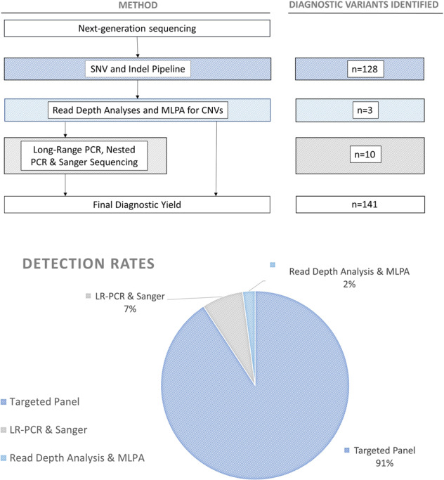
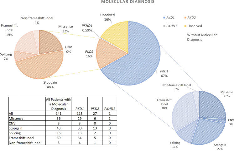
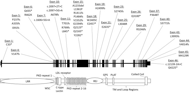
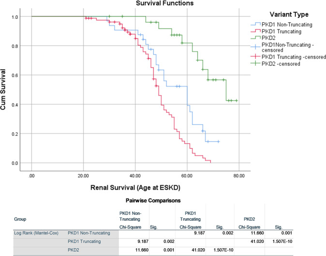
Polycystic kidney diseases (PKDs) comprise the most common Mendelian forms of renal disease. It is characterised by the development of fluid-filled renal cysts, causing progressive loss of kidney function, culminating in the need for renal replacement therapy or kidney transplant. Ireland represents a valuable region for the genetic study of PKD, as family sizes are traditionally large and the population relatively homogenous. Studying a cohort of 169 patients, we describe the genetic landscape of PKD in Ireland for the first time, compare the clinical features of patients with and without a molecular diagnosis and correlate disease severity with autosomal dominant pathogenic variant type. Using a combination of molecular genetic tools, including targeted next-generation sequencing, we report diagnostic rates of 71-83% in Irish PKD patients, depending on which variant classification guidelines are used (ACMG or Mayo clinic respectively). We have catalogued a spectrum of Irish autosomal dominant PKD pathogenic variants including 36 novel variants. We illustrate how apparently unrelated individuals carrying the same autosomal dominant pathogenic variant are highly likely to have inherited that variant from a common ancestor. We highlight issues surrounding the implementation of the ACMG guidelines for variant pathogenicity interpretation in PKD, which have important implications for clinical genetics.


Conflict of interest statement

The authors declare that they have no conflict of interest.

Figures

Fig. 1
Fig. 1. Detection rates of molecular causes of PKD.
Flow chart (above) describes the methodological workflow for the project alongside the number of AR and AD pathogenic variants identified at each stage. Chart (below) shows the rates of detection of pathogenic variants for each of the methods used. Variants displayed here were classified using Mayo Clinic pathogenicity guidelines.
Fig. 2
Fig. 2. Number and type of PKD1 and PKD2 AD pathogenic variants detected.
These charts describe the number and type of AD pathogenic variants identified in the Irish cohort (classified using Mayo Clinic pathogenicity guidelines). The central chart shows the gene in which variants were identified and the outer charts show the breakdown of variant types for each of those genes. Corresponding protein domains for the variants listed in this figure are shown in Supplementary Table 10.
Fig. 3
Fig. 3. Diagnostic PKD1 variants detected using custom next-generation sequencing pipeline.
Novel pathogenic PKD1 variants (classified using Mayo Clinic pathogenicity guidelines) detected in our Irish cohort shown across the PKD1 gene. The gene is divided into exons (1–46, as per NG_008617.1) above and key domains are shown below. LRR leucine-rich repeat; WSC cell wall integrity and stress response component; REJ receptor for egg jelly; GPS G protein-coupled receptor proteolytic site; PLAT polycystin-1, lipoxygenase, α toxin; TM transmembrane.
Fig. 4
Fig. 4. Kaplan Meier survival graph showing time to ESKD.
Kaplan Meier survival graph showing time to ESKD in those with pathogenic (classified using Mayo Clinic pathogenicity guidelines) truncating and non-truncating PKD1 variants and PKD2 variants. Results from the Log Rank (Mantel-Cox) are shown under the graph.

References

    1. Suwabe T, Shukoor S, Chamberlain AM, Killian JM, King BF, Edwards M, et al. Epidemiology of autosomal dominant polycystic kidney disease in olmsted county. Clin J Am Soc Nephrol. 2020;15:69–79. - PMC - PubMed
    1. ERA-EDTA Registry Committee. ERA-EDTA Registry Annual Report 2017. 2017.
    1. McEwan P, Bennett Wilton H, Ong ACM, Ørskov B, Sandford R, Scolari F, et al. A model to predict disease progression in patients with autosomal dominant polycystic kidney disease (ADPKD): the ADPKD Outcomes Model. BMC Nephrol. 2018;19:37. - PMC - PubMed
    1. Cornec-Le Gall E, Olson RJ, Besse W, Heyer CM, Gainullin VG, Smith JM, et al. Monoallelic mutations to DNAJB11 cause atypical autosomal-dominant polycystic kidney disease. Am J Hum Genet. 2018;102:832–44. - PMC - PubMed
    1. Audrézet MP, Cornec-Le Gall E, Chen JM, Redon S, Quéré I, Creff J, et al. Autosomal dominant polycystic kidney disease: comprehensive mutation analysis of PKD1 and PKD2 in 700 unrelated patients. Hum Mutat. 2012;33:1239–50. - PubMed

Publication types

LinkOut - more resources