Review
. 2021 Apr;101(4):412-422.
doi: 10.1038/s41374-020-00514-0. Epub 2021 Jan 16.

Artificial intelligence and computational pathology

Affiliations
Review

Artificial intelligence and computational pathology

Miao Cui et al. Lab Invest. 2021 Apr.

Abstract

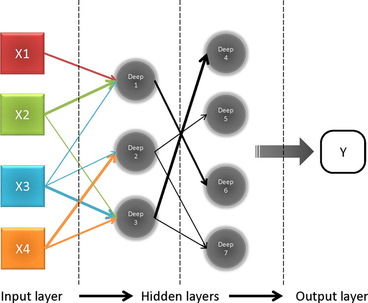
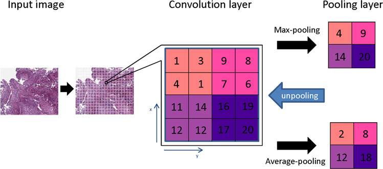
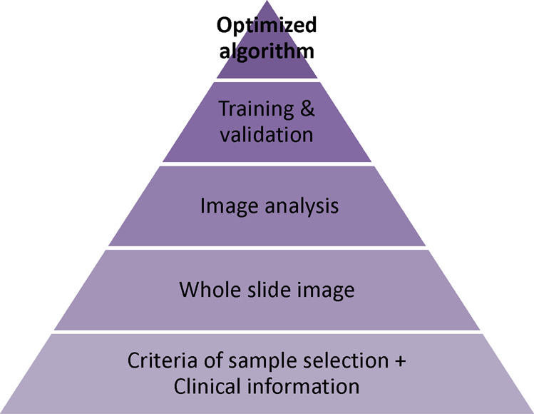
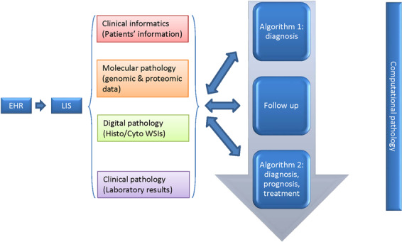
Data processing and learning has become a spearhead for the advancement of medicine, with pathology and laboratory medicine has no exception. The incorporation of scientific research through clinical informatics, including genomics, proteomics, bioinformatics, and biostatistics, into clinical practice unlocks innovative approaches for patient care. Computational pathology is burgeoning subspecialty in pathology that promises a better-integrated solution to whole-slide images, multi-omics data, and clinical informatics. However, computational pathology faces several challenges, including the ability to integrate raw data from different sources, limitation of hardware processing capacity, and a lack of specific training programs, as well as issues on ethics and larger societal acceptable practices that are still solidifying. The establishment of the entire industry of computational pathology requires far-reaching changes of the three essential elements connecting patients and doctors: the local laboratory, the scan center, and the central cloud hub/portal for data processing and retrieval. Computational pathology, unlocked through information integration and advanced digital communication networks, has the potential to improve clinical workflow efficiency, diagnostic quality, and ultimately create personalized diagnosis and treatment plans for patients. This review describes clinical perspectives and discusses the statistical methods, clinical applications, potential obstacles, and future directions of computational pathology.


Conflict of interest statement

The authors declare that they have no conflict of interest.

Figures

Fig. 1
Fig. 1. The basic structure of a deep neural network.
Each input value is assigned to each node from “X1–3” to “Xn” in the input layer (left). Nodes labeled “deep 1–7” are hidden layers behind the input layer. The values of hidden layers are not directly visible and are sent to the output layer after processed. Arrows connecting each node represent the direction and weight from previous layers. Both weights and nodes impact the network in generating an output (Y).
Fig. 2
Fig. 2. The principle of convolutional neural networks.
The input image is converted to numerical data (1–20) as the convolution layer. The convolutional neural network generates a pooling layer to reduce the dimensions of the image data as well as retain its characteristics for the statistic modeling. Several types of pooling methods including max pooling, which returns the maximum value from the portion of the image, and average pooling, which returns the average of all the values from the portion of the image. In addition, max pooling also performs de-noising along with dimensionality reduction, which improved analysis and accuracy.
Fig. 3
Fig. 3. Flow chart of algorithm training.
The process of creating an algorithm is divided into four necessary phases. The initial phase is to collect applicable samples along with clinical information. Next phase is to create whole-slide images with annotation. Based on the image analysis data, an algorithm was developed and trained by both the training set and the independent validation set.
Fig. 4
Fig. 4. Pathologist-centered medical system.
Clinical information from EHR, -omics data from molecular pathology, WSIs from digital pathology, and results from clinical laboratories aggregated into LIS to create “algorithm 1” for diagnosis. The updated disease-related data during follow-up are integrated into the previous data to build the “algorithm 2” over time for improved patient care.
Fig. 5
Fig. 5. The relation between different levels of artificial intelligence and the four bottlenecks that are facing currently.
The four challenges are experienced computational clinicians who are capable of developing algorithms of particular clinical issues, hardware limitations (i.e., cloud storage, computational capacity, network speed), qualified applicable data, and ethical issues.
Fig. 6
Fig. 6. Global pathology service model.
Each small green circle labeled “L” represents a local laboratory where the slides are scanned. Alternatively, slides can be scanned at a centralized scanning center (SC) where many local laboratories can send their slides for scanning. A central cloud laboratory with a large data storage and high capacity of computation will integrate, analyze, and store WSI data together with other medically related data.
Fig. 7
Fig. 7. Computational pathology team.
A computational pathology developer team includes pathologists to establish a clinically relevant issue, data scientists to develop and train the algorithm, and engineer to support the operating environment. During actual clinical practice, pathologists play a pivotal role in applying and monitoring the algorithm and relay feedback to the team to keep optimizing it.

References

    1. George MR, Johnson KA, Gratzinger DA, Brissette MD, McCloskey CB, Conran RM, et al. Will I need to move to get my first job?: geographic relocation and other trends in the pathology job market. Arch Pathol Lab Med. 2020;144:427–34. doi: 10.5858/arpa.2019-0150-CP. - DOI - PubMed
    1. Goldenberg SL, Nir G, Salcudean SE. A new era: artificial intelligence and machine learning in prostate cancer. Nat Rev Urol. 2019;16:391–403. doi: 10.1038/s41585-019-0193-3. - DOI - PubMed
    1. Parwani AV. Next generation diagnostic pathology: use of digital pathology and artificial intelligence tools to augment a pathological diagnosis. Diagn Pathol. 2019;14:138. doi: 10.1186/s13000-019-0921-2. - DOI - PMC - PubMed
    1. Liu Y, Gadepalli K, Norouzi M, Dahl GH, Kohlberger T, Boyko A, et al. Detecting cancer metastases on gigapixel pathology images. arXiv. 2017. https://arxiv.org/abs/1703.02442.
    1. McAlpine ED, Michelow P. The cytopathologist’s role in developing and evaluating artificial intelligence in cytopathology practice. Cytopathology. 2020;31:385–92. doi: 10.1111/cyt.12799. - DOI - PubMed