. 2021 Apr 12;180(2):383-394.
doi: 10.1093/toxsci/kfaa191.

Multiorgan Crystal Deposition of an Amphoteric Drug in Rats Due to Lysosomal Accumulation and Conversion to a Poorly Soluble Hydrochloride Salt

Affiliations

Multiorgan Crystal Deposition of an Amphoteric Drug in Rats Due to Lysosomal Accumulation and Conversion to a Poorly Soluble Hydrochloride Salt

Barbara Lenz et al. Toxicol Sci. .

Abstract

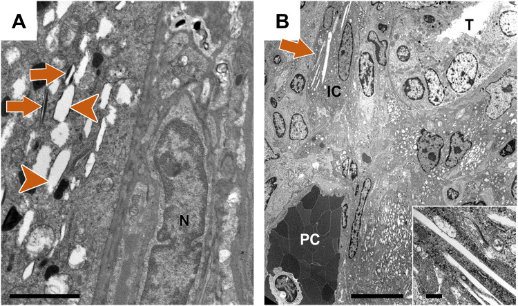
Poor solubility of drug candidates mainly affects bioavailability, but poor solubility of drugs and metabolites can also lead to precipitation within tissues, particularly when high doses are tested. RO0728617 is an amphoteric compound bearing basic and acidic moieties that has previously demonstrated good solubility at physiological pH but underwent widespread crystal deposition in multiple tissues in rat toxicity studies. The aim of our investigation was to better characterize these findings and their underlying mechanism(s), and to identify possible screening methods in the drug development process. Main microscopic features observed in rat RO0728617 toxicity studies were extensive infiltrates of crystal-containing macrophages in multiple organs. Matrix-assisted laser desorption/ionization Fourier transform ion cyclotron resonance mass spectrometry revealed that these crystals contained the orally administered parent compound, and locality was confirmed to be intracytoplasmic and partly intralysosomal by electron microscopic examination. Crystal formation was explained by lysosomal accumulation of the compound followed by precipitation of the hydrochloride salt under physiological conditions in the lysosomes, which have a lower pH and higher chloride concentration in comparison to the cytosol. This study demonstrates that risk of drug precipitation can be assessed by comparing the estimated lysosomal drug concentration at a given dose with the solubility of the compound at lysosomal conditions.

Keywords: crystal deposition; drug precipitation; lysosomal accumulation; macrophages; organ toxicity.


Figures

Figure 1.
Figure 1.
Chemical structure of RO0728617; an amphoteric compound. The acid dissociation constants (pKa) are given for the basic and acidic moieties. The molecular weight of RO0728617 is 453.6 g/mol, the melting point is 217°C and the LogD (pH7.4) is 1.5, as measured by the Carrier-Mediated Distribution System (Wagner et al., 2015). *Measured by photometric titration.
Figure 2.
Figure 2.
Histopathological examination. A, hematoxylin and eosin (H&E) stain of a lung from a female rat treated with RO0728617 at 90 mg/kg/day for 28 days showing enlarged intra-alveolar macrophages with vacuolated cytoplasms. Arrows show unchanged vessels. B, Lamp2 immunohistochemistry of a lung from a male rat treated with 90 mg/kg/day RO0728617 for 28 days in the mechanistic study, showing strong positive staining of cytoplasmic vacuoles in alveolar macrophages. Note: no sex differences were observed between (A and B). C, H&E stain of mesenteric lymph node from a female rat treated with 90 mg/kg/day for 28 days showing foamy macrophages in sinusoids (arrow), and single macrophages with intracytoplasmic crystal clefts (arrowhead). Scale bars = 50 µm.
Figure 3.
Figure 3.
Macrophage infiltrates of the bone marrow and parathyroid gland. A, hematoxylin and eosin (H&E) stain of bone marrow from the tibia of a male rat treated with RO0728617 at 90 mg/kg/day for 28 days showing macrophage infiltrates (arrowhead) with crystal clefts (arrow), and immunostaining with anti-CD68 confirming presence of macrophages in the bone marrow (B). Scale bars = 50 µm. C, H&E stain of parathyroid gland of a male rat treated with 90 mg/kg/day RO0728617 for 28 days shows normal parathyroid gland replaced by infiltrates of elongated macrophages containing crystal clefts, with occasional multinucleated cells (arrow). The surrounding thyroid gland is largely unchanged. Immunostaining with anti-CD68 confirms the presence of macrophages in the parathyroid gland (D). Scale bars = 200 µm.
Figure 4.
Figure 4.
Histopathological examination of the kidney. In the kidney of a female rat treated with RO0728617 at 90 mg/kg/day for 28 days, hematoxylin and eosin staining shows interstitial infiltrates of elongated, spindle-shaped macrophages (arrowhead) with crystal clefts (long arrows), and tubular cells with mitotic figures (short arrow, A). B, Tubules reveal degeneration and regeneration with mitoses (short arrow) and nuclear enlargement (long arrow); (dilated peritubular capillaries with increased number of mononuclear cells [arrowhead]). C, Normal cortex with unchanged glomeruli, arteries and capillaries. In the kidney of a male rat treated with RO0728617 at 90 mg/kg/day for 28 days, immunostaining with anti-CD68 confirms the presence of macrophages, with no crystals in the glomeruli (G) or tubules (D and E). Scale bars = 50 µm. Note: there were no sex differences between (A and E). F, In an unstained frozen section, massive crystal deposition in the renal medulla can be observed with polarized light. Note: no crystal deposits are present in the renal cortex. G, In the kidney of a male rat treated for 14 days (mechanistic study), immunostaining with anti-Lamp2 shows that crystal clefts within interstitial infiltrates are surrounded by positive membranes (encircled). Scale bar = 60 µm.
Figure 5.
Figure 5.
Histopathological examination of the eye. A, hematoxylin and eosin stain of the eye from a male rat treated with RO0728617 at 90 mg/kg/day for 28 days showing a subretinal cell cluster (likely to be macrophages) with crystal clefts (arrow) and retinal infolding. Note that adjacent tissue is unchanged. B, CD68 IHC of the eye from a female rat treated with RO0728617 at 90 mg/kg/day for 28 days showing the ciliary body with macrophage infiltrates and intracytoplasmic crystal clefts. Scale bars = 50 µm.
Figure 6.
Figure 6.
Molecular imaging of crystal deposits by matrix-assisted laser desorption/ionization Fourier transform ion cyclotron resonance (MALDI-FTICR) mass spectrometry (MS) in the eye of a male rat treated with RO0728617 at 90/70/90 mg/kg/day for 8 weeks. A, Polarized light microscopy image of the retina before MS image analysis visualizing crystal deposits (white arrows). B, Overlay of MALDI-FTICR MS image of the same retinal section with an optical scan to visualize spatial distribution of 454.1907 m/z, with a mass accuracy of <1 ppm, the protonated form of the parent compound orally administered to the animal.
Figure 7.
Figure 7.
Confirmation of crystal deposit presence by electron microscopy of the eyes and kidneys. A, Eye of a male rat treated with RO0728617 at 90/70/90 mg/kg/day for 8 weeks showing the pigment epithelium with intracellular crystal clefts surrounded by membranes (arrowheads) and single dark staining melanosomes with crystal clefts (arrows). Abbreviation: N, nucleus, scale bar = 2 µm. B, Kidney of a male rat treated with RO0728617 at 90 mg/kg/day for 28 days showing part of the tubular-interstitial space with interstitial cell infiltrates (macrophages). One Interstitial cell (IC) is shown with numerous intracytoplasmic crystals (arrow), peritubular capillary (PC) and tubules (T) are unremarkable. Scale bar = 10 µm. Intracellular crystals without clear-cut surrounding membrane are shown in the inset at 20-fold higher magnification. Scale bar = 500 nm.
Figure 8.
Figure 8.
Calculated pH-solubility profile of RO0728617 (free form) based on a measured solubility of 0.090 mg/ml at pH 7.96 (closed circle) and the compound’s basic pKa of 8.6 and acidic pKa of 7.2. The closed square represents the measured solubility of the RO0728617-hydrochloride (HCl) formed in 0.1 M hydrochloric acid. The open square shows the calculated pH of maximum solubility of the RO0728617-HCl salt (pHmax).
Figure 9.
Figure 9.
Schematic representation of the pH dependent partitioning of the amphoteric compound RO0728617 between blood plasma, the intracellular compartment and the lysosome. Abbreviations: Ccyto, u, free compound concentration in the cytosol; CLyso, u, free lysosomal concentration; Cplasma, u, free plasma concentration; Cplasma, t, total plasma concentration; fu, plasma, fraction unbound in plasma; Kp, uu Cytosol, the unbound drug partitioning coefficient of the cytosol; Kp, uu Lyso, unbound drug partitioning coefficient of the lysosome.
Figure 10.
Figure 10.
A, Flowchart describing the evaluation process of pre-clinical animal studies. When the ratio of Cmax, u in the cytosol/estimated solubility in the cytosol is higher or equal to 1, the likelihood that the compound precipitates within tissues is high, which may lead to a visible or invisible compound precipitation. Due to the accumulation of API in the lysosome, the CLyso, u at Cmax may exceed the estimated solubility in lysosome, which may also lead to API precipitation within the tissue. B, Flowchart describing the evaluation process at the level of the expected human therapeutic dose. In the preclinical phase, the human Cmax can be estimated with physiological-based pharmacokinetic modeling based on the available human in vitro data (Miller et al., 2019). When the ratio of Cmax, u in the cytosol/estimated solubility in the cytosol is ≥ 1, the likelihood that the compound precipitates within human tissues is high and the compound should be further optimized in the preclinical phase. If the ratio is < 1, it should be checked if CLyso, u at Cmax may exceed the estimated solubility in lysosome, which may to API precipitation within the human tissue. In this scenario, the compound should be optimized in the pre-clinical phase. Only if both ratios are < 1 should the compound proceed to the next development phase. Abbreviations: API, active pharmaceutical ingredient; Cmax (animal), maximal plasma concentration of the preclinical animal study; Cmax (human), maximal plasma concentration at the expected human therapeutic dose; Cmax, u, unbound concentration at Cmax; CLyso, u, unbound concentration in lysosomes at Cmax.

Similar articles

Cited by

References

    1. A, ziz R., Helms W. (2009). Pharmacology/toxicology review and evaluation: Pazopanib. Available at: https://www.accessdata.fda.gov/drugsatfda_docs/nda/2009/022465s000_Pharm....
    1. Baik J., Rosania G. (2012). Macrophages sequester clofazimine in an intracellular liquid crystal-like supramolecular organization. PLoS One 7, e47494. - PMC - PubMed
    1. Bhasin B., Urekli H., Atta M. (2015). Primary and secondary hyperoxaluria: Understanding the enigma. World J. Nephrol. 4, 235–244. - PMC - PubMed
    1. Bhatnagar V., Anjaiah S., Puri N., Darshanam B., Ramaiah A. (1993). pH of melanosomes of B 16 murine melanoma is acidic: Its physiological importance in the regulation of melanin biosynthesis. Arch. Biochem. Biophys. 307, 183–192. - PubMed
    1. Boyd B. J., Bergström C. A. S., Vinarov Z., Kuentz M., Brouwers J., Augustijns P., Brandl M., Bernkop-Schnürch A., Shrestha N., Préat V., et al. (2019). Successful oral delivery of poorly water-soluble drugs both depends on the intraluminal behavior of drugs and of appropriate advanced drug delivery systems. Eur. J. Pharm. Sci. 137, 104967. - PubMed

Publication types

Substances