. 2020 Sep 14;6(9):4851-4857.
doi: 10.1021/acsbiomaterials.0c01047. Epub 2020 Aug 24.

Gap Junction Liposomes for Efficient Delivery of Chemotherapeutics to Solid Tumors

Affiliations

Gap Junction Liposomes for Efficient Delivery of Chemotherapeutics to Solid Tumors

Andrea N Trementozzi et al. ACS Biomater Sci Eng. .

Abstract

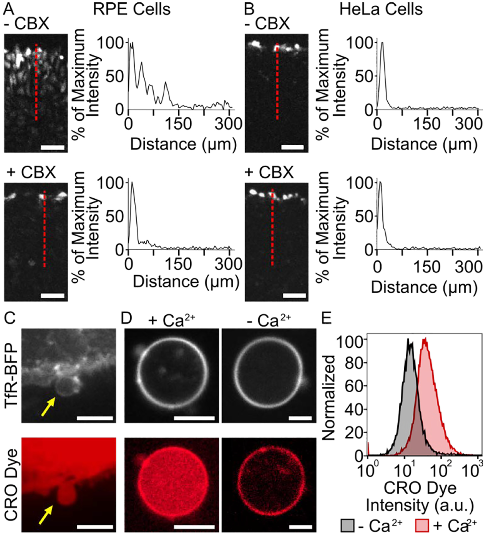
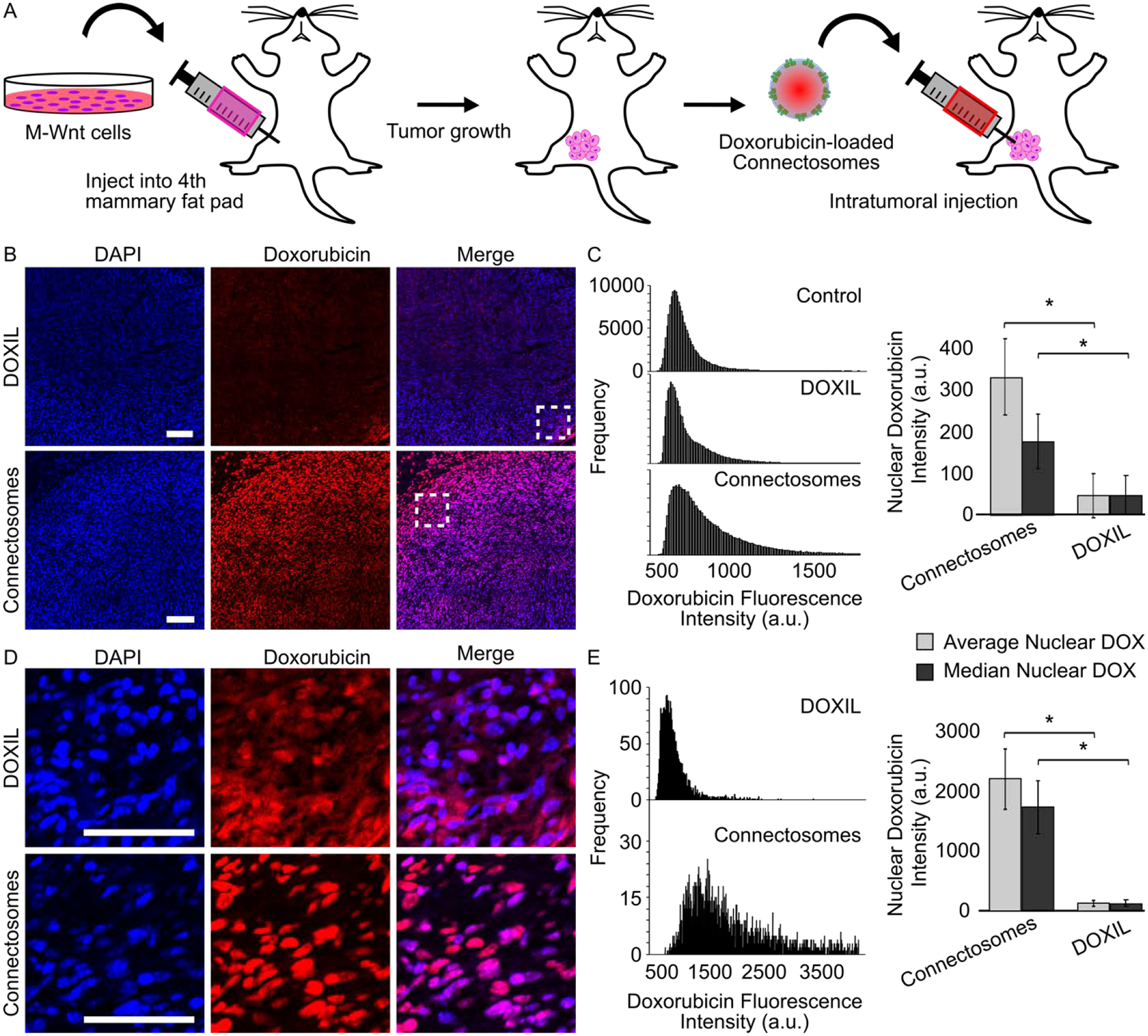
Chemotherapeutic delivery is limited by inefficient transport across cellular membranes. Here, we harness the cellular gap junction network to release therapeutic cargos directly into the cytosol. Specifically, cell-derived vesicles, termed connectosomes, contain gap junction transmembrane proteins that open a direct passageway to the cellular interior. Connectosomes were previously shown to substantially improve chemotherapeutic delivery in vitro. Here, we test connectosomes in vivo, using a murine breast tumor model. We demonstrate that connectosomes improve chemotherapeutic delivery to cellular targets within tumors by up to 16-fold, compared to conventional drug-loaded liposomes, suggesting an efficient alternative pathway for intracellular delivery.

Keywords: delivery; gap junction channels; intracellular; intratumoral; murine mammary tumor.


Conflict of interest statement

COMPETING INTERESTS

The authors declare no competing interest.

Figures

Figure 1.
Figure 1.
Characterization of Connectosome donor cells. Functional gap junctions between RPE donor cells (A) and HeLa cells (B) are evaluated based on lucifer yellow CH dye transport in a scratch loading assay. Cells were scratched in the absence and presence of 100 μM carbenoxolone (CBX) gap junction inhibitor. An intensity profile along the red dotted line indicates dye spreading. Scale bars indicate 50 μm. (C) Connectosomes can be extracted from RPE cells by a cellular membrane blebbing process and loaded with molecular dye molecules such as calcein red-orange (CRO) acetomethoxy (AM) dye. Scale bars indicate 10 μm. (D) Connectosomes retain CRO dye in the presence of calcium ions and release dye when calcium ions are removed by 5 mM of EDTA and EGTA as chelators. Upon removal of CRO dye from the lumen, the fluorescence of the dye is still visible in the membrane, most likely due to non-specific interactions of amphiphilic dye molecules with the membrane. Scale bars indicate 5 μm. (E) Flow cytometry histograms quantify CRO dye release from at least 10,000 connectosomes.
Figure 2.
Figure 2.
Doxorubicin-loaded Connectosomes and in vitro delivery to cells. (A) A schematic of passive loading of doxorubicin into Connectosomes by extrusion in a solution of doxorubicin. (B) A schematic of active loading of doxorubicin into Connectosomes by a transmembrane ammonium sulfate gradient, which drives precipitation of doxorubicin inside of vesicles. (C) A chart comparing encapsulation efficiency from passive loading and active loading. Bars indicate the mean encapsulation efficiency. A two-tailed t-test indicates p < 0.05. (D) Representative confocal fluorescence microscopy images of HeLa cells incubated for 2 h with 12 μg of doxorubicin in DOXIL or in Connectosomes. The dashed white lines outline the cells. Scale bars indicate 10 μm. (E) Flow cytometry histograms of doxorubicin fluorescence within HeLa cells after two-hour treatment with DOXIL, free doxorubicin, and doxorubicin-loaded Connectosomes. Median doxorubicin fluorescence intensity is quantified based on flow cytometry histograms. Error bars indicate standard deviation among three replicates. A two-tailed t-test indicates p < 0.05.
Figure 3.
Figure 3.
Intracellular delivery of doxorubicin to orthotopic mammary tumors in mouse models. (A) A schematic of the in vivo experimental process. M-Wnt cells were injected into the fourth mammary fat pad of nine mice at a 1×10⁶ cells/mouse to establish tumors. After tumors reached 4 – 7 mm in diameter, doxorubicin-loaded Connectosomes and DOXIL, each at 0.5 mg/kg, were injected directly into the tumors (n = 3). A third group of mice (n = 3) was left untreated. Mice were sacrificed 2 hours post intratumoral injection and tumors were analyzed. (B) Representative confocal fluorescence microscopy images of tumor sections after intratumoral injections of DOXIL and Connectosomes. DAPI staining was performed to visualize tumor cell nuclei. All images are adjusted to the same contrast settings and scale bars indicate 100 μm. (C) The nuclear doxorubicin fluorescence intensities from each entire tumor were plotted as histograms, and a bar chart quantifies the average nuclear doxorubicin intensity (light gray bar) and the median nuclear doxorubicin intensity (dark gray bar). Error bars indicate standard error of the mean, and a two-tailed t-test indicates p < 0.05. (D) Enlarged confocal microscopy images from the insets in (B) provide a closer view of nuclear doxorubicin fluorescence. (E) The nuclear doxorubicin fluorescence intensities from the brightest region within each tumor were plotted as histograms, and a bar chart quantifies the average nuclear doxorubicin intensity (light gray bar) and the median nuclear doxorubicin intensity (dark gray bar). Error bars indicate standard error of the mean, and a two tailed- t-test indicates p < 0.05.

Similar articles

Cited by

References

    1. Blanco E; Shen H; Ferrari M, Principles of nanoparticle design for overcoming biological barriers to drug delivery. Nat Biotechnol 2015, 33 (9), 941–51. - PMC - PubMed
    1. Wang AZ; Langer R; Farokhzad OC, Nanoparticle Delivery of Cancer Drugs. 10.1146/annurev-med-040210-1625442012, 63, 185–198. - DOI - PubMed
    1. Barenholz Y, Doxil(R)--the first FDA-approved nano-drug: lessons learned. J Control Release 2012, 160 (2), 117–34. - PubMed
    1. Ragelle H; Danhier F; Préat V; Langer R; Anderson DG, Nanoparticle-based drug delivery systems: a commercial and regulatory outlook as the field matures. 10.1080/17425247.2016.12441872016, 14 (7), 851–864. - DOI - PubMed
    1. Tran S; DeGiovanni PJ; Piel B; Rai P, Cancer nanomedicine: a review of recent success in drug delivery. Clin Transl Med 2017, 6 (1), 44. - PMC - PubMed

Publication types