Review
. 2021 Jan 18;15(1):13.
doi: 10.1186/s13256-020-02630-3.

An invasive mole with pulmonary metastases in a 55-year-old postmenopausal Syrian woman: a case report and review of the literature

Affiliations
Review

An invasive mole with pulmonary metastases in a 55-year-old postmenopausal Syrian woman: a case report and review of the literature

Sawsan Ismail et al. J Med Case Rep. .

Abstract

Background: Invasive mole is a subtype of gestational trophoblastic neoplasms (GTNs) that usually develops from the malignant transformation of trophoblastic tissue after molar evacuation. Invasive moles mostly occur in women of reproductive age, while they are extremely rare in postmenopausal women.

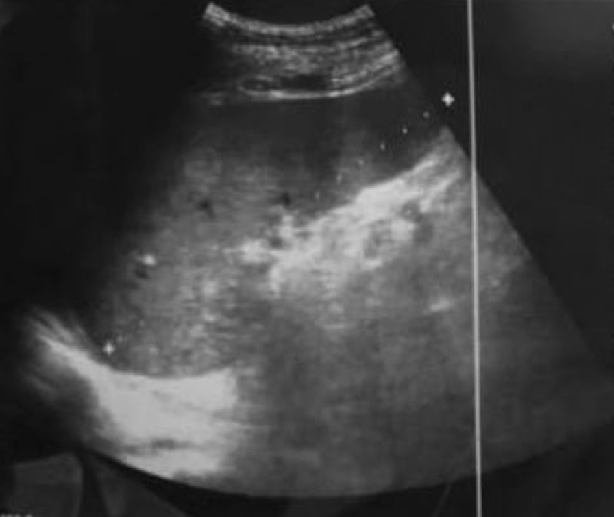
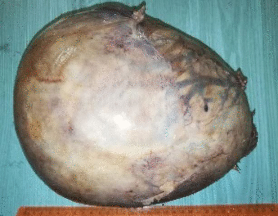
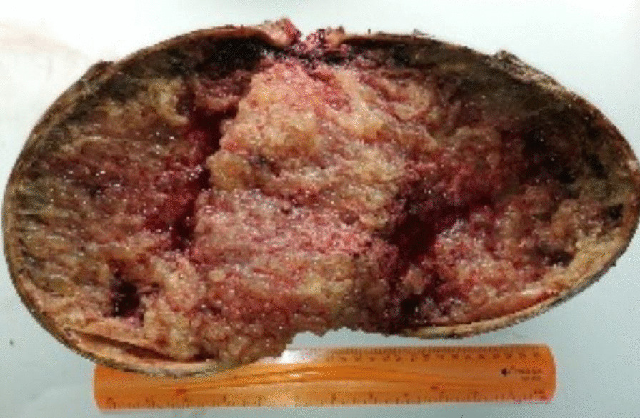
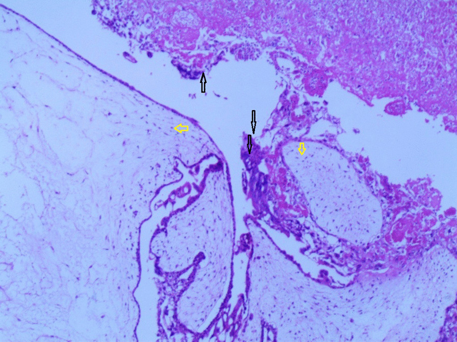
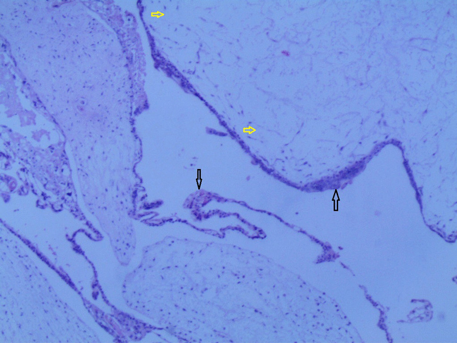
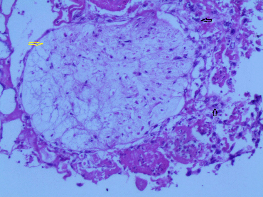
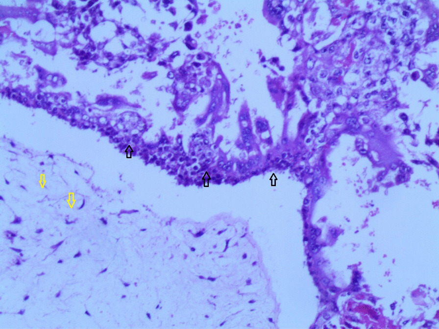
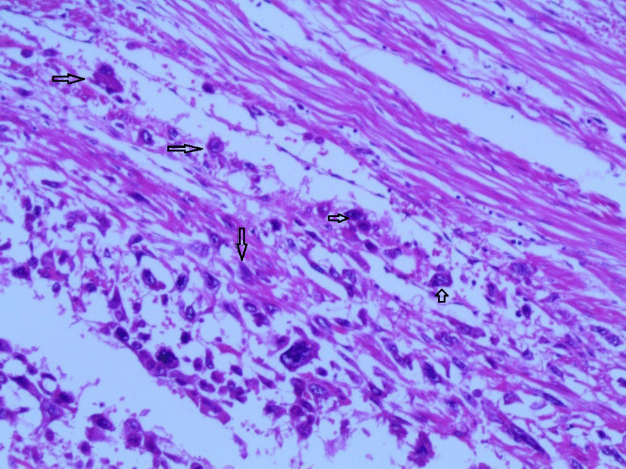
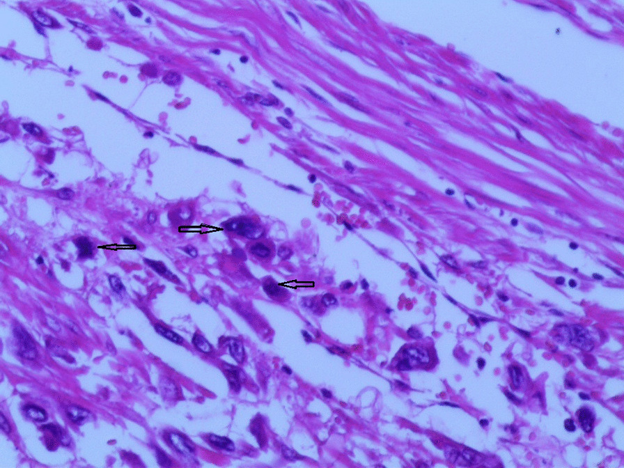
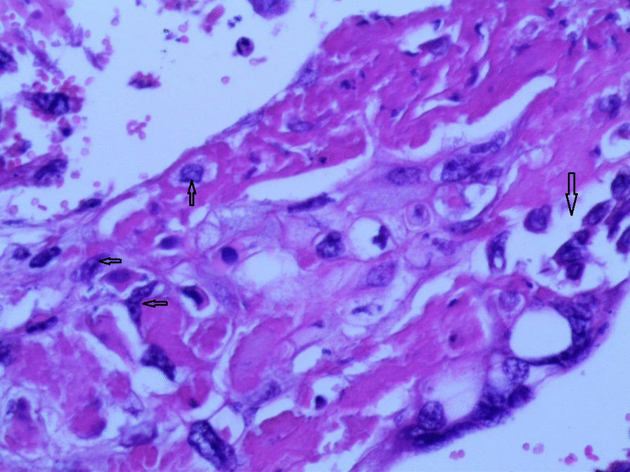
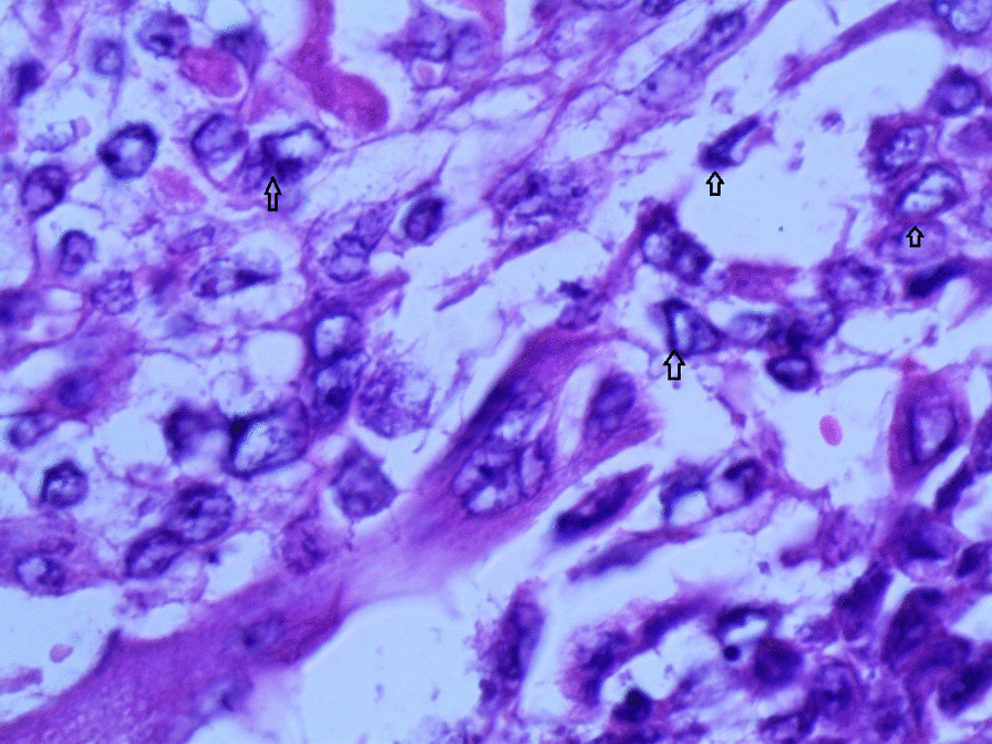
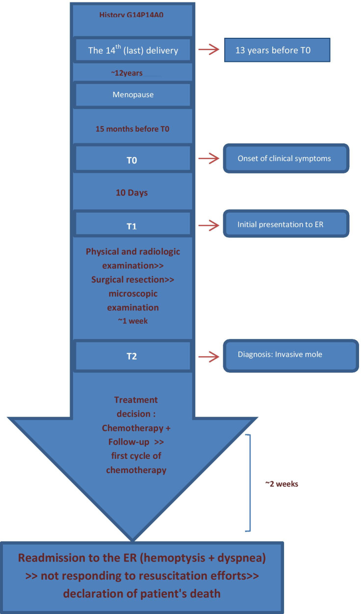
Case presentation: We present the case of a 55-year-old postmenopausal Syrian woman who was admitted to the emergency department at our hospital due to massive vaginal bleeding for 10 days accompanied by constant abdominal pain with diarrhea and vomiting. Following clinical, laboratory and radiological examination, total hysterectomy with bilateral salpingo-oophorectomy was performed. Histologic examination of the resected specimens revealed the diagnosis of an invasive mole with pulmonary metastases that were diagnosed by chest computed tomography (CT). Following surgical resection, the patient was scheduled for combination chemotherapy. However, 2 weeks later the patient was readmitted to the emergency department due to severe hemoptysis and dyspnea, and later that day the patient died in spite of resuscitation efforts.

Conclusion: Although invasive moles in postmenopausal women have been reported previously, we believe our case is the first reported from Syria. Our case highlights the difficulties in diagnosing invasive moles in the absence of significant history of gestational trophoblastic diseases. The present study further reviews the diagnostic methods, histological characteristics and treatment recommendations.

Keywords: Gestational trophoblastic neoplasms; Invasive mole; Postmenopausal woman; Pulmonary metastases.


Conflict of interest statement

The authors declare that they have no competing interests.

Figures

Fig. 1
Fig. 1
Pelvic ultrasound demonstrating an enlarged uterus with a heterogeneous mass obliterating the endometrial cavity, with a vesicular appearance
Fig. 2
Fig. 2
A macroscopic image of the resected mass measuring 25 × 20 × 13 cm
Fig. 3
Fig. 3
A macroscopic image of the endometrial cavity which was filled with hemorrhagic villi and edematous grape-like vesicles
Fig. 4
Fig. 4
A microscopic image demonstrating a circumferential proliferation of abnormal hyperchromatic trophoblastic cells (black arrows) surrounding edematous hydropic villi (yellow arrows) (hematoxylin and eosin [H&E] stain, original magnification ×40)
Fig. 5
Fig. 5
A microscopic image demonstrating edematous hydropic villi (yellow arrows) surrounded by abnormal trophoblastic cells (black arrows) (hematoxylin and eosin [H&E] stain, original magnification ×40)
Fig. 6
Fig. 6
A microscopic image with higher magnification demonstrating an edematous hydropic villus (a yellow arrow) surrounded by abnormal hyperchromatic trophoblastic cells (black arrows) (hematoxylin and eosin [H&E] stain, original magnification ×100)
Fig. 7
Fig. 7
A microscopic image with higher magnification demonstrating a circumferential proliferation of abnormal hyperchromatic trophoblastic cells (up to the right: black arrows) surrounding edematous hydropic villi (down to the left: yellow arrows) (hematoxylin and Eosin [H&E] stain, original magnification ×100)
Fig. 8
Fig. 8
A microscopic image demonstrating the abnormal trophoblastic cells (black arrows) invading the myometrium (hematoxylin and eosin [H&E] stain, original magnification ×100)
Fig 9
Fig 9
A microscopic image demonstrating the abnormal trophoblastic cells (black arrows) invading the myometrium with areas of necrosis and hemorrhage (hematoxylin and eosin [H&E] stain, original magnification ×100)
Fig 10
Fig 10
Microscopic images with higher magnification demonstrating the abnormal trophoblastic cells (black arrows) invading the myometrium (hematoxylin and eosin [H&E] stain, original magnification ×200)
Fig 11
Fig 11
Microscopic images with higher magnification demonstrating the abnormal trophoblastic cells (black arrows) invading the myometrium (hematoxylin and eosin [H&E] stain, original magnification ×200)
Fig. 12
Fig. 12
A microscopic image with high magnification demonstrating the atypical trophoblastic cells (black arrows) (hematoxylin and eosin [H&E] stain, original magnification ×400)
Fig. 13
Fig. 13
A microscopic image with high magnification demonstrating the atypical trophoblastic cells (black arrows) (hematoxylin and eosin [H&E] stain, original magnification ×600)
Fig. 14
Fig. 14
A timeline of the patient’s case

References

    1. Seckl MJ, Sebire NJ, Berkowitz RS. Gestational trophoblastic disease. Lancet. 2010;376(9742):717–729. doi: 10.1016/S0140-6736(10)60280-2. - DOI - PubMed
    1. Altieri A, Franceschi S, Ferlay J, et al. Epidemiology and aetiology of gestational trophoblastic diseases. Lancet Oncol. 2003;4(11):670–678. doi: 10.1016/S1470-2045(03)01245-2. - DOI - PubMed
    1. Tsukamoto N, Iwasaka T, Kashimura Y, et al. Gestational trophoblastic disease in women aged 50 or more. Gynecol Oncol. 1985;20:53–61. doi: 10.1016/0090-8258(85)90124-6. - DOI - PubMed
    1. Martínez Leocadio C, García Villayzán J, García-Foncillas López J, et al. Invasive mole in perimenopausal woman with lung and vaginal metastases: a case report. Clin Case Rep. 2019;7:2300–2305. doi: 10.1002/ccr3.2386. - DOI - PMC - PubMed
    1. de la Fouchardière A, Cassignol A, Benkiran L. Môle hydatiforme invasive chez une patiente ménopausée [Invasive hydatiform mole in a postmenopausal woman] Ann Pathol. 2003;1:443–446. - PubMed

MeSH terms