. 2021 Apr 21;67(2):89-97.
doi: 10.1262/jrd.2020-107. Epub 2021 Jan 15.

HGF/c-Met signaling regulates early differentiation of placental trophoblast cells

Affiliations

HGF/c-Met signaling regulates early differentiation of placental trophoblast cells

Yeling Ma et al. J Reprod Dev. .

Abstract

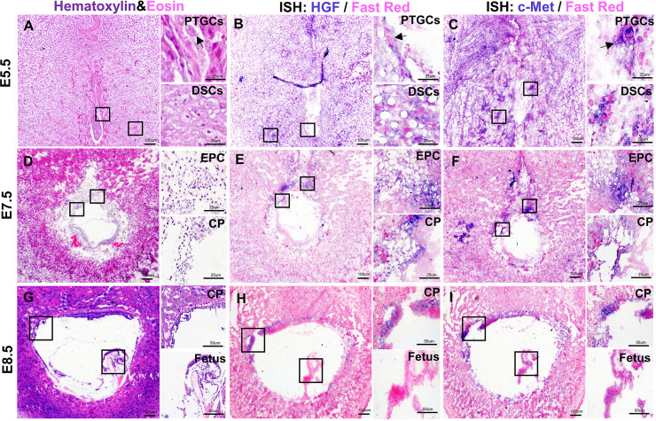
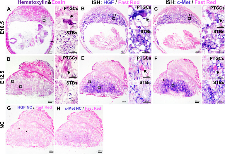
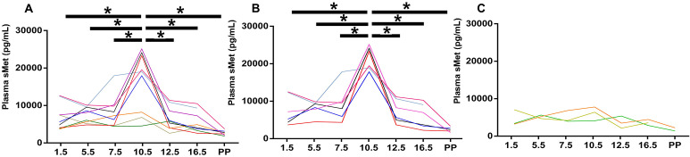
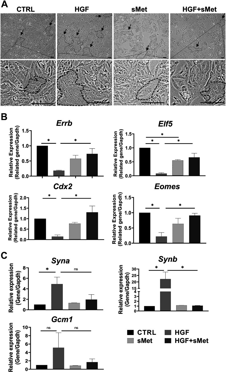
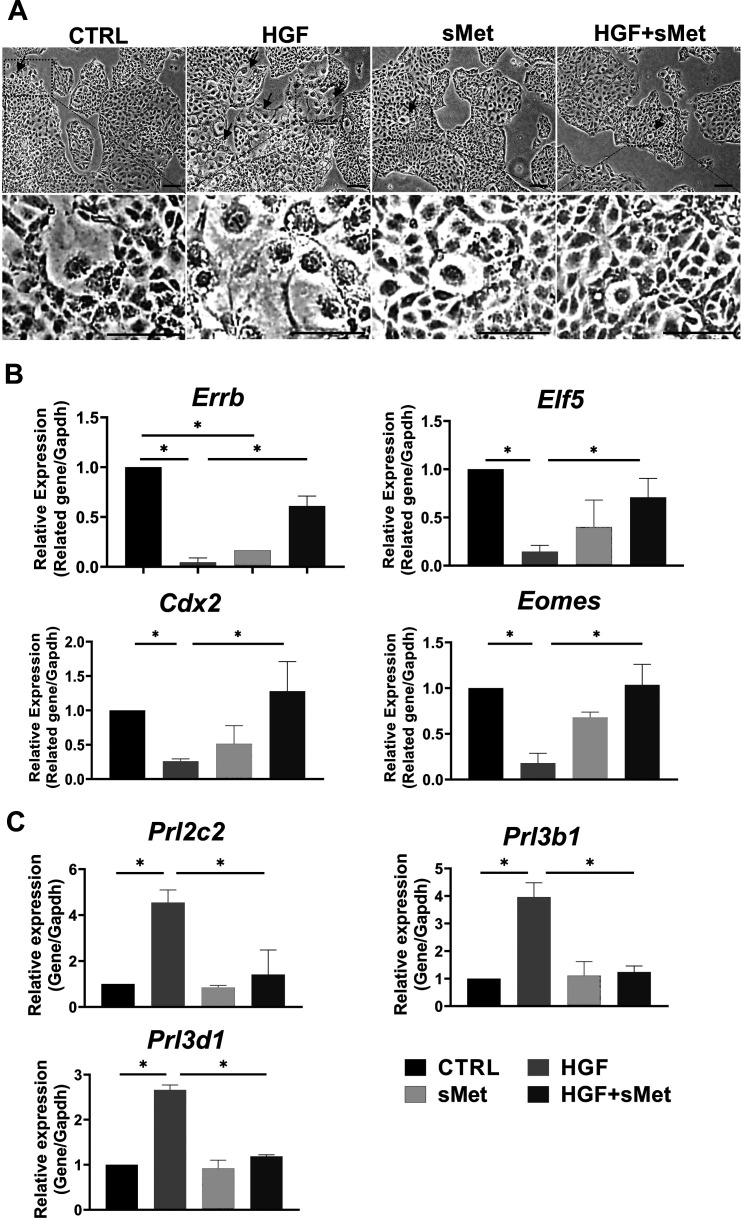
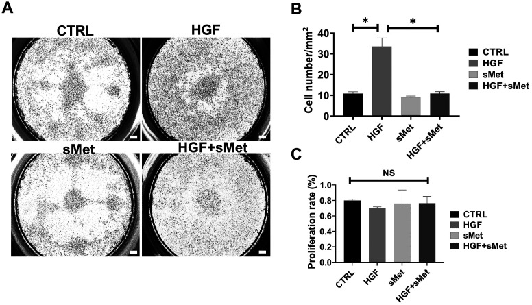
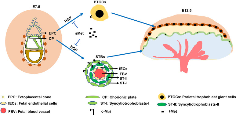
Depletion of hepatocyte growth factor (HGF) or mesenchymal-epithelial transition factor (c-Met) in mice leads to fetal lethality and placental maldevelopment. However, the dynamic change pattern of HGF/c-Met signaling during placental development and its involvement in the early differentiation of trophoblasts remain to be elucidated. In this study, using in situ hybridization assay, we elaborately demonstrated the spatial-temporal expression of Hgf and c-Met in mouse placenta from E5.5, the very early stage after embryonic implantation, to E12.5, when the placental structure is well developed. The concentration of the soluble form of c-Met (sMet) in maternal circulation peaked at E10.5. By utilizing the induced differentiation model of mouse trophoblast stem cells (mTSCs), we found that HGF significantly promoted mTSC differentiation into syncytiotrophoblasts (STBs) and invasive parietal trophoblast giant cells (PTGCs). Interestingly, sMet efficiently reversed the effect of HGF on mTSC differentiation. These findings indicate that HGF/c-Met signaling participates in regulating placental trophoblast cell fate at the early differentiation stage and that sMet acts as an endogenous antagonist in this aspect.

Keywords: Hepatocyte growth factor (HGF); Mesenchymal-epithelial transition factor (c-Met); Placenta; Soluble form of c-Met (sMet); Syncytiotrophoblast cells; Trophoblast giant cells.


Conflict of interest statement

The authors declare no conflict of interest.

Figures

Fig. 1.
Fig. 1.
The localization of Hgf and c-Met during early placental development in CD1 mice. A: H&E staining of E5.5 placenta. B: In situ hybridization for Hgf on E5.5 placenta. C: In situ hybridization for c-Met on E5.5 placenta. D: H&E staining of E7.5 placenta. E: In situ hybridization for Hgf on E7.5 placenta. F: In situ hybridization for c-Met on E7.5 placenta. G: H&E staining of E8.5 placenta. H: In situ hybridization for Hgf on E8.5 placenta. I: In situ hybridization for c-Met on E8.5 placenta. PTGCs, parietal trophoblast giant cells; EPC, ectoplacental cone; CP, chorionic plate. Arrows indicate PTGCs. Scale bars are shown in panels.
Fig. 2.
Fig. 2.
The localization of Hgf and c-Met during the development of mid-placenta in CD1 mice. A: H&E staining of E10.5 placenta. B: In situ hybridization for Hgf on E10.5 placenta. C: In situ hybridization for c-Met on E10.5 placenta. D: H&E staining of E12.5 placenta. E: In situ hybridization for Hgf on E12.5 placenta. F: In situ hybridization for c-Met on E12.5 placenta. G: In situ hybridization of negative controls for Hgf probes. H: In situ hybridization of negative controls for c-Met probes. STBs, syncytiotrophoblast cells; PTGCs, parietal trophoblast giant cells. Arrows indicate PTGCs. Scale bars are shown in panels.
Fig. 3.
Fig. 3.
ELISA for plasma sMet in CD1 pregnant mice. A: The concentration of plasma sMet during pregnancy. B: The concentration of plasma sMet in the sMet-high group. C: The concentration of plasma sMet in the sMet-low group. PP, postpartum. Comparison between groups was performed with one-way ANOVA multiple comparisons and analyzed with Tukey-Kramer test. * P < 0.05.
Fig. 4.
Fig. 4.
Met blocked HGF-induced differentiation of STBs. A: A typical result for morphological observation of the mTSCs among different groups. B: Relative mRNA expression of stemness markers after different treatments (n = 3). C: Relative mRNA expression of STB markers after different treatments. Arrows indicate STBs (n = 3). Scale bars: 100 μm. Data are presented as mean ± SD. Comparison between groups was performed with one-way ANOVA multiple comparisons and analyzed with Tukey-Kramer test. NS, no significance. * P < 0.05.
Fig. 5.
Fig. 5.
Met blocked HGF-induced differentiation of PTGCs. A: A typical result for morphological observation of the mTSCs among different groups. B: Relative mRNA expression of stemness markers after different treatments (n = 3). C: Relative mRNA expression of TGC markers after different treatments (n = 3). Arrows indicate TGCs. Scale bars: 100 μm. Data are presented as mean ± SD. Comparison between groups was performed with one-way ANOVA multiple comparisons and analyzed with Tukey-Kramer test. * P < 0.05.
Fig. 6.
Fig. 6.
Met blocked the cell invasion of HGF-induced PTGCs. A: Transwell assay among different groups. B: Statistical analysis of invasive cells among different groups (n = 3). C: Cell proliferation analysis among different groups (n = 3). Scale bars are shown in panels. Data are presented as mean ± SD. Comparison between groups was performed with one-way ANOVA multiple comparisons and analyzed with Tukey-Kramer test. * P < 0.05. NS, no significance. Scale bars: 100 μm.
Fig. 7.
Fig. 7.
A scheme of the role of HGF/c-Met signal in trophoblast differentiation. Autocrine HGF/c-Met signaling pathway for the regulation of trophoblast differentiation.

Similar articles

Cited by

References

    1. Ma WW, Adjei AA. Novel agents on the horizon for cancer therapy. CA Cancer J Clin 2009; 59: 111–137. - PubMed
    1. Bottaro DP, Rubin JS, Faletto DL, Chan AM, Kmiecik TE, Vande Woude GF, Aaronson SA. Identification of the hepatocyte growth factor receptor as the c-met proto-oncogene product. Science 1991; 251: 802–804. - PubMed
    1. Stoker M, Gherardi E, Perryman M, Gray J. Scatter factor is a fibroblast-derived modulator of epithelial cell mobility. Nature 1987; 327: 239–242. - PubMed
    1. Nakamura T, Nishizawa T, Hagiya M, Seki T, Shimonishi M, Sugimura A, Tashiro K, Shimizu S. Molecular cloning and expression of human hepatocyte growth factor. Nature 1989; 342: 440–443. - PubMed
    1. Jiang WG, Martin TA, Parr C, Davies G, Matsumoto K, Nakamura T. Hepatocyte growth factor, its receptor, and their potential value in cancer therapies. Crit Rev Oncol Hematol 2005; 53: 35–69. - PubMed

MeSH terms