

The complement system in primary Sjögren's syndrome: the expression of certain cascade and regulatory proteins in labial salivary glands - observational study
- PMID: 33456078
- PMCID: PMC7792541
- DOI: 10.5114/reum.2020.102000
The complement system in primary Sjögren's syndrome: the expression of certain cascade and regulatory proteins in labial salivary glands - observational study
Abstract
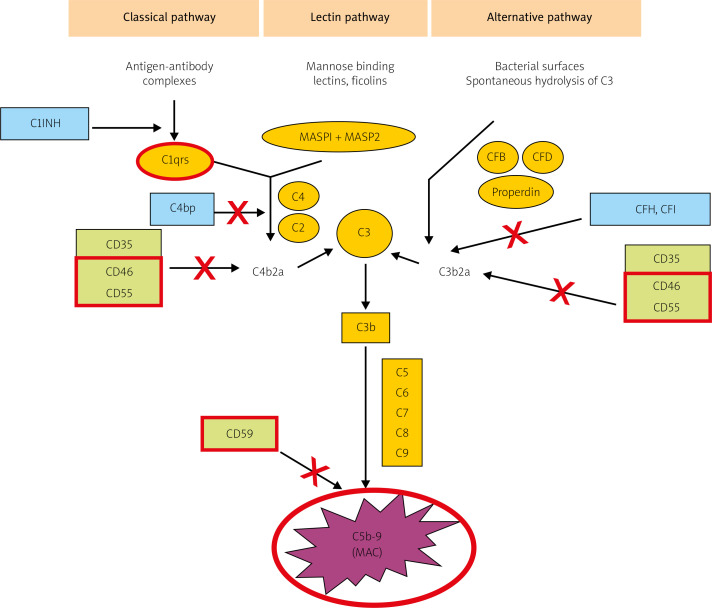
Introduction: The complement cascade and regulatory proteins are involved in the pathogenesis of the Sjögren's syndrome and other autoimmune diseases. The complement activation via the alternative pathway was recognized as a major pathogenic mechanism in autoimmune conditions. The aim of this study was to assess expression of complement cascade components and regulatory proteins in minor salivary glands in patients with primary Sjögren's syndrome (pSS).
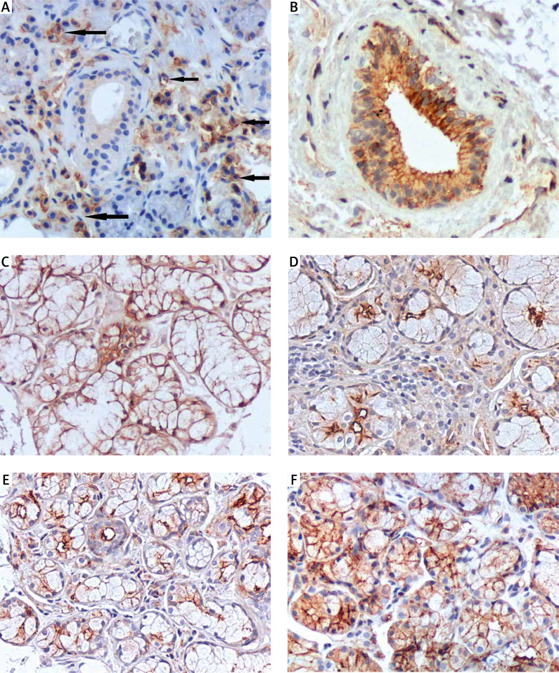
Materials and methods: The expression of C1q and C5b-9 - membrane attack complex and regulatory proteins such as: membrane cofactor protein (MCP), decay-accelerating factor (DAF) and protectin were examined using immunochemistry method in specimens from biopsy of minor salivary glands in pSS patients. The biopsy material was obtained from 20 pSS patients, 5 patients with non-specific sialadenitis and from 5 patients with suspicion of dryness syndrome without sialadenitis confirmation.
Results: None of the examined samples showed the expression of C1q or the effector C5b-9. Membrane cofactor protein expression was lower in pSS group than in both non-specific sialadenitis and noninflamed salivary glands. The inflammatory cells in pSS samples partially expressed MCP. There were differences in the sites and intensity of membrane protectin expression exclusively on the luminal surfaces in pSS; on the luminal and, partially, antiluminal surface in non-specific inflammation, and on the entire cell surface in unaffected salivary glands. There were no DAF expression in salivary gland tissue in biopsy specimens in all studied subjects.
Conclusions: The study demonstrated the absence of complement-cascade proteins (C1q, MAC) in the salivary glands of pSS patients, which may indicated a lack of local complement activation via the classical pathway and the observed gland tissue damage being due to a mechanism other than MAC-induced cytolysis. The differences in the expression of complement regulatory proteins between pSS, non-specific sialadenitis, and normal salivary glands may indicate that alternative functions of these regulatory proteins may be of greater significance in pSS. Low MCP expression in pSS in comparison with non-specific sialadenitis and normal salivary glands, may suggest altered modulation of cell-mediated immunity in pSS. The differences in the location and intensity of protectin (CD59) expression indicates a possibility of reducing the proinflammatory effect of protectin in pSS.
Keywords: complement system proteins; membrane cofactor protein CD46; primary Sjogren’s syndrome; protectin CD59.
Copyright: © 2020 Narodowy Instytut Geriatrii, Reumatologii i Rehabilitacji w Warszawie.
Conflict of interest statement
The authors declare no conflict of interest.
Figures
References
LinkOut - more resources
Full Text Sources
Other Literature Sources
Miscellaneous