. 2020 Dec 15:2020:7305392.
doi: 10.1155/2020/7305392. eCollection 2020.

The Effects of Hypoxia-Reoxygenation in Mouse Digital Flexor Tendon-Derived Cells

Affiliations

The Effects of Hypoxia-Reoxygenation in Mouse Digital Flexor Tendon-Derived Cells

Chen Chen et al. Oxid Med Cell Longev. .

Abstract

Objective: Ischemia-reperfusion injury refers to the exacerbated and irreversible tissue damage caused by blood flow restoration after a period of ischemia. The hypoxia-reoxygenation (H/R) model in vitro is ideal for studying ischemia-reperfusion injury at the cellular level. We employed this model and investigated the effects of cobalt chloride- (CoCl2-) induced H/R in cells derived from mouse digital flexor tendons.

Materials and methods: Various H/R conditions were simulated via treatment of tendon-derived cells with different concentrations of CoCl2 for 24 h, followed by removal of CoCl2 to restore a normal oxygen state for up to 96 h. Cell viability was measured using the Cell Counting Kit-8 (CCK-8) assay. Cell growth was determined via observation of cell morphology and proliferation. Oxidative stress markers and mitochondrial activity were detected. The expression levels of hypoxia-inducible factor- (HIF-) 1α, vascular endothelial growth factor-A (VEGF-A), collagen I, and collagen III were determined using Western blot (WB), real-time PCR, and immunofluorescence staining. Cellular apoptosis was analyzed via flow cytometry, and the expression of apoptosis-related proteins Bax and bcl-2 was examined using WB.

Results: The cells treated with low concentrations of CoCl2 showed significantly increased cell viability after reoxygenation. The increase in cell viability was even more pronounced in cells that had been treated with high concentrations of CoCl2. Under H/R conditions, cell morphology and growth were unchanged, while oxidative stress reaction was induced and mitochondrial activity was increased. H/R exerted opposite effects on the expression of HIF-1α mRNA and protein. Meanwhile, the expression of VEGF-A was upregulated, whereas collagen type I and type III were significantly downregulated. The level of cellular apoptosis did not show significant changes during H/R, despite the significantly increased Bax protein and reduced bcl-2 protein levels that led to an increase in the Bax/bcl-2 ratio during reoxygenation.

Conclusions: Tendon-derived cells were highly tolerant to the hypoxic environments induced by CoCl2. Reoxygenation after hypoxia preconditioning promoted cell viability, especially in cells treated with high concentrations of CoCl2. H/R conditions caused oxidative stress responses but did not affect cell growth. The H/R process had a notable impact on collagen production and expression of apoptosis-related proteins by tendon-derived cells, while the level of cellular apoptosis remained unchanged.


Conflict of interest statement

All authors have read the journal's policy on disclosure of potential conflicts of interest and declare no conflict of interest.

Figures

Figure 1
Figure 1
Protein expression of HIF-1α after treatment with different concentrations of CoCl2 for 24 h. P < 0.05 vs. control group; ∗∗P < 0.005 vs. control group.
Figure 2
Figure 2
Changes in cell viability of tendon-derived cells under H/R conditions induced by a concentration gradient of CoCl2. (a) Cell viability under CoCl2-induced hypoxia. (b–e) Cell viability after reoxygenation for 24 h, 48 h, 72 h, and 96 h. P < 0.05 vs. control group; ∗∗P < 0.005 vs. control group.
Figure 3
Figure 3
Changes in cell viability of tendon-derived cells under H/R at each concentration of CoCl2. (a–h) Cell viability increases along the CoCl2 concentration gradient (0.05–0.4 mM). P < 0.05 vs. control group; ∗∗P < 0.005 vs. control group; #P < 0.05 vs. H group; ##P < 0.005 vs. H group.
Figure 4
Figure 4
Cell morphology and growth curves of tendon-derived cells when treated with 0.3 mM CoCl2. (a) Cell morphology under H/R conditions. Scale bar: 200 μm. (b) Cell growth curves under H/R conditions.
Figure 5
Figure 5
(a) Cellular malondialdehyde (MDA) levels. (b) Superoxide dismutase (SOD) activity. (c) The intensity of mitochondria in different groups. Scale bar: 100 μm. P < 0.05 vs. control group; ∗∗P < 0.005 vs. control group; #P < 0.05 vs. H group; ##P < 0.005 vs. H group.
Figure 6
Figure 6
Protein expression of (a) HIF-1α, (b) VEGF-A, (c) collagen I, and (d) collagen III when cells were under H/R conditions at 0.3 mM CoCl2. P < 0.05 vs. control group; ∗∗P < 0.005 vs. control group; #P < 0.05 vs. H group; ##P < 0.005 vs. H group.
Figure 7
Figure 7
mRNA expression of (a) HIF-1α, (b) VEGF-A, (c) collagen I, and (d) collagen III when cells were under H/R conditions induced by 0.3 mM CoCl2. P < 0.05 vs. control group; ∗∗P < 0.005 vs. control group; #P < 0.05 vs. H group; ##P < 0.005 vs. H group.
Figure 8
Figure 8
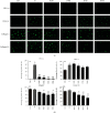
(a) Representative fluorescence micrographs of HIF-1α, VEGF-A, collagen I, and collagen III in cells under H/R conditions induced by 0.3 mM CoCl2. (b) Percentages of positive cells stained with HIF-1α (A), VEGF-A (B), collagen I (C), and collagen III (D). Scale bar: 100 μm. P < 0.05 vs. control group; ∗∗P < 0.005 vs. control group; #P < 0.05 vs. H group; ##P < 0.005 vs. H group.
Figure 9
Figure 9
Flow cytometric analysis of cellular apoptosis and ratios of Bax/bcl-2 proteins: (a) representative graphs of flow cytometric assays showing the apoptosis of different groups; (b) the histogram showing the ratio of apoptotic cells; (c) expression ratio of Bax/bcl-2 protein. P < 0.05 vs. control group; ∗∗P < 0.005 vs. control group. #P < 0.05 vs. H group; ##P < 0.005 vs. H group.

References

    1. Zhang Y., Wang B., Zhang W. J., Zhou G., Cao Y., Liu W. Enhanced proliferation capacity of porcine tenocytes in low O2 tension culture. Biotechnology Letters. 2010;32(2):181–187. doi: 10.1007/s10529-009-0137-8. - DOI - PubMed
    1. Zhang J., Wang J. H. Human tendon stem cells better maintain their stemness in hypoxic culture conditions. PLoS One. 2013;8(4, article e61424) doi: 10.1371/journal.pone.0061424. - DOI - PMC - PubMed
    1. Yu Y., Lin L., Zhou Y., et al. Effect of hypoxia on self-renewal capacity and differentiation in human tendon-derived stem cells. Medical Science Monitor. 2017;23:1334–1339. doi: 10.12659/MSM.903892. - DOI - PMC - PubMed
    1. Rempel D., Abrahamsson S. O. The effects of reduced oxygen tension on cell proliferation and matrix synthesis in synovium and tendon explants from the rabbit carpal tunnel: an experimental study in vitro. Journal of Orthopaedic Research. 2001;19(1):143–148. doi: 10.1016/S0736-0266(00)00005-X. - DOI - PubMed
    1. Fung F. K., Law B. Y., Lo A. C. Lutein attenuates both apoptosis and autophagy upon cobalt (II) chloride-induced hypoxia in rat Műller cells. PLoS One. 2016;11(12, article e0167828) doi: 10.1371/journal.pone.0167828. - DOI - PMC - PubMed

MeSH terms