

Review
doi: 10.1016/j.bjae.2017.11.003.
Epub 2017 Nov 21.
Anaesthesia and analgesia for knee joint arthroplasty
Affiliations
- PMID: 33456789
- PMCID: PMC7807845
- DOI: 10.1016/j.bjae.2017.11.003
Item in Clipboard
Review
Anaesthesia and analgesia for knee joint arthroplasty
BJA Educ.
2018 Jan.
No abstract available
Conflict of interest statement
None declared.
Figures
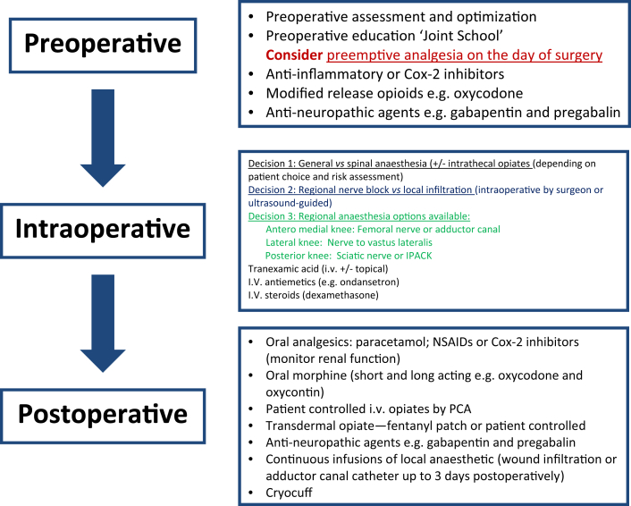
Preoperative, intraoperative and postoperative pathways for patients undergoing knee joint arthroplasty.
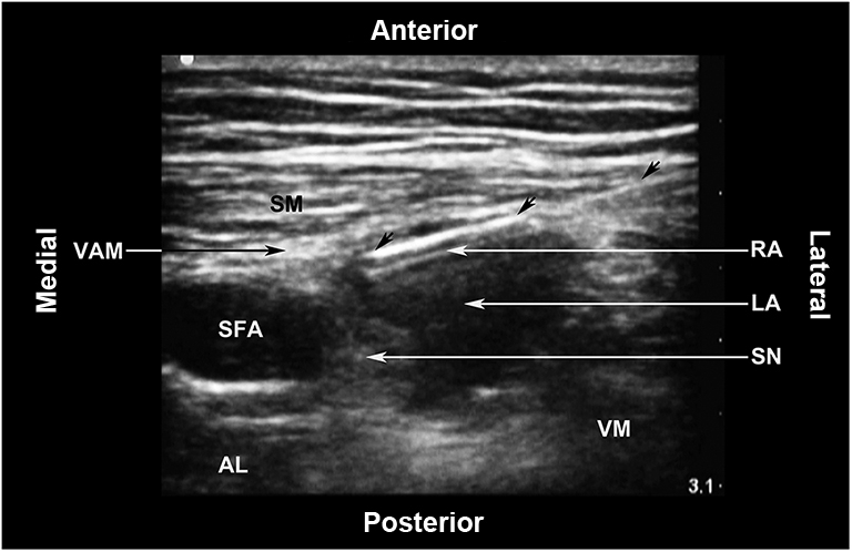
Ultrasound-guided adductor canal block. AL: Adductor longus muscle; LA: Local anaesthetic; RA: Reverberation artefact; SFA: Superficial femoral artery; SM: Sartorius muscle; SN: Saphenous nerve; VAM: Vastoadductor membrane; VM: Vastus medialis muscle. The unlabelled arrows indicate the block needle.
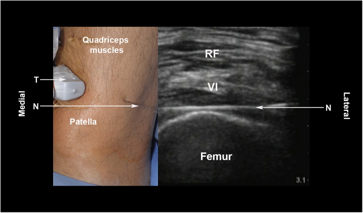
Ultrasound-guided local infiltration. N: Needle; RF: Rectus femoris muscle; T: Transducer (linear, high frequency); VI: Vastus intermedius muscle.
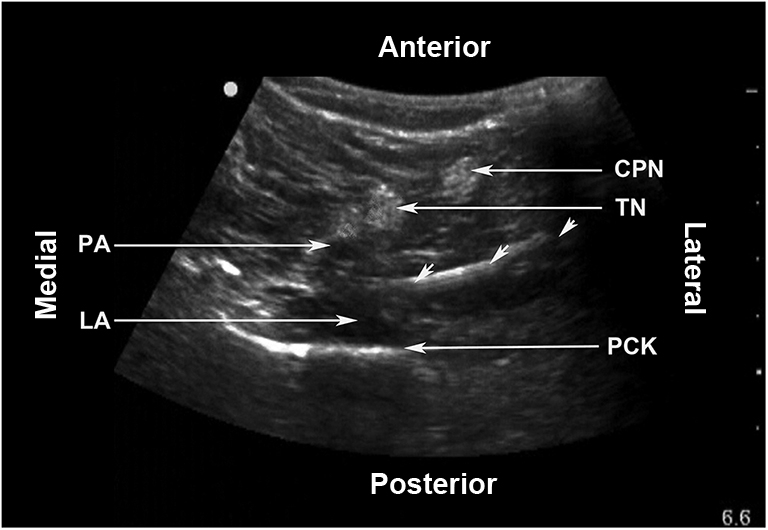
IPACK Block. CPN: Common peroneal nerve; LA: Local anaesthetic; PA: Popliteal artery; PCK: Posterior capsule of the knee; TN: Tibial nerve. The unlabelled arrows indicate the needle.
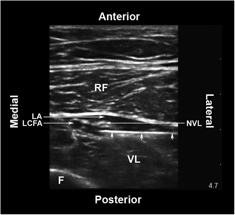
The distal branch of nerve to vastus lateralis in the thigh. F: Femur; LA: Local anaesthetic; LCFA: Descending branch of the lateral circumflex femoral artery; NVL: Nerve to vastus lateralis muscle; RF: Rectus femoris muscle; VL: Vastus lateralis muscle. The small unlabelled arrows indicate the block needle.
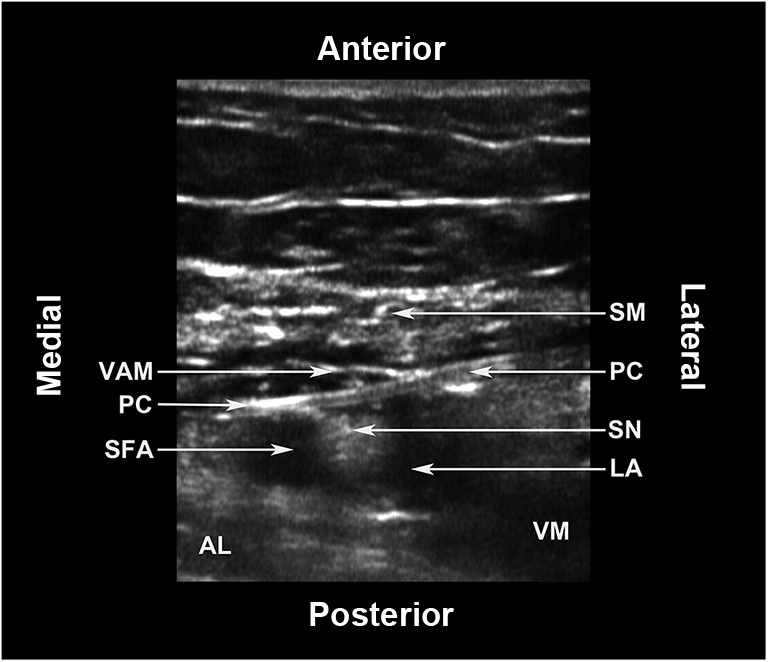
The adductor canal catheter. AL: Adductor longus muscle; LA: Local anaesthetic; PC: Perineural catheter; SFA: Superficial femoral artery; SM: Sartorius muscle; SN: Saphenous nerve; VAM: Vastoadductor membrane; VM: Vastus medialis muscle.
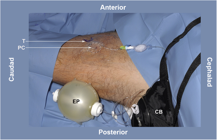
Continuous adductor canal infusion. CB: Carry bag for the elastomeric pump; EP: Elastomeric pump containing local anaesthetic; PC: Perineural catheter; T: Transducer position for inserting the catheter.
References
-
- Cheville A., Chen A., Oster G., McGarry L., Narcessian E. A randomized trial of controlled-release oxycodone during inpatient rehabilitation following unilateral total knee arthroplasty. J Bone Joint Surg Am. 2001;83:572–576. - PubMed
-
- Comfort V.K., Code W.F., Rooney M.E., Yip R.W. Naproxen premedication reduces postoperative tubal ligation pain. Can J Anaesth. 1992;4:349–352. - PubMed
Publication types
LinkOut - more resources
Full Text Sources
