. 2020 Dec 29;10(1):1854529.
doi: 10.1080/2162402X.2020.1854529.

Anti-NKG2D single domain-based antibodies for the modulation of anti-tumor immune response

Affiliations

Anti-NKG2D single domain-based antibodies for the modulation of anti-tumor immune response

Adeline Raynaud et al. Oncoimmunology. .

Abstract

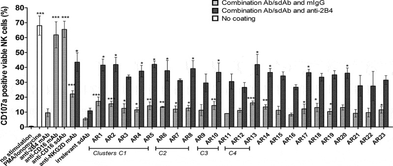
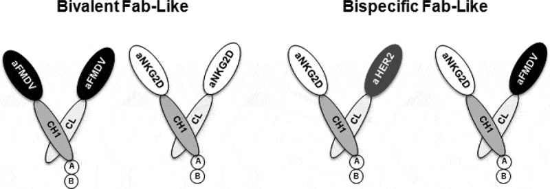
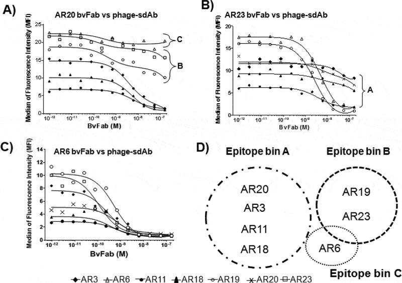
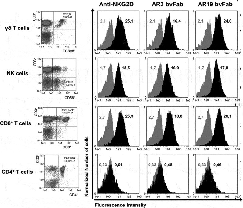
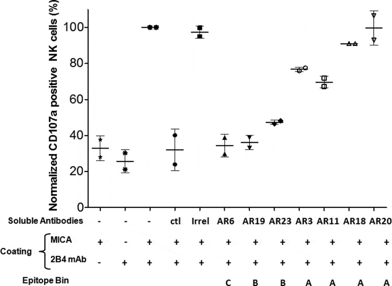
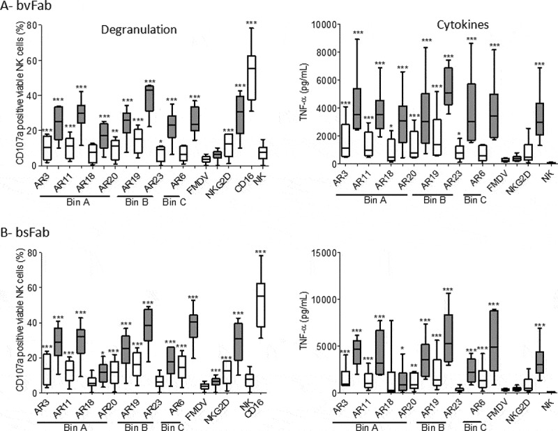
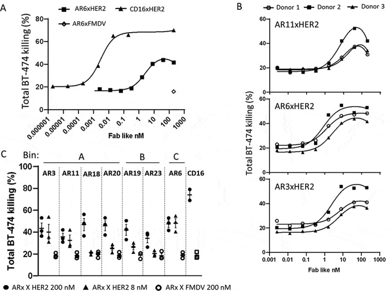
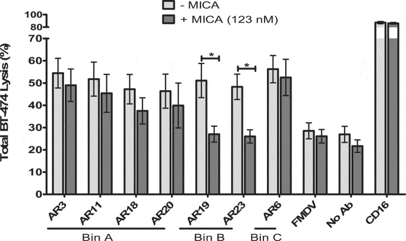
The natural killer group 2 member D (NKG2D) receptor is a C-type lectin-like activating receptor mainly expressed by cytotoxic immune cells including NK, CD8+ T, γδ T and NKT cells and in some pathological conditions by a subset of CD4+ T cells. It binds a variety of ligands (NKG2DL) whose expressions is finely regulated by stress-related conditions. The NKG2DL/NKG2D axis plays a central and complex role in the regulation of immune responses against diverse cellular threats such as oncogene-mediated transformations or infections. We generated a panel of seven highly specific anti-human NKG2D single-domain antibodies targeting various epitopes. These single-domain antibodies were integrated into bivalent and bispecific antibodies using a versatile plug-and-play Fab-like format. Depending on the context, these Fab-like antibodies exhibited activating or inhibitory effects on the immune response mediated by the NKG2DL/NKG2D axis. In solution, the bivalent anti-NKG2D antibodies that compete with NKG2DL potently blocked the activation of NK cells seeded on immobilized MICA, thus constituting antagonizing candidates. Bispecific anti-NKG2DxHER2 antibodies that concomitantly engage HER2 on tumor cells and NKG2D on NK cells elicited cytotoxicity of unstimulated NK in a tumor-specific manner, regardless of their apparent affinities and epitopes. Importantly, the bispecific antibodies that do not compete with ligands binding retained their full cytotoxic activity in the presence of ligands, a valuable property to circumvent immunosuppressive effects induced by soluble ligands in the microenvironment.

Keywords: HER2; NK cells; NKG2D/NKG2DL axis; cell engagers; single-domain antibody.


Figures

Figure 1.
Figure 1.
NK cell activation by immobilized anti-NKG2D ISVDs. Bacterial supernatants containing soluble ISVDs were added onto 96-well microplates pre-coated with a 1:1 mix of either anti-HA tag mAb and mIgG1 (gray bars) or anti-HA tag mAb and anti-2B4 mAb (dark gray bars). IL-2 pre-stimulated NK cells were incubated onto plates for 2 h, harvested and the percentage of CD107a-positive cells was quantified by flow cytometry. PMA/Ionomycin was used as positive control (white bar). Control conditions with anti-CD16 ISVD, anti-CD16, anti-2B4, anti-NKG2D mAbs were included. Each bar represents the mean value ± SEM of 3 independent assays (different donors). One-way analysis of variance with repeated measures and Bonferroni-Holm adjustment were used to compare each compound with irrelevant ISVD. * P < .05, ** p < .001, ***P < .0001
Figure 2.
Figure 2.
Schematic representation of bivalent and bispecific Fab-like antibodies. The CH1 constant domain of human IgG1 (dark gray) was fused to anti-NKG2D ISVD (white) whereas the CL constant domain (light gray) was fused to the same anti-NKG2D ISVD to generate bivalent Fab-like antibodies or to an anti-HER2 (C7b) or anti-FMDV ISVDs (gray and black respectively) to build bispecific Fab-like antibodies. (a): HA tag, (b): hexahistidine tag
Figure 3.
Figure 3.
Epitope binning experiments. NKG2D/DAP10-transfected FreeStyle 293-T cells were incubated at 4°C for 90 min with ISVDs-on-phage added at their predetermined EC90 concentration and increasing concentrations of NKG2D bvFab. Phage binding was detected by flow cytometry using an Alexa-647 conjugated anti-M13 mAb. Representative profiles obtained with bvFab AR20 (a), bvFab AR23 (b) and bvFab AR6 (c) are shown. (d) Schematic representation of NKG2D ISVD epitope bins
Figure 4.
Figure 4.
Binding of anti-NKG2D bvFabs on different immune cell populations. Cell subsets (CD3CD56+ NK cells, CD3+CD8+ and CD3+ CD4+ T cells, CD3+ TCRγδ+ T cells) of human PBMC were identified by a combination of surface markers (left panel). Binding of anti-NKG2D bvFabs on the different cell subsets was detected by flow cytometry using PE-conjugated anti-HA mAb. PE-conjugated anti-human NKG2D mAb was used as positive control. Representative histograms (black) of AR3 and AR19 bvFabs binding are shown and compared to PE-isotype binding (gray). Numbers displayed in each histogram are the median of fluorescence intensity of 2 independent experiments
Figure 5.
Figure 5.
Blockade of MICA-mediated NK cell activation by soluble NKG2D bvFab. Human recombinant MICA protein and/or anti-2B4 mAb (ratio 1/1) were immobilized on microplates. IL2-stimulated human NK cells and soluble antibodies (150 nM) were then added. After 2 h of incubation, NK cell activation was assessed by quantification of cell surface CD107a by flow cytometry. The percentage of CD107a-positive NK cells obtained in the presence of MICA and anti-2B4 mAb without any soluble anti-NKG2D mAb was normalized to 100%. Ctl = soluble anti-NKG2D mAb. Irrel = FMDV bvFab. Each bar represents the mean value ± SD of 2 independent experiments
Figure 6.
Figure 6.
NK cell activation by NKG2D bvFabs or bsFabs. IL2-prestimulated human NK cells were added to 96-well microplates pre-coated with a mix of (a) bvFab or (b) bsFab either with mIgG1 (white bars) or anti-2B4 mAb (grey bars). Monoclonal anti-NKG2D and anti-CD16 antibodies, and FMDV bvFab were used as positive and negative controls, respectively. After 2 h of incubation, NK cell activation was evaluated by quantification of cell surface CD107a by flow cytometry (left panel) and of secreted TNF-α levels by ELISA (right panel). Assays were performed 6 times (n = 6 donors). Data are represented as box-and-whisker plots with min and max. Two-way ANOVA with repeated measures on rank-transformed data with two-tailed Dunnett’s test was used to compare CD107a values for each compound versus FMDV bvFab. Two-way ANOVA with repeated measures on log-transformed data with two-tailed Dunnett’s test was used to compare TNF-α levels for each compound with FMDV bvFab. * P < .05, **p < .01, ***P < .0001
Figure 7.
Figure 7.
Redirected NK cell lysis against BT-474 tumor cells by NKG2DxHER2 bsFabs. CFSE-stained BT-474 cells (3.4x10⁵ cells/well) were incubated for 2 h with unstimulated human NK cells (E/T:6/1) in the presence or absence of serial dilutions of NKG2DxHER2, NKG2DxFMDV or HER2xCD16 bsFabs. TO-PRO®-3 was used as dead cell indicator. Percentage of total BT-474 cell lysis was determined by flow cytometry as the percentage of double positive CFSE/TO-PRO-3 cells. (a) Representative curves for CD16XHER2 and AR6xHER2 bsFabs (1 donor). (b) Tumor cell lysis curves for the 3 most potent HER2xNKG2D bsFabs. (c) Killing of BT-474 tumor cells mediated by 200 nM and 8 nM HER2xNKG2D bsFabs (resp. black circle and triangle) and 200 nM of their cognate FMDVxNKG2D (open circle) bsFabs. (b) and c) represent data of 3 independent experiments (3 donors)
Figure 8.
Figure 8.
Effect of soluble NKG2D ligand on redirected NK cell lysis mediated by bsFabs. CFSE-stained BT-474 cells were incubated for 2 h with unstimulated human NK cells (E/T:6/1) and 40 nM bsFabs in the presence (dark gray bar) or absence (light gray bar) of 123 nM human His-tagged MICA. TO-PRO®-3 was used as dead cell indicator. BT-474 cell killing was quantified by flow cytometry as the percentage of double positive CFSE/TO-PRO-3 cells. Each bar represents the mean value ± SEM of 4 independent experiments. The p values were calculated with one-tailed Student’s t-test for bsFab in the presence or absence of soluble MICA. * P < .05

Similar articles

Cited by

References

    1. Hegmans JP, Aerts JG.. Immunomodulation in cancer. Curr Opin Pharmacol. 2014;17:17–14. doi:10.1016/j.coph.2014.06.007. - DOI - PubMed
    1. Fridman WH, Zitvogel L, Sautès–Fridman C, Kroemer G.The immune contexture in cancer prognosis and treatment. Nat Rev Clin Oncol. 2017;14(12):717–734. doi:10.1038/nrclinonc.2017.101. - DOI - PubMed
    1. Chen DS, Mellman I. Elements of cancer immunity and the cancer-immune set point. Nature. 2017;541:321–330. - PubMed
    1. Mahoney KM, Rennert PD, Freeman GJ.. Combination cancer immunotherapy and new immunomodulatory targets. Nat Rev Drug Discov. 2015;14:561–584. - PubMed
    1. Pardoll DM. The blockade of immune checkpoints in cancer immunotherapy. Nat Rev Cancer. 2012;12(4):252–264. doi:10.1038/nrc3239. - DOI - PMC - PubMed

Publication types

Substances