

Bronchogenic cyst or lung cancer. Only biopsy can tell
- PMID: 33457198
- PMCID: PMC7797910
- DOI: 10.1016/j.rmcr.2020.101328
Bronchogenic cyst or lung cancer. Only biopsy can tell
Abstract
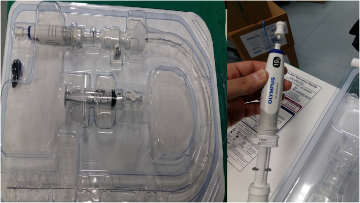
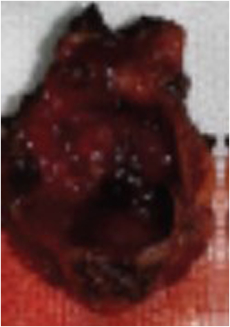
Bronchogenic cysts are rare congenital malformations which derive from primitive ventral foregut. They are usually observed in intrathoracically. A fifty year old male was admitted for the investigation of a three month chest pain. Computed tomography scan of the thorax revealed a lesion around the esophagus and left stem bronchus. Endobronchial ultrasound with convex probe and a 19G needle biopsy revealed a bronchogenic cystic which was removed with video assisted thoracic surgery. Initial radiologic assessment although was thought to be lung cancer because of the smoking habit it turned out to be benignancy. EBUS-TBNAB with 10G needle is safe and absolutely necessary for these lesions, as they take large samples.
Keywords: 19G needle; Bronchogenic cyst; EBUS; Lung cancer.
© 2020 The Author(s).
Conflict of interest statement
None to declare.
Figures


References
-
- Bukamur H.S., Alkhankan E., Mezughi H.M., Munn N.J., Shweihat Y.R. The role and safety of endobronchial ultrasound-guided transbronchial needle aspiration in the diagnosis and management of infected bronchogenic mediastinal cysts in adults. Respiratory medicine case reports. 2018;24:46–49. - PMC - PubMed
-
- Jiang C., Wang H., Chen G., Jiang G., Zhang P. Intradiaphragmatic bronchogenic cyst. Ann. Thorac. Surg. 2013;96(2):681–683. - PubMed
Publication types
LinkOut - more resources
Full Text Sources
Other Literature Sources
