Review
. 2020 Dec;9(6):2890-2907.
doi: 10.21037/tau-19-671.

Role of magnetic resonance imaging in tumor staging and follow-up for bladder cancer

Affiliations
Review

Role of magnetic resonance imaging in tumor staging and follow-up for bladder cancer

Ki Choon Sim et al. Transl Androl Urol. 2020 Dec.

Abstract

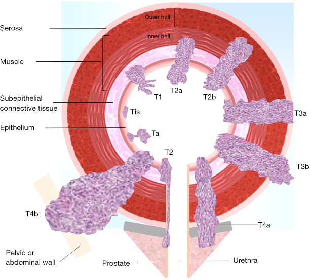
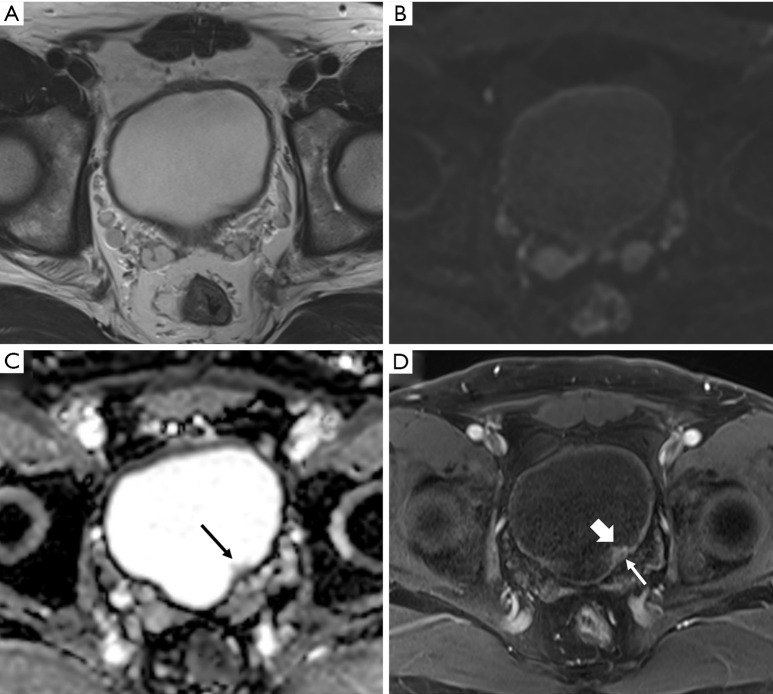
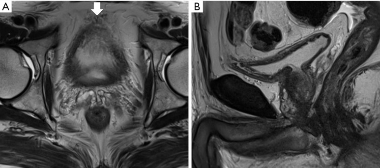
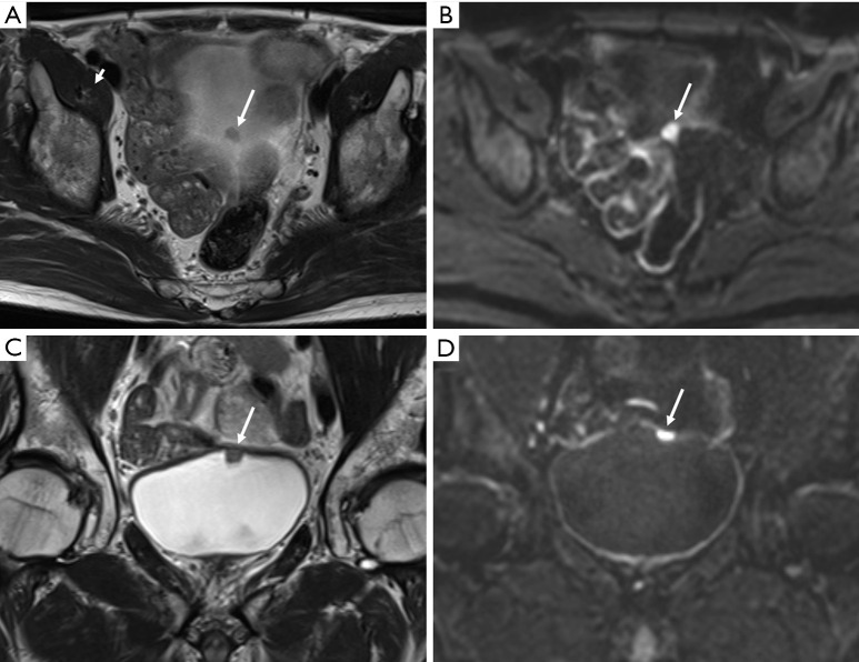
Urothelial carcinoma of the bladder is a common urologic malignancy. Complex factors, such as local stage, tumor grade, biologic potential, and various conditions, can affect the treatment strategy for bladder cancer. However, the local stage-in particular, the presence or absence of muscle invasion-significantly influences decisions regarding treatment strategy. The role of cystoscopy for screening, diagnosis, and transurethral resection cannot be overlooked. The importance of local staging with magnetic resonance imaging is increasing; magnetic resonance imaging of the bladder is considered a useful staging modality. Moreover, a radiologic reporting system for evaluating and scoring muscle invasion of bladder cancer was recently released. This system is based on multiparametric magnetic resonance imaging and is also expected to be feasible for post-treatment follow-up of bladder cancer. In this review, we discuss the role of magnetic resonance imaging in the local staging of urothelial carcinoma of the urinary bladder and post-treatment imaging. In addition, several technical aspects for obtaining appropriate quality magnetic resonance images of the bladder will be discussed.

Keywords: TNM; Urothelial carcinoma; magnetic resonance imaging; stage; urinary bladder neoplasms.


Conflict of interest statement

Conflicts of Interest: Both authors have completed the ICMJE uniform disclosure form (available at http://dx.doi.org/10.21037/tau-19-671). The series “Muscle-Invasive Bladder Cancer” was commissioned by the editorial office without any funding or sponsorship. The authors have no other conflicts of interest to declare.

Figures

Figure 1
Figure 1
Diagram showing bladder wall anatomy and local tumor staging of urinary bladder cancer based on invasion depth (see Tables 1,2 for T stage definitions).
Figure 2
Figure 2
Urothelial carcinoma (stage Ta) in a 74-year-old man. Axial T2-weighted magnetic resonance image (T2WI) (A) showing the normal bladder wall as a hypointense line. Axial diffusion-weighted image (DWI) with a high b value (B) showing the normal bladder wall as an intermediate signal intensity line. Axial apparent diffusion coefficient map (C) showing the bladder wall with low signal intensity. Small papillary lesion with intermediate signal intensity (arrow) in the left posterior wall that was not clearly visible on previous sequences. Axial three-dimensional T1-weighted spoiled gradient echo image (D) obtained 60 seconds after the administration of contrast material shows intense enhancement of the bladder tumor (thick arrow) unlike the muscle layer (thin arrow).
Figure 3
Figure 3
Magnetic resonance imaging (MRI) of a 74-year-old man who underwent transurethral resection of a bladder tumor previously. Axial (A) and sagittal high-resolution T2-weighted magnetic resonance image (B) showing a poorly distended urinary bladder. The bladder lumen is barely visible on the axial image because of insufficient distension. The vascular structures (thick arrow) between the anterior and perivesical fat mimic tumor infiltration. Evaluation of the bladder wall and lumen on the sagittal image was limited yet marginally feasible; MRI recall or additional cystoscopy should be considered in such cases.
Figure 4
Figure 4
Urothelial carcinoma (stage T1) in a 63-year-old man. Axial T2-weighted image (T2WI) (A) showing a small tumor in the bladder dome (arrow). In the diffusion-weighted image (DWI) with a high b value (B), the tumor shows markedly increased signal intensity (arrow). However, the localization of bladder tumors solely based on these images is difficult. The lesions located on the bladder dome are often not clearly visualized on axial images because of the persistence of partial volume artifacts. In such cases, coronal images (C, coronal T2WI; D, coronal DWI with high b value) are extremely helpful for tumor localization and T stage evaluation.
Figure 5
Figure 5
Vulnerability on a coronal diffusion-weighted imaging. Additional slice of the coronal plane image of the patient described in Figure 4. (A) T2-weighted image (T2WI), (B) b =0, (C) b =1,000, and (D) apparent diffusion coefficient map. Coronal T2WI showing no abnormality in the left dome portion, and an air-filled bowel structure (asterisk) with dark signal intensity is located immediately adjacent to it. However, in the diffusion-weighted images, distortion is observed in the left dome of the bladder (arrow). It is believed that motion artifacts caused by bowel peristalsis and susceptibility artifacts caused by air existed simultaneously.
Figure 6
Figure 6
Urothelial carcinoma (stage T1) in a 65-year-old man highlighting the importance of magnetic resonance (MR) image acquisition order. (A) A small tumor (about 1 cm) is visible in the right posterior wall (thick arrow), while another tiny polypoid lesion is seen in the left anterior wall (thin arrow) on an axial T2-weighted image. Diffusion-weighted image (B, b =0; C, b =800) and apparent diffusion coefficient map (D) obtained after delayed contrast enhancement shows a new heterogeneous low signal in the bladder-dependent portion (asterisk) that differs from the signal of urine in the nondependent portion. This represents the change in the signal intensity of urine because of excretion of the MR contrast agent that, to some extent, influences the contrast between the right posterior tumor and urine in the low b value image (B).
Figure 7
Figure 7
Urothelial carcinoma in a 68-year-old woman. Axial T2-weighted image (T2WI) (A) showing two papillary masses in the left posterior wall of bladder (arrow). Axial three-dimensional T1-weighted spoiled gradient echo image (B) obtained 300 seconds after contrast agent administration showing marked hyperintensity due to excreted magnetic resonance contrast agent in the urinary bladder (asterisk). The urine, which has been filling the bladder, has a dark signal intensity. In this case, the tumor is localized on the left posterior wall and cannot be visualized on delayed contrast-enhanced images (arrow).
Figure 8
Figure 8
Urothelial carcinoma (stage T3b) in a 79-year-old man. Axial T2-weighted image (A) shows a T2 heterogeneous mass with intermediate to low signal intensity in the left wall. If the dotted line is considered the boundary between the bladder wall and the perivesical fat, a tumorous lesion extending into the perivesical fat area is observed, which can be staged as T3b. A high b value image (B) and apparent diffusion coefficient maps (C) also show diffusion restriction in the tumors extending to the bladder wall (asterisk). Axial three-dimensional T1-weighted spoiled gradient echo image (D) obtained 40 seconds after contrast agent administration shows early peripheral enhancement (thin arrow) and central necrosis (thick arrow) of the large tumor. This case is classified as vesicle imaging-reporting and data system grade 5.
Figure 9
Figure 9
Urothelial carcinoma (stage T1) in a 60-year-old man. Axial (A) T2-weighted images showing a tumor in the left bladder wall. The muscle layer with a T2 hypointensity line is suspicious of focal disruption because of the presence of tumors with T2 intermediate signal intensity (thin arrow). In the high b value (b =800) diffusion-weighted image (B) and apparent diffusion coefficient map (C), the peripheral region of the tumor shows marked diffusion restriction (thick arrow). However, diffusion restriction is not seen at the base of the tumor, including the stalk (asterisk). Additionally, disruption of the muscle layer with intermediate signal intensity is not observed even in high b value images. An axial three-dimensional T1-weighted spoiled gradient echo image (D) obtained 300 seconds after contrast agent administration shows bright delayed enhancement of the tumor stalk (asterisk). This case was preoperatively classified as vesicle imaging-reporting and data system grade 2. The patient underwent transurethral resection of the bladder tumor. Appropriate tumor specimens with muscle layer were obtained and it was diagnosed as high-grade stage T1 cancer with invasion only up to the subepithelial connective tissue.
Figure 10
Figure 10
Correlation of apparent diffusion coefficient (ADC) value and tumor grade. (A,B) Urothelial carcinoma (stage T1) in an 82-year-old man. High b value axial diffusion-weighted magnetic resonance image (A) and apparent diffusion coefficient (ADC) map (B) with an ADC value of 0.9×10⁻³ mm2/s showing a papillary mass (arrow) in the left wall with marked diffusion restriction. This tumor was staged as a high-grade urothelial carcinoma after the histopathological examination of transurethral resection of bladder tumor (TURBT) specimens. (C,D) Urothelial carcinoma (stage Ta) in a 64-year-old man. High b value axial diffusion-weighted magnetic resonance image (C) and ADC map with an ADC value of 1.45×10⁻³ mm2/s (D) showing a papillary mass in right wall with mild to moderate diffusion restriction. The tumor was confirmed as a low-grade urothelial carcinoma on the histopathological examination of TURBT specimens. Both patients underwent diffusion-weighted imaging with the same 3T magnetic resonance imaging equipment (Skyra, Siemens) and parameters; moreover, the window level and width were similar. Although direct comparisons may be inaccurate owing to differences in the cases, a more pronounced diffusion restriction is observed in the diffusion-weighted images of (A) and (B), which were confirmed as high-grade urothelial carcinoma.
Figure 11
Figure 11
Urothelial carcinoma (stage Ta) in a 56-year-old man. Axial (A), coronal (B), and sagittal (C) T2-weighted images showing an approximately 3-cm papillary mass (asterisk) in the left posterior wall. In axial and coronal images, the tumor appears to float on the bladder. However, the tumor stalk (arrow) is clearly observed in the sagittal image. A high b value diffusion-weighted image (D) and apparent diffusion coefficient map (E) showing the papillary mass with marked diffusion restriction (asterisk). There is no disruption of the muscle layer in the T2WI. Axial three-dimensional T1-weighted spoiled gradient echo image (F) obtained 180 seconds after contrast agent administration showing preserved mild delayed enhancement of the muscle layer (arrow). Therefore, the case can be classified as vesicle imaging-reporting and data system grade 2.
Figure 12
Figure 12
Urothelial carcinoma (stage T2) in an 80-year-old man. Axial (A) and coronal (B) T2-weighted images showing an intermediate signal intensity ovoid-shaped mass (arrow) in the bladder dome. A high b value axial diffusion-weighted magnetic resonance image (C) and apparent diffusion coefficient map (D) showing marked diffusion restriction of the whole tumor area. There is no clear disruption of the low signal intensity muscle layer. However, category 2 findings are lacking on T2-weighted images and diffusion-weighted images. Axial (E) and coronal (F) three-dimensional T1-weighted spoiled gradient echo image obtained 180 seconds after contrast agent administration also show contrast-enhanced category 3. This case should be scored as vesicle imaging-reporting and data system grade 3. The patient underwent transurethral resection of the bladder tumor. Appropriate tumor specimens with muscle tissue were examined and the patient was diagnosed with high-grade stage T2 cancer.
Figure 13
Figure 13
Follow-up magnetic resonance imaging (MRI) of an 82-year-old man who underwent transurethral resection of bladder tumor (TURBT) for urothelial carcinoma. Axial T2-weighted image (T2WI) (A), high b value diffusion-weighted image (DWI) (B), and apparent diffusion coefficient (ADC) map (C) showing two masses in the bladder anterior wall. MRI shows a T2 hypointense bladder cancer without muscle invasion that was diagnosed as high-grade T1 urothelial carcinoma on TURBT specimens including proper muscle tissue. In follow-up MRI, axial T2WI (D), high b value DWI (E), and ADC map (F) obtained 8 months after TURBT showing new perivesical infiltration with hair-like projection (arrow) in the left anterior perivesical area. There was no remarkable diffusion restriction on DWI, and histopathological examination of repeat TURBT specimens containing proper muscle tissue showed acute and chronic inflammation in the adjacent bladder wall.

Similar articles

Cited by

References

    1. Bray F, Ferlay J, Soerjomataram I, et al. Global cancer statistics 2018: GLOBOCAN estimates of incidence and mortality worldwide for 36 cancers in 185 countries. CA Cancer J Clin 2018;68:394-424. 10.3322/caac.21492 - DOI - PubMed
    1. Wong-You-Cheong JJ, Woodward PJ, Manning MA, et al. Neoplasms of the urinary bladder: radiologic-pathologic correlation. Radiographics 2006;26:553-80. 10.1148/rg.262055172 - DOI - PubMed
    1. Wang H, Luo C, Zhang F, et al. Multiparametric MRI for bladder cancer: validation of VI-RADS for the detection of detrusor muscle invasion. Radiology 2019;291:668-74. 10.1148/radiol.2019182506 - DOI - PubMed
    1. Soukup V, Čapoun O, Cohen D, et al. Prognostic performance and reproducibility of the 1973 and 2004/2016 World Health Organization Grading classification systems in non–muscle-invasive bladder cancer: a European Association of Urology non-muscle invasive bladder cancer guidelines panel systematic review. Eur Urol 2017;72:801-13. 10.1016/j.eururo.2017.04.015 - DOI - PubMed
    1. Witjes J, Bruins M, Compérat E, et al. EAU guidelines muscle invasive bladder cancer 2018. EAU Annual Congress Copenhagen, 2018.