miR-1929-3p Overexpression Alleviates Murine Cytomegalovirus-Induced Hypertensive Myocardial Remodeling by Suppressing Ednra/NLRP3 Inflammasome Activation
- PMID: 33457411
- PMCID: PMC7787724
- DOI: 10.1155/2020/6653819
miR-1929-3p Overexpression Alleviates Murine Cytomegalovirus-Induced Hypertensive Myocardial Remodeling by Suppressing Ednra/NLRP3 Inflammasome Activation
Abstract
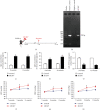
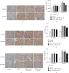
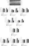
MicroRNAs (miRNAs) play crucial roles in the development of essential hypertension (EH). Previously, we found that the expression of miR-1929-3p was decreased in C57BL/6 mice with hypertension induced by murine cytomegalovirus (MCMV). In this study, we explored the role of miR-1929-3p in hypertension myocardial remodeling in MCMV-infected mice. First, we measured MCMV DNA and host IgG and IgM after infection and determined the expression of miR-1929-3p and its target gene endothelin A receptor (Ednra) mRNA in the myocardium of mice. Then, we performed invasive blood pressure (BP) monitoring. Heart-to-body weight ratio (HW/BW%), along with mRNA levels of B-type natriuretic peptide (BNP) and beta myosin heavy chain (β-MHC), revealed myocardial remodeling. Hematoxylin/eosin and Masson's trichrome staining indicated morphological changes in the myocardium. Cardiac function was assessed via echocardiography. Moreover, MCMV-infected mice were injected with recombinant adeno-associated virus- (rAAV-) miR-1929-3p overexpression vector. Immunohistochemistry and western blotting showed the expression of Ednra and the activation of NOD-like receptor pyrin domain containing 3 (NLRP3) inflammasome. And enzyme-linked immunosorbent assay (ELISA) revealed the concentrations of endothelin-1 (ET-1), interleukin-1β (IL-1β), and interleukin-18 (IL-18). In this study, we found that decreased expression of miR-1929-3p in MCMV-infected mice induced high BP and further development of myocardial remodeling cardiac function injury through increased expression of Ednra. Strikingly, overexpression of miR-1929-3p ameliorated these pathological changes of the heart. The positive effect was shown to be associated with inhibition of NLRP3 inflammasome activation and decreased expression of key proinflammatory cytokine IL-1β. Collectively, these results indicate that miR-1929-3p overexpression may effectively alleviate EH myocardial remodeling by suppressing Ednra/NLRP3 inflammasome activation in MCMV-infected mice.
Copyright © 2020 YongJia Wang et al.
Conflict of interest statement
The authors declare that there are no conflicts of interest regarding the publication of this paper.
Figures


Similar articles
-
Low expression of miR-1929-3p mediates murine cytomegalovirus-induced fibrosis in cardiac fibroblasts via targeting endothelin a receptor/NLRP3 inflammasome pathway.In Vitro Cell Dev Biol Anim. 2023 Mar;59(3):179-192. doi: 10.1007/s11626-022-00742-2. Epub 2023 Mar 31. In Vitro Cell Dev Biol Anim. 2023. PMID: 37002490
-
MicroRNA‑1929‑3p participates in murine cytomegalovirus‑induced hypertensive vascular remodeling through Ednra/NLRP3 inflammasome activation.Int J Mol Med. 2021 Feb;47(2):719-731. doi: 10.3892/ijmm.2020.4829. Epub 2020 Dec 22. Int J Mol Med. 2021. PMID: 33416142 Free PMC article.
-
Murine Cytomegalovirus Infection Induced miR-1929-3p Down-Regulation Promotes the Proliferation and Apoptosis of Vascular Smooth Muscle Cells in Mice by Targeting Endothelin A Receptor and Downstream NLRP3 Activation Pathway.Mol Biotechnol. 2023 Dec;65(12):1954-1967. doi: 10.1007/s12033-023-00720-3. Epub 2023 Apr 6. Mol Biotechnol. 2023. PMID: 37022597
-
MicroRNAs as important regulators of the NLRP3 inflammasome.Prog Biophys Mol Biol. 2020 Jan;150:50-61. doi: 10.1016/j.pbiomolbio.2019.05.004. Epub 2019 May 15. Prog Biophys Mol Biol. 2020. PMID: 31100298 Review.
-
Role of NLRP3 Inflammasome in Cardiac Inflammation and Remodeling after Myocardial Infarction.Biol Pharm Bull. 2019;42(4):518-523. doi: 10.1248/bpb.b18-00369. Biol Pharm Bull. 2019. PMID: 30930410 Review.
Cited by
-
Identification of marker genes associated with oxidative stress in hypertrophic cardiomyopathy using bioinformatics analysis and experimental validation.Sci Rep. 2025 Aug 6;15(1):28817. doi: 10.1038/s41598-025-14313-4. Sci Rep. 2025. PMID: 40770404 Free PMC article.
-
The Role of Melatonin on NLRP3 Inflammasome Activation in Diseases.Antioxidants (Basel). 2021 Jun 24;10(7):1020. doi: 10.3390/antiox10071020. Antioxidants (Basel). 2021. PMID: 34202842 Free PMC article. Review.
-
How do pre-operative intra-articular injections impact periprosthetic joint infection risk following primary total hip arthroplasty? A systematic review and meta-analysis.Arch Orthop Trauma Surg. 2023 Mar;143(3):1627-1635. doi: 10.1007/s00402-022-04375-8. Epub 2022 Feb 12. Arch Orthop Trauma Surg. 2023. PMID: 35150302
-
MicroRNAs in cardiovascular diseases.Med Rev (2021). 2022 Apr 26;2(2):140-168. doi: 10.1515/mr-2021-0001. eCollection 2022 Apr. Med Rev (2021). 2022. PMID: 37724243 Free PMC article. Review.
-
Low expression of miR-1929-3p mediates murine cytomegalovirus-induced fibrosis in cardiac fibroblasts via targeting endothelin a receptor/NLRP3 inflammasome pathway.In Vitro Cell Dev Biol Anim. 2023 Mar;59(3):179-192. doi: 10.1007/s11626-022-00742-2. Epub 2023 Mar 31. In Vitro Cell Dev Biol Anim. 2023. PMID: 37002490
References
-
- Xue H., Wang S. X., Wang X. J., et al. Variants of tumor necrosis factor-induced protein 3 gene are associated with left ventricular hypertrophy in hypertensive patients. Chinese Medical Journal. 2011;124(10):1498–1503. - PubMed
MeSH terms
Substances
LinkOut - more resources
Full Text Sources
Research Materials
Miscellaneous

