

In vitro performance of echoPIV for assessment of laminar flow profiles in a carotid artery stent
- PMID: 33457445
- PMCID: PMC7804295
- DOI: 10.1117/1.JMI.8.1.017001
In vitro performance of echoPIV for assessment of laminar flow profiles in a carotid artery stent
Abstract
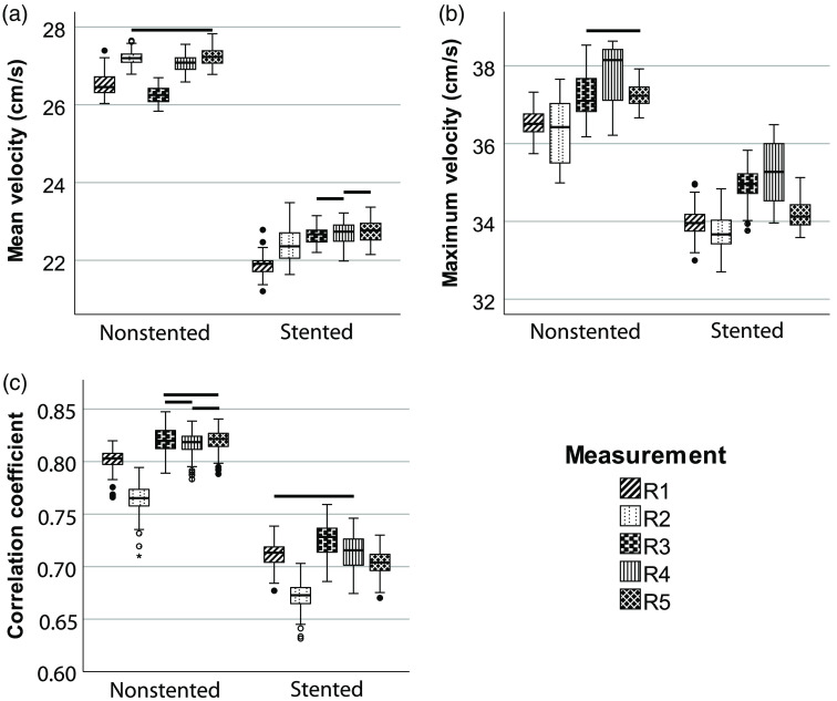
Purpose: Detailed blood flow studies may contribute to improvements in carotid artery stenting. High-frame-rate contrast-enhanced ultrasound followed by particle image velocimetry (PIV), also called echoPIV, is a technique to study blood flow patterns in detail. The performance of echoPIV in presence of a stent has not yet been studied extensively. We compared the performance of echoPIV in stented and nonstented regions in an in vitro flow setup. Approach: A carotid artery stent was deployed in a vessel-mimicking phantom. High-frame-rate contrast-enhanced ultrasound images were acquired with various settings. Signal intensities of the contrast agent, velocity values, and flow profiles were calculated. Results: The results showed decreased signal intensities and correlation coefficients inside the stent, however, PIV analysis in the stent still resulted in plausible flow vectors. Conclusions: Velocity values and laminar flow profiles can be measured in vitro in stented arteries using echoPIV.
Keywords: carotid; high-frame-rate ultrasound; in vitro; particle image velocimetry; stent.
© 2021 The Authors.
Figures


Similar articles
-
High-frame-rate contrast-enhanced ultrasound particle image velocimetry in patients with a stented superficial femoral artery: a feasibility study.Eur Radiol Exp. 2022 Jul 6;6(1):32. doi: 10.1186/s41747-022-00278-w. Eur Radiol Exp. 2022. PMID: 35790584 Free PMC article.
-
Blood Flow Quantification with High-Frame-Rate, Contrast-Enhanced Ultrasound Velocimetry in Stented Aortoiliac Arteries: In Vivo Feasibility.Ultrasound Med Biol. 2022 Aug;48(8):1518-1527. doi: 10.1016/j.ultrasmedbio.2022.03.016. Epub 2022 May 13. Ultrasound Med Biol. 2022. PMID: 35577661
-
Development of an in vitro setup for flow studies in a stented carotid artery bifurcation.Med Biol Eng Comput. 2024 Apr;62(4):1165-1176. doi: 10.1007/s11517-023-02977-x. Epub 2023 Dec 29. Med Biol Eng Comput. 2024. PMID: 38155315
-
Left ventricular high frame rate echo-particle image velocimetry: clinical application and comparison with conventional imaging.Cardiovasc Ultrasound. 2022 Apr 26;20(1):11. doi: 10.1186/s12947-022-00283-4. Cardiovasc Ultrasound. 2022. PMID: 35473581 Free PMC article.
-
High-Frame-Rate Ultrasound Velocimetry in the Healthy Femoral Bifurcation: A Comparative Study Against 4-D Flow Magnetic Resonance Imaging.Ultrasound Med Biol. 2024 Dec;50(12):1755-1763. doi: 10.1016/j.ultrasmedbio.2024.05.013. Epub 2024 Sep 7. Ultrasound Med Biol. 2024. PMID: 39244482
Cited by
-
High-frame-rate contrast-enhanced ultrasound particle image velocimetry in patients with a stented superficial femoral artery: a feasibility study.Eur Radiol Exp. 2022 Jul 6;6(1):32. doi: 10.1186/s41747-022-00278-w. Eur Radiol Exp. 2022. PMID: 35790584 Free PMC article.
References
LinkOut - more resources
Full Text Sources
Other Literature Sources
