

Case Reports
doi: 10.1080/23320885.2020.1769481.
An unusual and delayed complication of hyaluronic acid filler injection: a case report
Affiliations
- PMID: 33457452
- PMCID: PMC7782608
- DOI: 10.1080/23320885.2020.1769481
Item in Clipboard
Case Reports
An unusual and delayed complication of hyaluronic acid filler injection: a case report
Case Reports Plast Surg Hand Surg.
.
Abstract
48-year-old female with facial granulomatous nodules and fungal/bacterial infection after hyaluronic acid injection. She underwent anti-fungal/antibacterial therapy and local excision. The proposed mechanisms include inflammatory foreign body reaction and pathogen contamination. Providers must exercise caution with the use of facial fillers and demonstrate expertise in avoiding and managing potential complications.
Keywords: Filler; adverse reaction; hyaluronic acid; nasolabial folds.
© 2020 University of Mississippi Medical Center. Published by Informa UK Limited, trading as Taylor & Francis Group.
Conflict of interest statement
No potential conflict of interest was reported by the author(s).
Figures

One month after the hyaluronic acid filler injection, patient developed a facial hypersensitivity reaction consisting in facial edema, erythema, itchiness and mild fever.
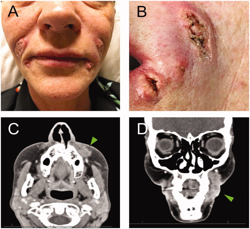
A. Verrucous ulcerative skin lesions on nasolabial folds, marionette lines and glabellar region (not shown). B. Detail of the lesions on the left side of the face. C and D. Arrowheads show the abnormal heterogeneous enhancing soft tissue extending along the lateral left maxilla (1.6 cm AP by 1.0 cm transverse by 2.6 cm craniocaudal dimensions).
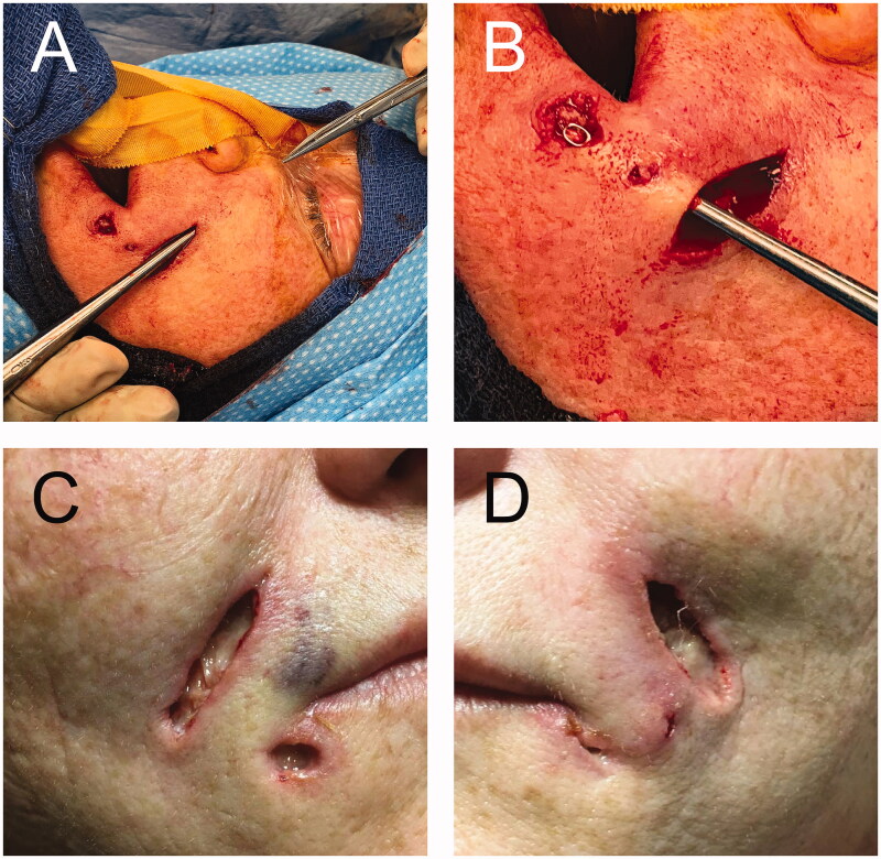
A and B. Intraoperative images showing the communication between nearby lesions and extension up to the infraorbital rim. C and D. Postoperative wounds that healed with regular dressing by secondary intention.
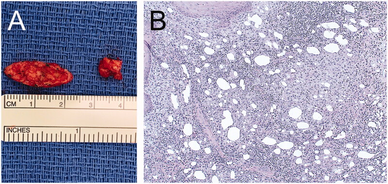
A. Intraoperative image showing the specimens from the left nasolabial fold and left marionette line. B. Hematoxylin-eosin stain showing lipogranulomas with granulomatous inflammation within the connective tissue stroma (20× magnification).
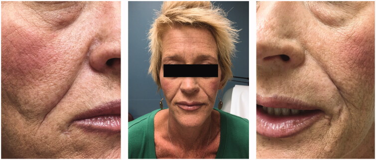
Eight months after surgical excision of verrucous skin lesions. Patient showed no signs of ongoing infection and wounds are well healed. Deep scarring in the nasolabial folds has made the folds more prominent.
References
-
- Bass LS. Injectable filler techniques for facial rejuvenation, volumization, and augmentation. Facial Plast Surg Clin North Am. 2015;23(4):479–488. - PubMed
-
- Greene JJ, Sidle DM.. The hyaluronic acid fillers: current understanding of the tissue device interface. Facial Plast Surg Clin North Am. 2015;23(4):423–432. - PubMed
-
- Monheit GD, Coleman KM.. Hyaluronic acid fillers. Dermatol Ther. 2006;19(3):141–150. - PubMed
-
- Small K, Kelly KM, Spinelli HM.. Are nurse injectors the new norm? Aesth Plast Surg. 2014;38(5):946–955. - PubMed
-
- Vidič M, Bartenjev I.. An adverse reaction after hyaluronic acid filler application: a case report. Acta Dermatovenerol Alp Pannonica Adriat. 2018;27(3):165–167. - PubMed
Publication types
LinkOut - more resources
Full Text Sources