

Automatic segmentation of cardiac structures for breast cancer radiotherapy
- PMID: 33458294
- PMCID: PMC7807574
- DOI: 10.1016/j.phro.2019.11.007
Automatic segmentation of cardiac structures for breast cancer radiotherapy
Abstract
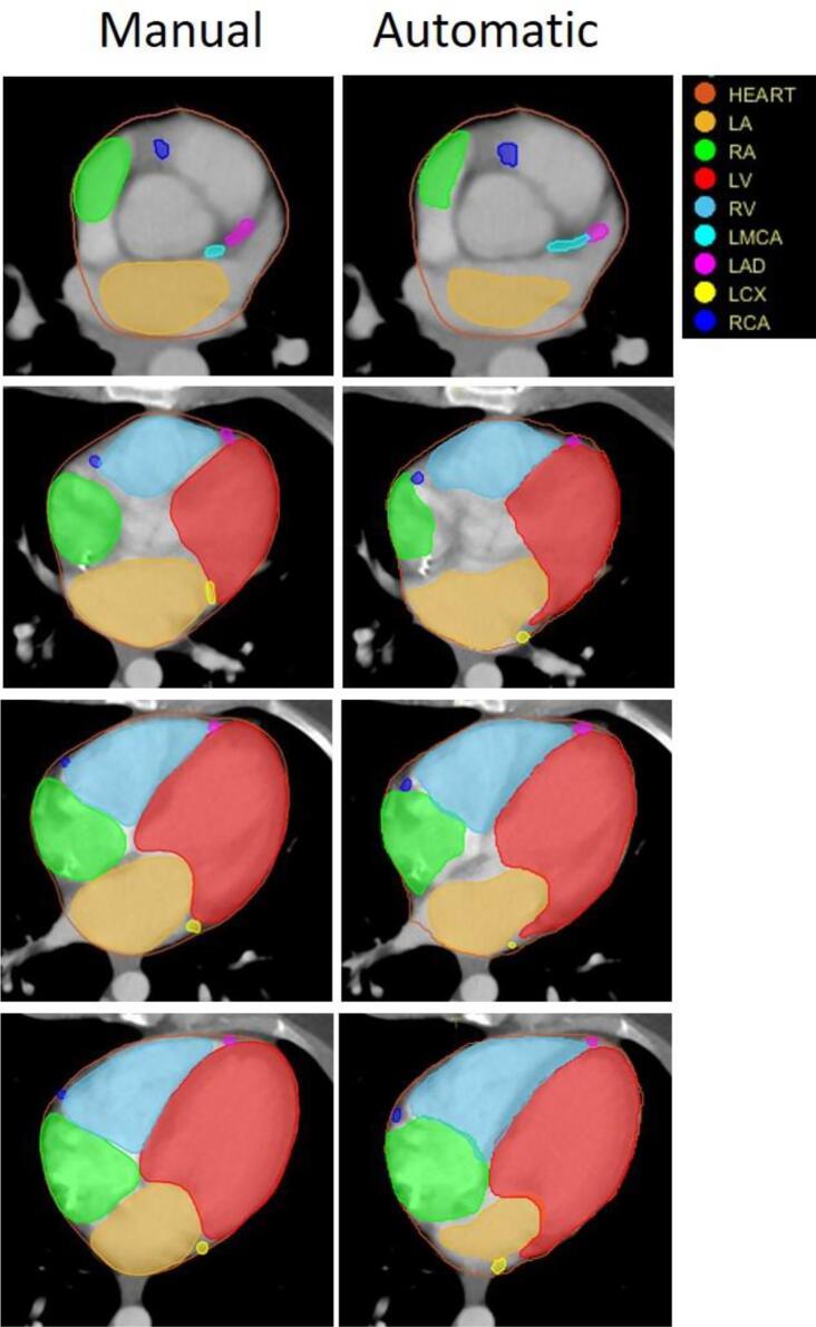
Background and purpose: We developed an automatic method to segment cardiac substructures given a radiotherapy planning CT images to support epidemiological studies or clinical trials looking at cardiac disease endpoints after radiotherapy.
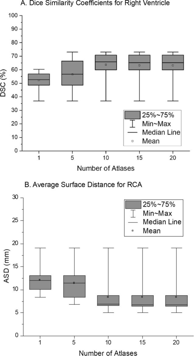
Material and methods: We used a most-similar atlas selection algorithm and 3D deformation combined with 30 detailed cardiac atlases. We cross-validated our method within the atlas library by evaluating geometric comparison metrics and by comparing cardiac doses for simulated breast radiotherapy between manual and automatic contours. We analyzed the impact of the number of cardiac atlas in the library and the use of manual guide points on the performance of our method.
Results: The Dice Similarity Coefficients from the cross-validation reached up to 97% (whole heart) and 80% (chambers). The Average Surface Distance for the coronary arteries was less than 10.3 mm on average, with the best agreement (7.3 mm) in the left anterior descending artery (LAD). The dose comparison for simulated breast radiotherapy showed differences less than 0.06 Gy for the whole heart and atria, and 0.3 Gy for the ventricles. For the coronary arteries, the dose differences were 2.3 Gy (LAD) and 0.3 Gy (other arteries). The sensitivity analysis showed no notable improvement beyond ten atlases and the manual guide points does not significantly improve performance.
Conclusion: We developed an automated method to contour cardiac substructures for radiotherapy CTs. When combined with accurate dose calculation techniques, our method should be useful for cardiac dose reconstruction of a large number of patients in epidemiological studies or clinical trials.
Keywords: Automatic segmentation; Breast radiotherapy; Cardiac structures; Deformation.
Conflict of interest statement
The authors declare that they have no known competing financial interests or personal relationships that could have appeared to influence the work reported in this paper.
Figures
References
LinkOut - more resources
Full Text Sources
Research Materials
