. 2020 May 25:14:24-31.
doi: 10.1016/j.phro.2020.04.002. eCollection 2020 Apr.

A single neural network for cone-beam computed tomography-based radiotherapy of head-and-neck, lung and breast cancer

Affiliations

A single neural network for cone-beam computed tomography-based radiotherapy of head-and-neck, lung and breast cancer

Matteo Maspero et al. Phys Imaging Radiat Oncol. .

Abstract

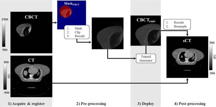
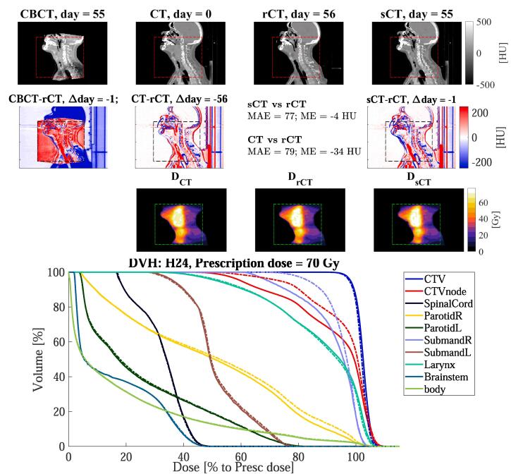
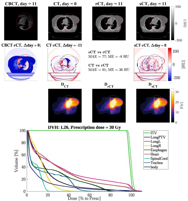
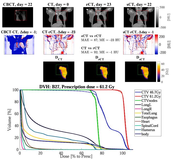
Background and purpose Adaptive radiotherapy based on cone-beam computed tomography (CBCT) requires high CT number accuracy to ensure accurate dose calculations. Recently, deep learning has been proposed for fast CBCT artefact corrections on single anatomical sites. This study investigated the feasibility of applying a single convolutional network to facilitate dose calculation based on CBCT for head-and-neck, lung and breast cancer patients. Materials and Methods Ninety-nine patients diagnosed with head-and-neck, lung or breast cancer undergoing radiotherapy with CBCT-based position verification were included in this study. The CBCTs were registered to planning CT according to clinical procedures. Three cycle-consistent generative adversarial networks (cycle-GANs) were trained in an unpaired manner on 15 patients per anatomical site generating synthetic-CTs (sCTs). Another network was trained with all the anatomical sites together. Performances of all four networks were compared and evaluated for image similarity against rescan CT (rCT). Clinical plans were recalculated on rCT and sCT and analysed through voxel-based dose differences and γ -analysis. Results A sCT was generated in 10 s. Image similarity was comparable between models trained on different anatomical sites and a single model for all sites. Mean dose differences < 0.5 % were obtained in high-dose regions. Mean gamma (3%, 3 mm) pass-rates > 95 % were achieved for all sites. Conclusion Cycle-GAN reduced CBCT artefacts and increased similarity to CT, enabling sCT-based dose calculations. A single network achieved CBCT-based dose calculation generating synthetic CT for head-and-neck, lung, and breast cancer patients with similar performance to a network specifically trained for each anatomical site.

Keywords: Adaptive radiotherapy; Artificial intelligence; CBCT; Deep learning; Dose calculation; Image-guided radiotherapy; Image-to-image translation; Machine learning.


Conflict of interest statement

The authors declare that they have no known competing financial interests or personal relationships that could have appeared to influence the work reported in this paper.

Figures

Fig. 1
Fig. 1
Schematic of the image workflow for applying the trained generator on a new 2D transverse slice of the CBCT of a breast cancer patient to create a sCT. After image acquisition, registration (1) and pre-processing (2) the trained network is deployed producing converted CBCT (CBCTconv, 3) which substituted the original CT within MaskCBCT obtaining the so-called synthetic CT (sCT).
Fig. 2
Fig. 2
Sagittal views for the head-and-neck cancer patient H24 of: (1st row) CBCT (1st column), CT (2nd column), rescan CT (rCT, 3rd column) and synthetic CT (sCT, 4th column), along with (2nd row) the respective difference to rCT, and the doses (3rd row). The red, black, or green dotted rectangles indicate the position of MaskCBCT. The days refer to the acquisition date relative of the planning CT. In the 4th row, the DVH is shown for target and OARs of sCT (solid lines) and rCT (dashed lines). Note that for the clinical target volume (CTV) of the node (CTVnode) and the right (R) submandibular, the DVH differed between rCT and sCT. This is due to anatomical differences between sCT and rCT.
Fig. 3
Fig. 3
Axial views for the lung cancer patient L26 of: (1st row) CBCT (1st column), CT (2nd column), rescan CT (rCT, 3rd column) and synthetic CT (sCT, 4th column), along with (2nd row) the respective difference to rCT, and the doses (3rd row). The red, black, or green dotted rectangles indicate the position of MaskCBCT. The days refer to the acquisition date relative of the planning CT. In the 4th row, the DVH is shown for target and OARs of sCT (solid lines) and rCT (dashed lines).
Fig. 4
Fig. 4
Coronal views for the breast cancer patient B27 of: (1st row) CBCT (1st column), CT (2nd column), rescan CT (rCT, 3rd column) and synthetic CT (sCT, 4th column), along with (2nd row) the respective difference to rCT, and the doses (3rd row). The red, black, or green dotted rectangles indicate the position of MaskCBCT. The days refer to the acquisition date relative of the planning CT. In the 4th rows, the DVH is shown for target and OARs of sCT (solid lines) and rCT (dashed lines).

Similar articles

Cited by

References

    1. Boda-Heggemann J., Lohr F., Wenz F., Flentje M., Guckenberger M. kV cone-beam CT-based IGRT. Strahlen Onkol. 2011;187:284–291. doi: 10.1007/s00066-011-2236-4. - DOI - PubMed
    1. Wu Q.J., Li T., Wu Q., Yin F.F. Adaptive Radiation Therapy. Cancer J. 2011;17:182–189. doi: 10.1097/PPO.0b013e31821da9d8. - DOI - PubMed
    1. Schulze R., Heil U., Groß D., Bruellmann D., Dranischnikow E., Schwanecke U. Artefacts in CBCT: a review. Dentomax Radiol. 2011;40:265–273. doi: 10.1259/dmfr/30642039. - DOI - PMC - PubMed
    1. Ramella S., Fiore M., Silipigni S., Zappa M.C., Jaus M., Alberti A.M. Local control and toxicity of adaptive radiotherapy using weekly CT imaging: results from the LARTIA trial in stage III NSCLC. J Thor Oncol. 2017;12:1122–1130. doi: 10.1016/J.JTHO.2017.03.025. - DOI - PubMed
    1. Dunlop A., McQuaid D., Nill S., Murray J., Poludniowski G., Hansen V.N. Comparison of CT number calibration techniques for CBCT-based dose calculation. Strahl Onkol. 2015;191:970–978. doi: 10.1007/s00066-015-0890-7. - DOI - PMC - PubMed