

Measuring eye deformation between planning and proton beam therapy position using magnetic resonance imaging
- PMID: 33458341
- PMCID: PMC7807689
- DOI: 10.1016/j.phro.2020.09.010
Measuring eye deformation between planning and proton beam therapy position using magnetic resonance imaging
Abstract
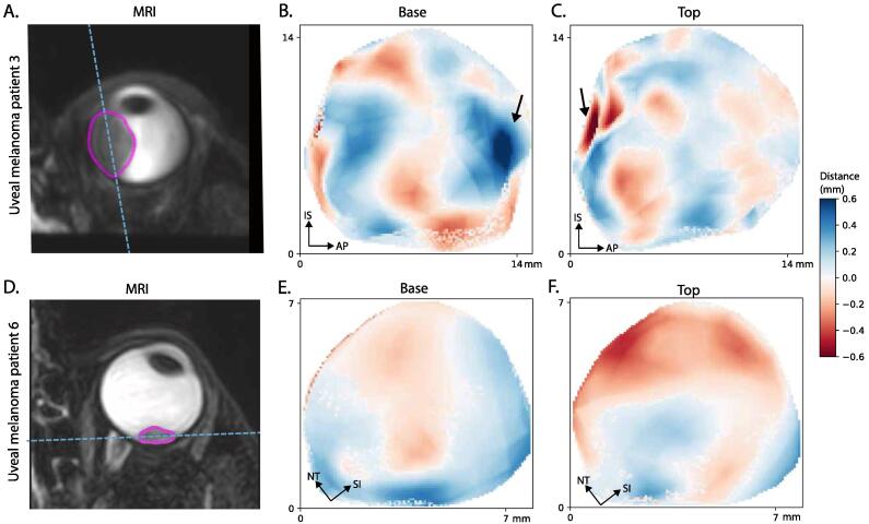
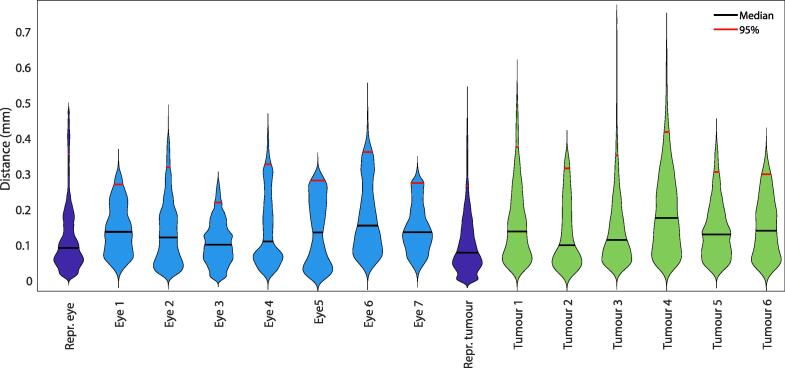
Proton beam therapy (PBT) for uveal melanoma (UM) is performed in sitting position, while the acquisition of the Magnetic resonance (MR)-images for treatment planning is performed in supine position. We assessed the effect of this difference in position on the eye- and tumour- shape. Seven subjects and six UM-patients were scanned in supine and a seating mimicking position. The distances between the tumour/sclera in both positions were calculated. The median distance between both positions was 0.1 mm. Change in gravity direction produced no substantial changes in sclera and tumour shape, indicating that supinely acquired MR-images can be used to plan ocular-PBT.
Keywords: Eye deformation; Gravity; MRI; Ocular imaging; Therapy planning; Uveal melanoma.
© 2020 The Authors.
Conflict of interest statement
The authors declare that they have no known competing financial interests or personal relationships that could have appeared to influence the work reported in this paper.
Figures
References
-
- Blum E.S., Yang J., Komatsubara K.M., Carvajal R.D. Clinical Management of Uveal and Conjunctival Melanoma. Oncology (Williston Park) 2016;30(1):29–43. - PubMed
-
- Koopmans A.E., de Klein A., Naus N.C., Kilic E. Diagnosis and Management of Uveal Melanoma. Eur Ophthalmic Rev. 2013
LinkOut - more resources
Full Text Sources
