Review
. 2020 Oct 17:16:69-73.
doi: 10.1016/j.phro.2020.09.007. eCollection 2020 Oct.

Quantitative magnetic resonance imaging on hybrid magnetic resonance linear accelerators: Perspective on technical and clinical validation

Affiliations
Review

Quantitative magnetic resonance imaging on hybrid magnetic resonance linear accelerators: Perspective on technical and clinical validation

Daniela Thorwarth et al. Phys Imaging Radiat Oncol. .

Abstract

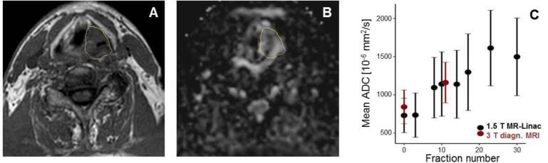
Many preclinical and clinical observations support that functional magnetic resonance imaging (MRI), such as diffusion weighted (DW) and dynamic contrast enhanced (DCE) MRI, might have a predictive value for radiotherapy. The aim of this review was to assess the current status of quantitative MRI on hybrid MR-Linacs. In a literature research, four publications were identified, investigating technical feasibility, accuracy, repeatability and reproducibility of DW and DCE-MRI in phantoms and first patients. Accuracy and short term repeatability was < 5% for DW-MRI in current MR-Linac systems. Consequently, quantitative imaging providing accurate and reproducible functional information seems possible in MR-Linacs.

Keywords: Diffusion-weighted MRI; MR-Linac; MR-guided radiotherapy; Quantitative MRI.


Conflict of interest statement

The authors declare that they have no known competing financial interests or personal relationships that could have appeared to influence the work reported in this paper.

Figures

Fig. 1
Fig. 1
Assessment of accuracy and repeatability with respect to ADC determination based on DW MRI at the 1.5 T MR-Linac using a dedicated diffusion phantom. (A) Diffusion Phantom (Model 128, Qalibre MD, Boulder, USA) containing inserts prepared with different known diffusion coefficients. (B) Regions of interest used to analyze ADC values in the different probe areas, acquired with an EPI-based DW MRI sequence (b = 0, 200, 500 s/mm2; TE/TR = 117/6683 ms; voxel size: 1.53 × 1.53 × 4 mm3). (C,D) Bland-Altman plots showing accuracy and short term repeatability of ADC assessed in four measurements on two different days, including bias (full line) and LoA (limits of agreement, dashed line). (C) Accuracy of ADC assessment with a bias of −2.5·10-6 mm2/s and LoA ± 26.8·10-6 mm2/s. (D) Repeatability analysis presents a bias of 0.6%, LoA = ±7.6%. Median accuracy and reproducibility were −0.31% and 0.31%, respectively.
Fig. 2
Fig. 2
Sequential DWI measurements acquired on the 1.5 T MR-Linac (Unity, Elekta AB, Stockholm, Sweden) in a HNC patient. This patient was treated in the context of a phase 2 feasibility trial (NCT04172753), which had been approved by the ethics committee of the University Hospital Tübingen, and gave written informed consent. (A) Anatomical T1-weighted MRI acquired on the 1.5 T MR-Linac after two weeks of RT (fraction 10) with gross tumor volume (GTV) delineated. (B) ADC map derived from EPI-based DW MRI (b = 200, 500, 800 s/mm2, TE/TR = 107/10392 ms, voxel size: 3 × 3 × 4.8 mm3). (C) Mean ADC values in the tumor region sequentially assessed during the course of fractionated RT, in comparison to two DW-MRI scans acquired at a diagnostic 3 T MR scanner (Vida, Siemens Healthineers, Erlangen, Germany). The error bars visualize the standard deviation of the ADC distribution inside the GTV. ADC values are continuously increasing during RT, which may be a sign of individual treatment response.

References

    1. Gurney-Champion O.J., Kieselmann J.P., Wong K.H., Ng-Cheng-Hin B., Harrington K., Oelfke U. A convolutional neural network for contouring metastatic lymph nodes on diffusion-weighted magnetic resonance images for assessment of radiotherapy response. Phys Imaging Radiation Oncol. 2020;15:1–7. - PMC - PubMed
    1. Ligtenberg H, Schakel T, Dankbaar JW, Ruiter LN, Peltenburg B, Willems SM, et al. Target Volume Delineation Using Diffusion-weighted Imaging for MR-guided Radiotherapy: A Case Series of Laryngeal Cancer Validated by Pathology. Cureus. 2018;10:e2465. - PMC - PubMed
    1. Leibfarth S., Winter R.M., Lyng H., Zips D., Thorwarth D. Potentials and challenges of diffusion-weighted magnetic resonance imaging in radiotherapy. Clin Transl Radiation Oncol. 2018;13:29–37. - PMC - PubMed
    1. Martens R.M., Noij D.P., Ali M., Koopman T., Marcus J.T., Vergeer M.R., de Vet H., de Jong M.C., Leemans C.R., Hoekstra O.S., de Bree R., de Graaf P., Boellaard R., Castelijns J.A. Functional imaging early during (chemo)radiotherapy for response prediction in head and neck squamous cell carcinoma; a systematic review. Oral Oncol. 2019;88:75–83. - PubMed
    1. Iima M., Le Bihan D. Clinical Intravoxel Incoherent Motion and Diffusion MR Imaging: Past, Present, and Future. Radiology. 2016;278(1):13–32. - PubMed

LinkOut - more resources