Case Reports
. 2019 Mar 4:9:65-68.
doi: 10.1016/j.phro.2019.02.004. eCollection 2019 Jan.

Prostate-specific membrane antigen positron emission tomography/computed tomography as a potential tool to assess and guide salivary gland irradiation

Affiliations
Case Reports

Prostate-specific membrane antigen positron emission tomography/computed tomography as a potential tool to assess and guide salivary gland irradiation

Matthijs H Valstar et al. Phys Imaging Radiat Oncol. .

Abstract

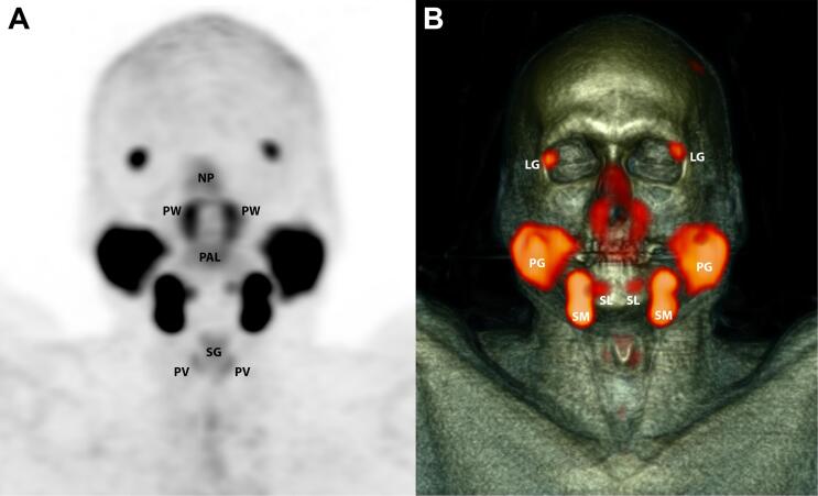
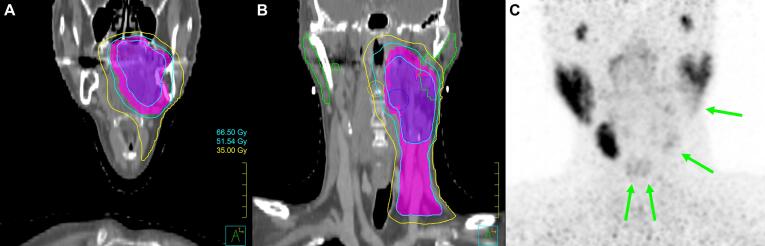
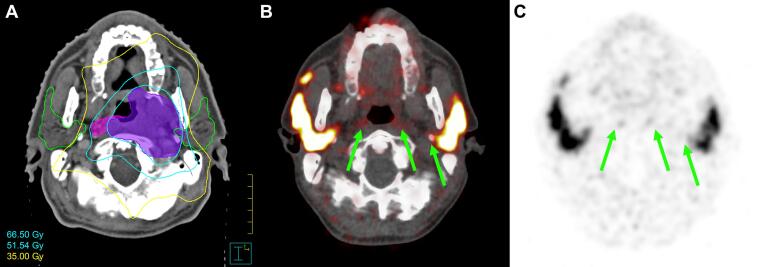
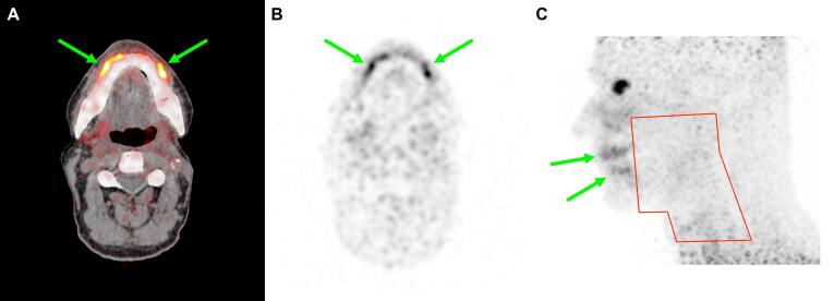
Evaluation of salivary gland damage after head and neck radiotherapy (RT) is difficult with current tools, such as subjective patient-reported outcome measures. We demonstrate the use of prostate-specific membrane antigen positron emission tomography/computed tomography (PSMA PET/CT) as an objective non-invasive tool to visualize damage to salivary glands resulting from RT. In three clinical cases, the PSMA-ligand distribution correlates to the RT dose distribution including intra-gland dose gradients and matches patient-reported toxicity, suggesting a dose-response relation. These findings support further exploration of PSMA PET/CT to guide and evaluate RT, with the ultimate aim to reduce salivary gland toxicity.

Keywords: Case series; Head and neck neoplasms; PSMA; Radiotherapy; Salivary glands; Toxicity.


Figures

Fig. 1
Fig. 1
Example PSMA PET/CT of the head and neck with healthy salivary glands. Coronal maximum intensity projection of normal PSMA distribution in a 60-year-old male with no history of disease in the head and neck, who was scanned for staging of prostate cancer using PET (A) in combination with CT (B). NP = nasal and nasopharyngeal mucosa; PW = posterior pharyngeal wall; PAL = soft palate; SG = supraglottic larynx; PV = plica vocalis; LG = lacrimal gland; PG = parotid gland; SM = submandibular gland; SL = sublingual gland.
Fig. 2
Fig. 2
Case A. Coronal slices of radiotherapy dose distribution at the level of the oral cavity (A) and parotid glands (B). Coronal maximum intensity projection of PSMA PET (C), showing the correlation between dose distribution and reduced PSMA uptake in salivary glands: left parotid gland, left submandibular gland, and both sublingual glands (green arrows). (For interpretation of the references to colour in this figure legend, the reader is referred to the web version of this article.)
Fig. 3
Fig. 3
Case B. Transverse slices of radiotherapy dose distribution (A), fused PET/CT (B) and PSMA PET (C) at the level of the parotid and retropharyngeal glands, illustrating complete loss of PSMA uptake in pharyngeal wall glands and in the deep lobe of the left parotid gland (green arrows), which received a high dose. (For interpretation of the references to colour in this figure legend, the reader is referred to the web version of this article.)
Fig. 4
Fig. 4
Case C. Transverse slice of fused PET/CT (A) and PSMA PET (B), and lateral maximum intensity projection of PSMA PET with indication of prior radiotherapy field in red (C). Complete loss of PSMA uptake is seen in most salivary glands after radiotherapy. Remarkably, PSMA PET indicates hypertrophy of the spared minor salivary glands in the lips (green arrows). (For interpretation of the references to colour in this figure legend, the reader is referred to the web version of this article.)

References

    1. Pinna R., Campus G., Cumbo E., Mura I., Milia E. Xerostomia induced by radiotherapy: an overview of the physiopathology, clinical evidence, and management of the oral damage. Ther Clin Risk Manag. 2015;11:171–188. doi: 10.2147/TCRM.S70652. - DOI - PMC - PubMed
    1. Jellema A., Slotman B., Doornaert P., Leemans C., Langendijk J. Impact of radiation-induced xerostomia on quality of life after primary radiotherapy among patients with head and neck cancer. Int J Radiat Oncol Biol Phys. 2007;69:751–760. doi: 10.1016/j.ijrobp.2007.04.021. - DOI - PubMed
    1. Cheng S., Wu V., Kwong D., Ying M. Assessment of post-radiotherapy salivary glands. Br J Radiol. 2011;84:393–402. doi: 10.1259/bjr/66754762. - DOI - PMC - PubMed
    1. Oliveira J., Gomes C., Faria D., Vieira T., Silva F., Vale J. 68Ga-prostate-specific membrane antigen positron emission tomography/computed tomography for prostate cancer imaging: a narrative literature review. World J Nucl Med. 2017;16:3–7. doi: 10.4103/1450-1147.198237. - DOI - PMC - PubMed
    1. Perera M., Papa N., Christidis D., Wetherell D., Hofman M., Murphy D. Sensitivity, specificity, and predictors of positive 68ga-prostate-specific membrane antigen positron emission tomography in advanced prostate cancer: a systematic review and meta-analysis. Eur Urol. 2016;70:926–937. doi: 10.1016/j.eururo.2016.06.021. - DOI - PubMed

Publication types

LinkOut - more resources