. 2020 Dec 28;6(1):358-366.
doi: 10.1021/acsomega.0c04677. eCollection 2021 Jan 12.

Systematic Evaluations of Doxorubicin-Induced Toxicity in Rats Based on Metabolomics

Affiliations

Systematic Evaluations of Doxorubicin-Induced Toxicity in Rats Based on Metabolomics

Chunmei Geng et al. ACS Omega. .

Abstract

Doxorubicin (DOX) is widely used to treat solid tumors, but its use is limited by its severe cardiotoxicity, nephrotoxicity, hepatotoxicity, and neurotoxicity. Metabolomic studies on DOX-induced toxicity are mainly focused on alterations in the heart and kidney, but systematic research on multiple matrices (serum, heart, liver, brain, and kidney) is rare. Thus, in our study, gas chromatography-mass spectrometry analysis of main targeted tissues (serum, heart, liver, brain, and kidney) was used to systemically evaluate the toxicity of DOX. Multivariate analyses, including orthogonal projections to the latent structure and t-test, revealed 21 metabolites in the serum, including cholesterol, d-glucose, d-lactic acid, glycine, l-alanine, l-glutamic acid, l-isoleucine, l-leucine, l-proline, l-serine, l-tryptophan, l-tyrosine, l-valine, MG (0:0/18:0/0:0), MG (16:0/0:0/0:0), N-methylphenylethanolamine, oleamide, palmitic acid, pyroglutamic acid, stearic acid, and urea. In the heart, perturbed metabolites included 3-methyl-1-pentanol, cholesterol, d-glucose, d-lactic acid, glycerol, glycine, l-alanine, l-valine, MG (16:0/0:0/0:0), palmitic acid, phenol, propanoic acid, and stearic acid. For the liver, DOX exposure caused alterations of acetamide, acetic acid, d-glucose, glycerol, l-threonine, palmitic acid, palmitoleic acid, stearic acid, and urea. In the brain, metabolic changes involved 2-butanol, carbamic acid, cholesterol, desmosterol, d-lactic acid, l-valine, MG (16:0/0:0/0:0), palmitic acid, and stearic acid. In the kidney, disturbed metabolites were involved in cholesterol, glycerol, glycine, l-alanine, MG (0:0/18:0/0:0), MG (16:0/0:0/0:0), and squalene. Complementary evidence by multiple matrices revealed disturbed pathways concerning amino acid metabolism, energy metabolism, and lipid metabolism. Our results may help to systematically elucidate the metabolic changes of DOX-induced toxicity and clarify the underlying mechanisms.


Conflict of interest statement

The authors declare no competing financial interest.

Figures

Figure 1
Figure 1
Representative GC–MS total ion current chromatograms of the serum (A), heart tissue (B), liver tissue (C), brain tissue (D), and kidney tissue (E) samples from a mixture of the control and DOX-treated rats.
Figure 2
Figure 2
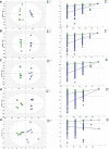
OPLS scores and 200 permutation tests for the OPLS-DA models: serum (A), heart tissue (B), liver tissue (C), brain tissue (D), and kidney tissue (E).
Figure 3
Figure 3
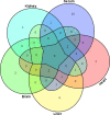
Venn diagram of the metabolite distribution in the serum, heart, liver, brain, and kidney between the control and DOX groups. Note: the numbers in the figure represent the same metabolites among different matrices (serum, heart, liver, brain, or kidney).
Figure 4
Figure 4
Summary of pathway analysis using MetaboAnalyst 4.0. Serum (A): (a) phenylalanine, tyrosine, and tryptophan biosynthesis, (b) alanine, aspartate, and glutamate metabolism, (c) aminoacyl-tRNA biosynthesis, (d) glyoxylate and dicarboxylate metabolism, (e) arginine biosynthesis, and (f) glutathione metabolism; heart tissue (B): (g) primary bile acid biosynthesis and (h) galactose metabolism; liver tissue (C): (h) galactose metabolism; brain tissue (D): (i) steroid biosynthesis; kidney tissue (E): (i) steroid biosynthesis, (g) primary bile acid biosynthesis, and (j) glycerolipid metabolism.

Similar articles

Cited by

References

    1. Kalyanaraman B. Teaching the basics of the mechanism of doxorubicin-induced cardiotoxicity: Have we been barking up the wrong tree?. Redox Biol. 2019, 29, 101394.10.1016/j.redox.2019.101394. - DOI - PMC - PubMed
    1. Chen Y.; Tang Y.; Zhang Y.-C.; Huang X.-H.; Xie Y.-Q.; Xiang Y. A metabolomic study of rats with doxorubicin-induced cardiomyopathy and Shengmai injection treatment. PLoS One 2015, 10, e012520910.1371/journal.pone.0125209. - DOI - PMC - PubMed
    1. Damiani R. M.; Moura D. J.; Viau C. M.; Caceres R. A.; Henriques J. A. P.; Saffi J. Pathways of cardiac toxicity: comparison between chemotherapeutic drugs doxorubicin and mitoxantrone. Arch. Toxicol. 2016, 90, 2063–2076. 10.1007/s00204-016-1759-y. - DOI - PubMed
    1. Liu Y.; Xu W.; Xiong Y.; Du G.; Qin X. Evaluations of the effect of HuangQi against heart failure based on comprehensive echocardiography index and metabonomics. Phytomedicine 2018, 50, 205–212. 10.1016/j.phymed.2018.04.027. - DOI - PubMed
    1. Prša P.; Karademir B.; Biçim G.; Mahmoud H.; Dahan I.; Yalçın A. S.; Mahajna J.; Milisav I. The potential use of natural products to negate hepatic, renal and neuronal toxicity induced by cancer therapeutics. Biochem. Pharmacol. 2020, 173, 113551.10.1016/j.bcp.2019.06.007. - DOI - PubMed