

Neurobiology of SARS-CoV-2 interactions with the peripheral nervous system: implications for COVID-19 and pain
- PMID: 33458558
- PMCID: PMC7803673
- DOI: 10.1097/PR9.0000000000000885
Neurobiology of SARS-CoV-2 interactions with the peripheral nervous system: implications for COVID-19 and pain
Abstract
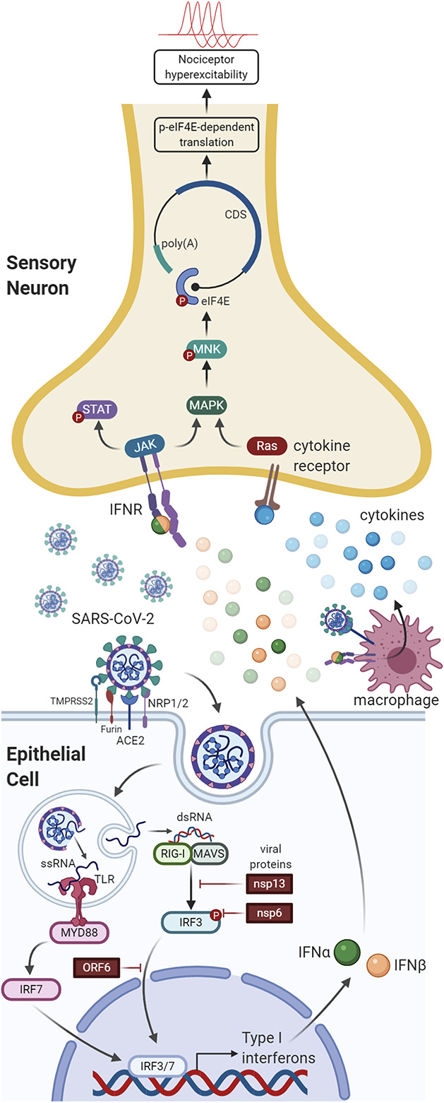
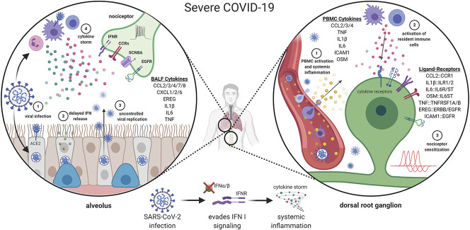
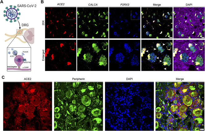
SARS-CoV-2 is a novel coronavirus that infects cells through the angiotensin-converting enzyme 2 receptor, aided by proteases that prime the spike protein of the virus to enhance cellular entry. Neuropilin 1 and 2 (NRP1 and NRP2) act as additional viral entry factors. SARS-CoV-2 infection causes COVID-19 disease. There is now strong evidence for neurological impacts of COVID-19, with pain as an important symptom, both in the acute phase of the disease and at later stages that are colloquially referred to as "long COVID." In this narrative review, we discuss how COVID-19 may interact with the peripheral nervous system to cause pain in the early and late stages of the disease. We begin with a review of the state of the science on how viruses cause pain through direct and indirect interactions with nociceptors. We then cover what we currently know about how the unique cytokine profiles of moderate and severe COVID-19 may drive plasticity in nociceptors to promote pain and worsen existing pain states. Finally, we review evidence for direct infection of nociceptors by SARS-CoV-2 and the implications of this potential neurotropism. The state of the science points to multiple potential mechanisms through which COVID-19 could induce changes in nociceptor excitability that would be expected to promote pain, induce neuropathies, and worsen existing pain states.
Keywords: COVID-19; Neuropathy; Nociceptor; SARS-CoV-2.
Copyright © 2021 The Author(s). Published by Wolters Kluwer Health, Inc. on behalf of The International Association for the Study of Pain.
Conflict of interest statement
The authors have no conflicts of interest to declare. This work was supported by NIH grants NS065926 and NS111929 to T.J. Price. T.J. Price is a co-founder of 4E Therapeutics, a company developing MNK inhibitors for neuropathic pain.Sponsorships or competing interests that may be relevant to content are disclosed at the end of this article.
Figures
References
-
- Andoh T, Maki T, Li S, Uta D. β2-Microglobulin elicits itch-related responses in mice through the direct activation of primary afferent neurons expressing transient receptor potential vanilloid 1. Eur J Pharmacol 2017;810:134–40. - PubMed
-
- Arunachalam PS, Wimmers F, Mok CKP, Perera R, Scott M, Hagan T, Sigal N, Feng Y, Bristow L, Tak-Yin Tsang O, Wagh D, Coller J, Pellegrini KL, Kazmin D, Alaaeddine G, Leung WS, Chan JMC, Chik TSH, Choi CYC, Huerta C, Paine McCullough M, Lv H, Anderson E, Edupuganti S, Upadhyay AA, Bosinger SE, Maecker HT, Khatri P, Rouphael N, Peiris M, Pulendran B. Systems biological assessment of immunity to mild versus severe COVID-19 infection in humans. Science 2020;369:1210–20. - PMC - PubMed
-
- Balachandran S, Barber GN. PKR in innate immunity, cancer, and viral oncolysis. Methods Mol Biol 2007;383:277–301. - PubMed
Publication types
LinkOut - more resources
Full Text Sources
Other Literature Sources
Miscellaneous