. 2021 Jan 6;24(1):101968.
doi: 10.1016/j.isci.2020.101968. eCollection 2021 Jan 22.

Dopamine induces functional extracellular traps in microglia

Affiliations

Dopamine induces functional extracellular traps in microglia

Ishan Agrawal et al. iScience. .

Abstract

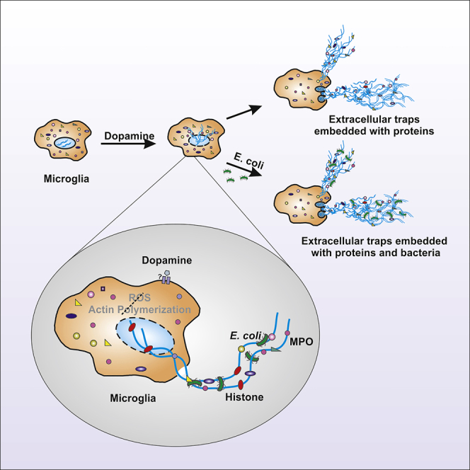
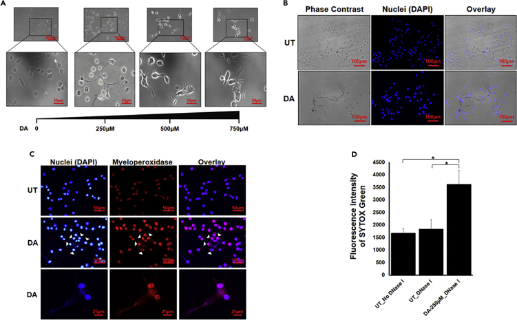
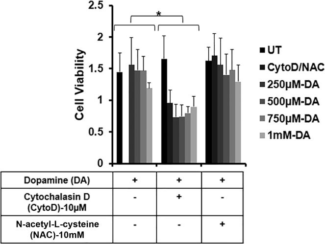
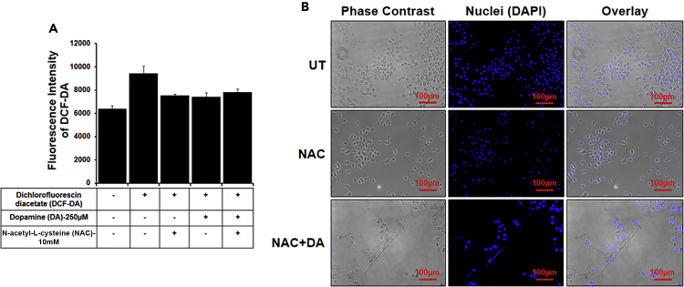
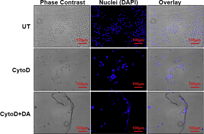
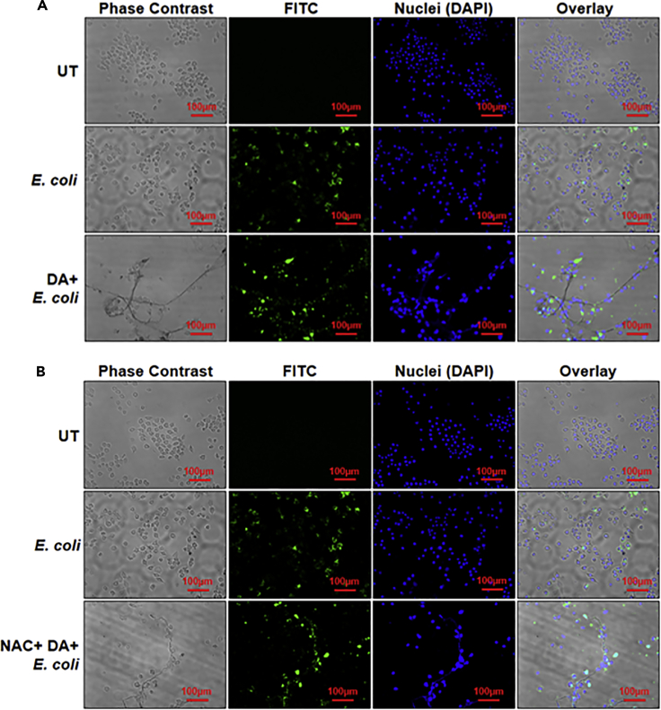
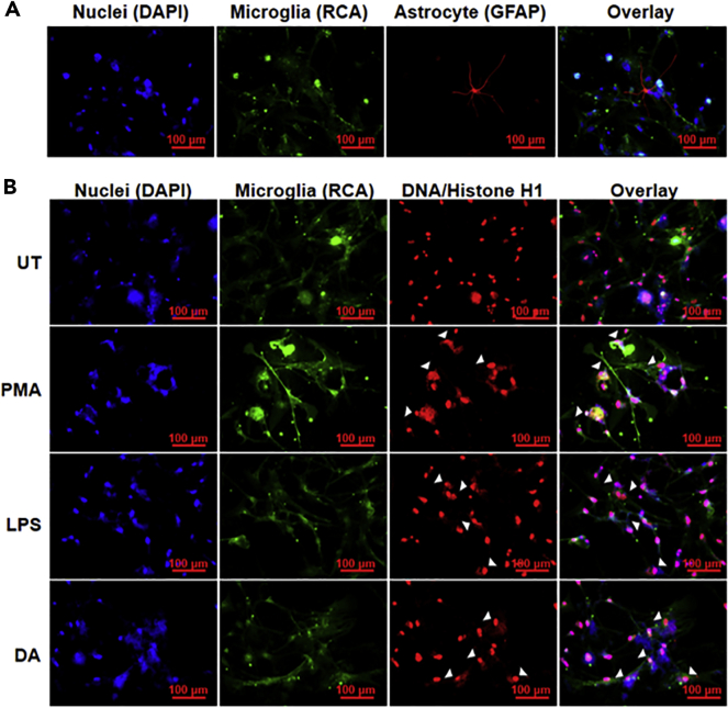
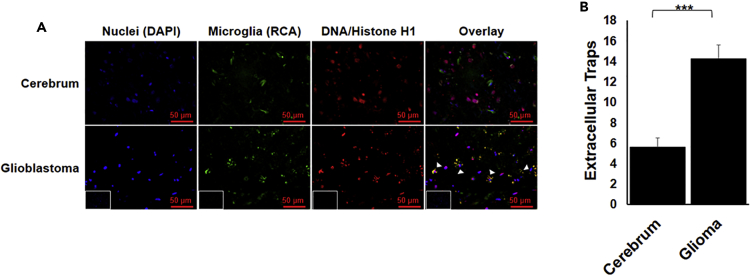
Dopamine (DA) plays many roles in the brain, especially in movement, motivation, and reinforcement of behavior; however, its role in regulating innate immunity is not clear. Here, we show that DA can induce DNA-based extracellular traps in primary, adult, human microglia and BV2 microglia cell line. These DNA-based extracellular traps are formed independent of reactive oxygen species, actin polymerization, and cell death. These traps are functional and capture fluorescein (FITC)-tagged Escherichia coli even when reactive oxygen species production or actin polymerization is inhibited. We show that microglial extracellular traps are present in Glioblastoma multiforme. This is crucial because Glioblastoma multiforme cells are known to secrete DA. Our findings demonstrate that DA plays a significant role in sterile neuro-inflammation by inducing microglia extracellular traps.

Keywords: Cell Biology; Cellular Neuroscience; Immunology; Molecular Biology.


Conflict of interest statement

The authors declare no competing interests.

Figures

None
Graphical abstract
Figure 1
Figure 1
Visualization of DA-induced extracellular traps in BV2 microglia (A) BV2 microglia were incubated with different concentrations of dopamine (DA) for 24 hr. ETs were clearly visible at all the concentrations of DA. Scale bars, 100μm and 50μm. (B and C) BV2 microglia were incubated with 250μM of DA for 24 hr. ETs were stained with just DAPI (blue) (B) or DAPI and MPO (C). The images are representative of three experiments. At least 7 frames were imaged per well in a two well chamber slide. Scale bars, 100μm, 50μm, and 25μm. (D) DNase I was added after 24 hr in DA-treated wells and untreated (UT) wells. Fluorescence was measured in the collected supernatant with the help of SYTOX Green. Graph is representative of 4 experiments. Data are represented as mean ± SEM. ∗p < 0.05 (Student's t-test).
Figure 2
Figure 2
Dopamine induces extracellular traps formation independent of cell death BV2 microglia were seeded in a 96-well microplate and were pretreated with 10μM cytochalasin D (CytoD) for 20 min and 10mM N-acetyl-L-cysteine (NAC) for 3 hr. Pretreatment was followed by dopamine (DA) treatment for 24 hr. Cell viability was observed by taking the absorbance at 570 nm. The graph is representative of 3 experiments. Data are represented as mean ± SEM. ∗p < .05 (Student's t-test).
Figure 3
Figure 3
Dopamine induces extracellular traps independent of ROS Cells were pretreated with 10mM N-acetyl-L-cysteine (NAC) for 3 hr followed by 250μM dopamine (DA) treatment for 24 hr. (A) ROS production was checked with the help of dichlorofluorescin diacetate (DCF-DA). The graph is representative of three experiments. Data are represented as mean ± SEM. (B) ET formation was induced by DA even in the presence of NAC. ETs were stained with DAPI (blue). At least 7 frames were imaged per well. Scale bars, 100μm.
Figure 4
Figure 4
Extracellular trap formation is independent of actin polymerization Cells were pretreated for 20 min with 10μM cytochalasin D (CytoD) followed by treatment with 250μM dopamine (DA) for 24 hr. Inhibition of actin polymerization had no effect on ET formation. ETs were stained with DAPI (blue). The images are the representative of two experiments. At least 7 frames were imaged per well. Scale bars, 100μm.
Figure 5
Figure 5
Dopamine induces formation of functional extracellular traps (A) BV2 microglia were incubated with dopamine (DA) for 3 hr. FITC-tagged E. coli (green) was added followed by DA incubation, and cells were further incubated for 21 hours. ETs were stained with DAPI (blue). In the overlay image, green dots are overlapping with the blue ETs, suggesting that ETs are trapping E. coli. Images are representative of two experiments. At least 7 frames were imaged per well. Scale bars, 100μm. (B) BV2 microglia were pretreated with N-acetyl-L-cysteine (NAC) and were incubated with dopamine (DA) for 3 hr. FITC-tagged E. coli (green) was added followed by DA incubation, and cells were further incubated for 21 hours. ETs were stained with DAPI (blue). In the overlay image, green dots are overlapping with the blue ETs, suggesting that ETs are trapping E. coli. Images are representative of two experiments. At least 7 frames were imaged per well. Scale bars, 100μm.
Figure 6
Figure 6
Dopamine induces extracellular traps in primary human microglia (A) Primary human microglia isolated from adult human brain tissues. RCA (green) was used as a microglia marker, and glial fibrillary acidic protein (GFAP) (red) was used as an astrocyte marker. About 80% of the cells isolated were microglia. (B) Isolated microglia were treated with 2.5μM dopamine (DA) for 12 hr. RCA (green) was used as a microglia marker. ETs (arrow heads) were visualized using DAPI (blue) and DNA/Histone H1 antibodies (red). At least 7 frames were imaged per well of the two well chamber slide. Scale bars, 100μm.
Figure 7
Figure 7
Presence of microglia extracellular traps in GBM (A) GBM tissue was stained with DAPI (blue), RCA (green) for microglia, and DNA/Histone H1 (red) for ETs. The formation of ETs by microglia can be visualized by the punctate red staining overlapping with green (arrow heads). Inset represents secondary antibody control. Scale bars, 50μm. (B) The punctate red staining overlapping with green was quantified. The graph represents average of the quantified value. The result is representative of two experiments. At least 7 frames were imaged per section and quantified. Data are represented as mean ± SEM. ∗∗∗p < .0001 (Student's t-test).

Similar articles

Cited by

References

    1. Abi Abdallah D.S., Lin C., Ball C.J., King M.R., Duhamel G.E., Denkers E.Y. Toxoplasma gondii triggers release of human and mouse neutrophil extracellular traps. Infect. Immun. 2012;80:768–777. - PMC - PubMed
    1. Agrawal I., Saxena S., Nair P., Jha D., Jha S. Obtaining Human Microglia from Adult Human Brain Tissue. JoVE. 2020;162:e61438. - PubMed
    1. Arreola R., Alvarez-Herrera S., Pérez-Sánchez G., Becerril-Villanueva E., Cruz-Fuentes C., Flores-Gutierrez E.O., Garcés-Alvarez M.E., De La Cruz-Aguilera D.L., Medina-Rivero E., Hurtado-Alvarado G. Immunomodulatory effects mediated by dopamine. J. Immunol. Res. 2016;2016:3160486. - PMC - PubMed
    1. Basu S., Nagy J.A., Pal S., Vasile E., Eckelhoefer I.A., Susan Bliss V., Manseau E.J., Dasgupta P.S., Dvorak H.F., Mukhopadhyay D. The neurotransmitter dopamine inhibits angiogenesis induced by vascular permeability factor/vascular endothelial growth factor. Nat. Med. 2001;7:569–574. - PubMed
    1. Beninger R.J. The role of dopamine in locomotor activity and learning. Brain Res. 1983;287:173–196. - PubMed