Review
. 2021 Mar 1;50(4):2224-2243.
doi: 10.1039/d0cs01261k.

Targeting RNA with small molecules: from fundamental principles towards the clinic

Affiliations
Review

Targeting RNA with small molecules: from fundamental principles towards the clinic

James P Falese et al. Chem Soc Rev. .

Abstract

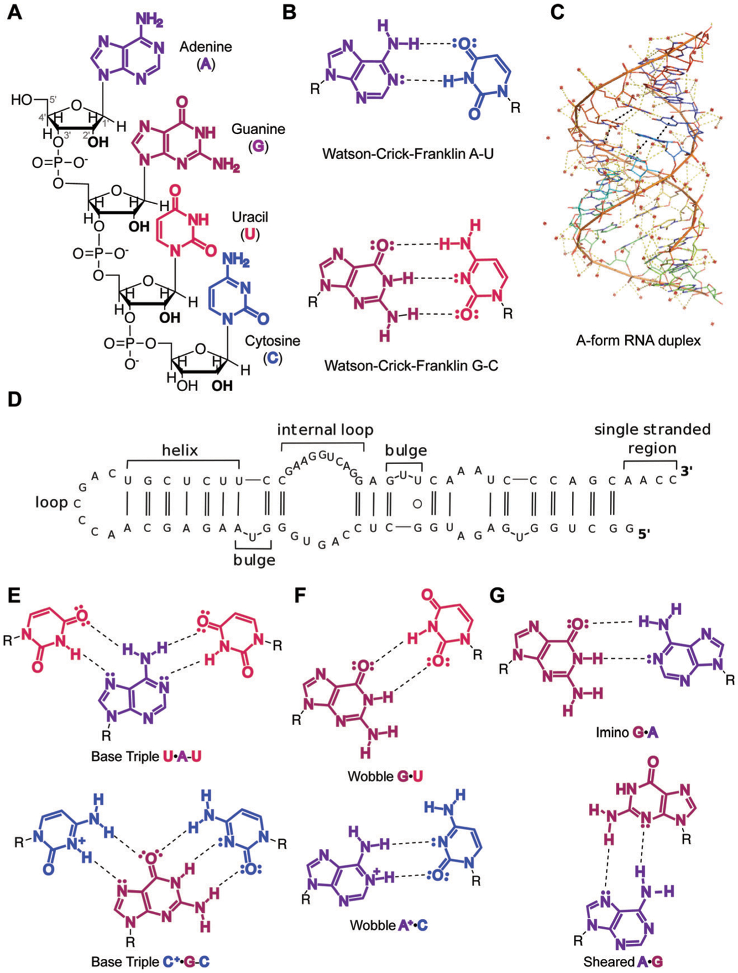
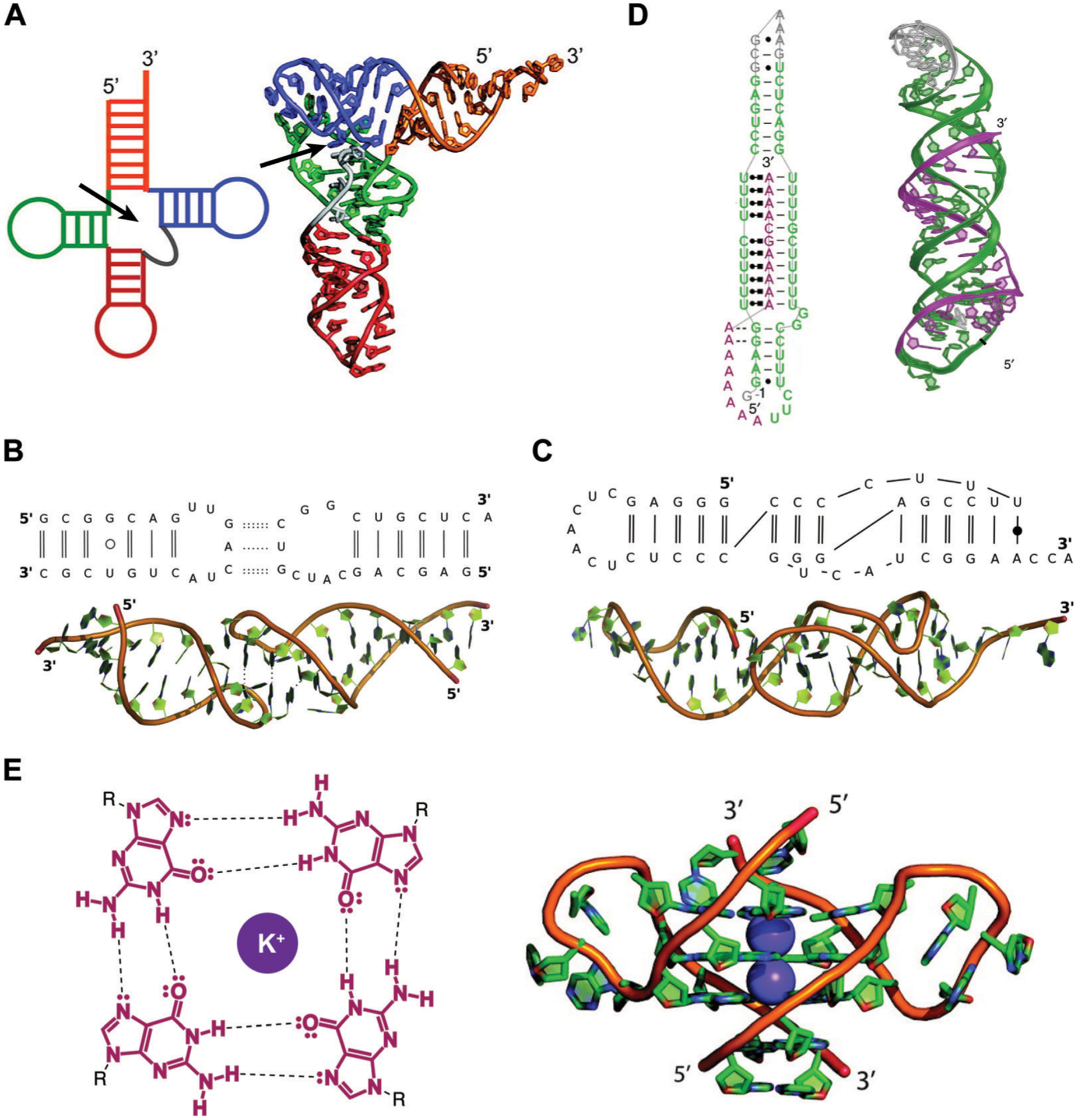
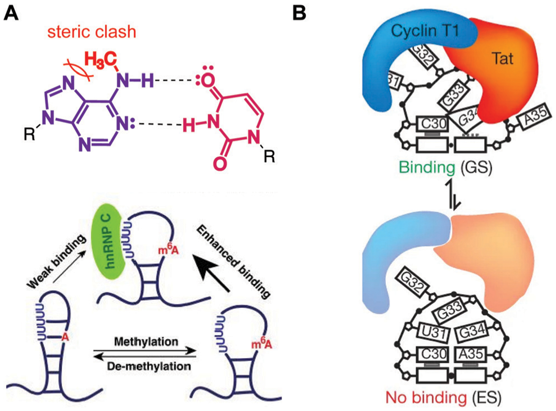
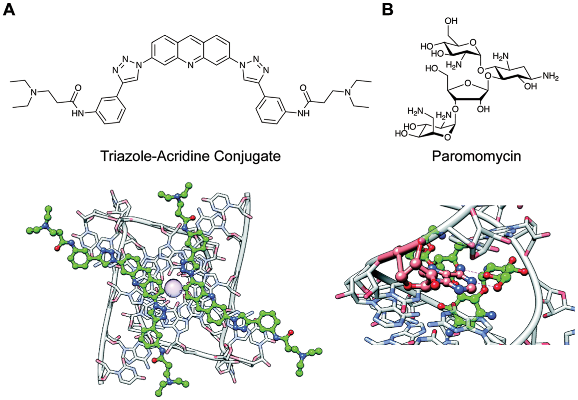
Recent advances in our understanding of RNA biology have uncovered crucial roles for RNA in multiple disease states, ranging from viral and bacterial infections to cancer and neurological disorders. As a result, multiple laboratories have become interested in developing drug-like small molecules to target RNA. However, this development comes with multiple unique challenges. For example, RNA is inherently dynamic and has limited chemical diversity. In addition, promiscuous RNA-binding ligands are often identified during screening campaigns. This Tutorial Review overviews important considerations and advancements for generating RNA-targeted small molecules, ranging from fundamental chemistry to promising small molecule examples with demonstrated clinical efficacy. Specifically, we begin by exploring RNA functional classes, structural hierarchy, and dynamics. We then discuss fundamental RNA recognition principles along with methods for small molecule screening and RNA structure determination. Finally, we review unique challenges and emerging solutions from both the RNA and small molecule perspectives for generating RNA-targeted ligands before highlighting a selection of the "Greatest Hits" to date. These molecules target RNA in a variety of diseases, including cancer, neurodegeneration, and viral infection, in cellular and animal model systems. Additionally, we explore the recently FDA-approved small molecule regulator of RNA splicing, risdiplam, for treatment of spinal muscular atrophy. Together, this Tutorial Review showcases the fundamental role of chemical and molecular recognition principles in enhancing our understanding of RNA biology and contributing to the rapidly growing number of RNA-targeted probes and therapeutics. In particular, we hope this widely accessible review will serve as inspiration for aspiring small molecule and/or RNA researchers.


Conflict of interest statement

Conflicts of interest

The authors declare no conflicts of interest.

Figures

Fig. 1
Fig. 1
Overview of RNA primary and secondary structure and base pairing patterns. (A) Nucleotide building blocks of RNA primary structure. Each consists of a ribose sugar, containing a 2′ hydroxyl unique to RNA over DNA; phosphodiester groups, which connect the 5′ and 3′ hydroxyl groups of the ribose sugars; and adenine, guanine, uracil and cytosine nucleobases, which define the sequence of an RNA molecule. (B) Canonical Watson–Crick–Franklin hydrogen bonding patterns. Dashed lines represent hydrogen bonds. (C) An A-form RNA duplex secondary structure (PDB ID: 1LNT). Red dots represent metal ions. Yellow dashes indicate electrostatic interactions, including hydrogen bonds. Black dashes indicate stacking interactions. (D) Example of canonical RNA secondary structures as observed in 5′-end of long non-coding RNA (lncRNA) B2-SINE. Single and double solid lines represent A–U and G–C Watson–Crick–Franklin base pairs, respectively, while circles represent wobble base pairs. Adapted from ref. with permission from Elsevier B. V., Copyright 2015. (E) Higher-order base triple interactions. (F) Non-canonical wobble interactions. (G) Purine–purine interactions, including imino and sheared hydrogen bonding patterns.
Fig. 2
Fig. 2
Examples of RNA tertiary structures. (A) Yeast phenylalanine t-RNA (PDB ID: 6TNA). The individual helices arising from the multi-way junction (arrow) that form the cloverleaf-shaped secondary structure (left) were found to stack on each other to determine the L-shaped three-dimensional fold (right). Adapted from ref. with permission from American Chemical Society, Copyright 2011. (B) Example of a kissing loop tertiary interaction formed between substrate and catalytic domain stem-loops of the Neurospora Varkud satellite ribozyme (PDB ID: 2MIO). Dashed lines indicate hydrogen bonding. Wobble and non-WCF base pairs are shown as unfilled and filled circles, respectively. Adapted from ref. with permission from Elsevier B. V., Copyright 2015. (C) Example of a pseudoknot tertiary interaction in the turnip yellow mosaic virus RNA (PDB ID: 1A60). Non-WCF base pair is shown as a filled circle. Adapted from ref. with permission from Elsevier B. V., Copyright 2015. (D) 3′-Triple helix in lncRNA MALAT1 (PDB ID: 4PLX). Uridine-rich stem loop (green) form WCF (simple lines) and Hoogsteen (dot and square lines) base pairs with the A-rich tail (purple). Adapted from ref. with permission from Springer Nature, Copyright 2014. (E) G-quartets are stabilized by potassium cations through interactions with the guanine’s oxygens (left), and stack on top of each other to form G-quadruplex structures (right), as seen in the human telomeric (TERRA) RNA (PDB ID: 3IBK). Dashed lines represent hydrogen bonds, dots represent lone pairs of electrons, and purple circles represent the potassium ion. Adapted from ref. with permission from American Chemical Society, Copyright 2011.
Fig. 3
Fig. 3
Examples of RNA dynamic regulation and associated functional consequences. (A) N6-methyladenosine (m6A) modification disfavors WCF base pairing (top), promoting single-stranded hairpin formation in MALAT1 and consequent enhanced binding of the heterogeneous nuclear ribonucleoprotein C (hnRNP C, bottom). Adapted from ref. with permission from Springer Nature, Copyright 2015. (B) The ground state (GS) of HIV-1 TAR RNA is more strongly bound by the Cyclin T1-Tat protein complex than the less energetically stable excited state. Adapted from ref. with permission from Springer Nature, Copyright 2012.
Fig. 4
Fig. 4
Examples of common noncovalent interactions in RNA–small molecule recognition. Adapted from ref. with permission from The Royal Society of Chemistry, Copyright 2020. (A) Binding of two acridine-based ligands to the Telomeric Repeat-Containing RNA (TERRA) via stacking interactions (PDB ID: 3MIJ). Ball denotes a K+ ion. (B) Hydrogen bonding interactions (dashed lines) between paromomycin and HIV-1 Dimerization Initiation Site (DIS) (PDB ID: 3C44).
Fig. 5
Fig. 5
Common screening methods utilized for detecting RNA–small molecule binding. (A) Fluorescence Indicator Displacement (FID) assays detect changes in fluorescence intensity of an indicator as a result of small molecule binding. This method enables detection of binding events if a single small molecule concentration is used, and relative affinities or Competitive Displacement (CD50) values if multiple concentrations are used. Adapted from ref. with permission from Elsevier B.V., Copyright 2019. (B) Small Molecule Microarrays (SMM) enable detection of binding as a result of detecting fluorescent wells upon irradiating a microarray plate. The plate contains immobilized small molecule ligands and is incubated with a fluorescently labeled RNA construct prior to irradiation. (C) Isothermal Titration Calorimetry (ITC) functions through directly measuring changes in heat as a result of small molecule (titrant) binding to the RNA construct (sample cell) relative to buffer (reference cell). As such, this method enables determination of binding affinity (Kd), stoichiometry, as well as Gibbs free energy (ΔG), enthalpy (ΔH), and entropy (ΔS) values. Adapted from ref. with permission from American Chemical Society, Copyright 2019. (D) Surface Plasmon Resonance (SPR) directly measures changes in resonance angle or wavelength of a glass surface containing the immobilized RNA of interest upon addition of a small molecule. This method can be utilized to obtain both the association (kon) and dissociation (koff) constants that comprise binding affinities (Kd), providing insights into the kinetics of a binding event. Adapted from ref. with permission from American Chemical Society, Copyright 2019.
Fig. 6
Fig. 6
Chemical probing for interrogation of RNA structure. (A) Mechanism for covalent modification by SHAPE reagent 1-methyl-7-nitroisatoic anhydride (1M7, top) and dimethylsulfate (DMS, bottom). In SHAPE probing, deprotonation of the 2′-hydroxyl by a general base facilitates its nucleophilic attack of the anhydride moiety, resulting in the ring opening of 1M7. While this attack is an equilibrium process, the subsequent decarboxylation, generating the final adduct as depicted, is irreversible. In DMS probing, the nitrogen lone pair on A (depicted) or C attack the electrophilic methoxy position, resulting in irreversible covalent modification of the base. (B) Workflow for integration of chemical probing information into a structure model. Reagent modifications are incorporated as mutations during reverse transcription in the presence of manganese, and mutations and detected via sequencing. Reactivities for each nucleotide are incorporated as restraints in a structure model. Adapted from ref. with permission from Springer Nature, Copyright 2014. (C) Chemical probing with HIV TAR following small molecule treatment reveals changes in reactivity for small molecule binding site. Larger scale changes in secondary structure were not observed. Scale shows changes in reactivity between control and small molecule treatment. PBS: primer binding site. Adapted from ref. with permission American Chemical Society, Copyright 2014.
Fig. 7
Fig. 7
Structural dynamics of HIV-1 TAR are modulated with small molecules. (A) TAR oscillates between a ground state (GS) and excited state (ES) characterized by base flipping and angular rearrangement of the helix. Several ligands, including argininamide and DMA-169, were shown to alter excited state accessibility, whereas other ligands such as neomycin were not. Adapted from ref. with permission from Elsevier, Copyright 2020. (B) The structures of argininamide, DMA-169, and neomycin.
Fig. 8
Fig. 8
Approaches towards discovering privileged properties and chemotypes of RNA-binding small molecules. (A) Comparison of 20 physicochemical properties of bioactive, RNA-binding small molecules (R-BIND) and FDA ligands (mostly protein-targeted). R-BIND members were found to have increased nitrogen atom count, decreased number of sp3-hybridized carbons, among other properties. Statistically significant differences determined by Mann Whitney U test are indicated by P values of <0.05 in light blue and highly significant differences as <0.001 in dark blue. MW = molecular weight, HBA = hydrogen bond acceptors, HBD = hydrogen bond donors, log P = n-octanol/water partition coefficient, RotB = number of rotatable bonds, tPSA = topological polar surface area, log D = n-octanol/water partition coefficient, N = number of nitrogen atoms, O = number of oxygen atoms, rings = number of rings, ArRings = number of aromatic rings, HetRings = number of heteroatom-containing rings, SysRings = number of ring systems, SysRR = ring complexity, Fsp3 = fraction of sp3-hybridized carbons, nStereo = number of stereocenters, ASA = accessible surface area, RelPSA = relative polar surface area, TC = total charge, VWSA = van der Waals surface area. Adapted from ref. with permission from John Wiley & Sons, Copyright 2017. (B) Nearest Neighbor algorithm utilized to select R-BIND-like molecules as developed by Hargrove and co-workers. Each cheminformatic parameter from (A) is defined as a “dimension”, quantified along its dimension, and the Euclidian distance (smallest distance in space) between each library member is measured and averaged (purple line). The user’s input molecule is considered “R-BIND-like” (turquoise dots) or enriched in RNA-binding properties if it is found to be within the measured average distance to an R-BIND member (blue dots). Adapted from ref. with permission from American Chemical Society, Copyright 2019. (C) Summary of infoRNA platform utilized to select potential binders of an RNA target. By inputting an RNA sequence of interest, infoRNA is mined for similar RNA motifs to output lead small molecules that can be tested and optimized for specific targeting. Adapted from ref. with permission from American Chemical Society, Copyright 2016. (D) Examples of privileged chemotypes for binding of RNA hairpin or bulge secondary structures as discovered by infoRNA mining. Adapted from ref. with permission from American Chemical Society, Copyright 2016.
Fig. 9
Fig. 9
Small molecule targeting of the MALAT1 triple helix. (A) Structures of DPFp8 and SM5, which selectively bind the MALAT1 triple helix over controls. (B) Treatment of breast cancer organoids with SM5 significantly reduces organoid size (upper panel and lower panel differ by magnification). Adapted from ref. with permission from American Chemical Society, Copyright 2019. (C) SM5 and SM16 (not shown) significantly reduce branching morphogenesis of breast cancer organoids. Adapted from ref. with permission from American Chemical Society, Copyright 2019. ASO: antisense oligonucleotide.
Fig. 10
Fig. 10
Targeting CUG repeat expansions with a multivalent ligand in DM1. (A) Structure of 2H-K4NMeS. (B) 2H-K4NMeS treatment reduces CUG:MBNL1 foci. RNA was detected with Fluorescence In Situ Hybridization (FISH), where fluorescently-labeled oligonucleotides complementary to an RNA of interest are incubated in cells to show their subcellular localization. Nuclei are labeled with DAPI (4′,6-diamidino-2-phenylindole). Adapted from ref. with permission from Springer Nature, Copyright 2016. MBNL1 was detected using immunofluorescence, where a fluorescently-labeled antibody for MBNL1 was incubated in cells to show its subcellular localization. White regions in the “Overlay” show where FISH, MBNL1, and DAPI signals overlap. (C) Treatment with 2H-K4NMeS shows dose-dependent rescue of MBNL1 splicing defect. Adapted from ref. with permission from Springer Nature, Copyright 2016.
Fig. 11
Fig. 11
Small molecule allosteric modulation of EV71 SLII. (A) The structure of DMA-135. (B) DMA-135 results in a 77° interhelical shift in SLII as shown by NMR. Adapted from ref. with permission from Springer Nature, Copyright 2020. (C) A luciferase-based reporter assay shows reduction of FLuc translation from 5′ UTR upon DMA-135 treatment as compared to RLuc control. Adapted from ref. with permission from Springer Nature, Copyright 2020. (D) AUF1 protein was isolated via immunoprecipitation and bound RNA was detected via qPCR. Dose-dependent increase in bound SL2 RNA was observed upon DMA-135 treatment as compared to GAPDH control RNA. N. I. non-immune antibody. Adapted from ref. from Springer Nature, 2020 under Creative Commons CC-BY 4.0 License.
Fig. 12
Fig. 12
Small molecule disruption of foci containing G-quadruplex GGGGCC repeat expansions in ALS/FTD. (A) Structure of DB1273. (B) DB1273 significantly reduces GGGGCC RNA foci (red) in cellular nuclei (blue). Adapted from ref. , Copyright 2017, John Wiley and Sons in accordance to Creative Commons CC BY 4.0 License.
Fig. 13
Fig. 13
Small molecule targeting of the riboflavin riboswitch. (A) The structures of Ribocil-B and Riboflavin. (B) Crystal structure of Ribocil-B (blue sticks) in complex with riboflavin riboswitch. Ribocil binding is stabilized by stacking interactions with A48 and A85 and hydrogen bonding between the oxygen and A48 and A99. Additional stacking interactions and a methyl hydrogen bond are observed on the other face of the molecule. Adapted from ref. with permission from Springer Nature, Copyright 2015.
Fig. 14
Fig. 14
Small molecule modulation of splicing in SMA. (A) Schematic of SMN2 splicing. Inclusion of Exon 7 results in a functional SMN2 protein. (B) Structures of two documented SMN2 splicing modulators. (C) SMN-C5 (yellow) stabilizes an adenine residue (red) between Exon 7 and a spliceosomal U1 small nuclear ribonucleoprotein (snRNP), facilitating Exon 7 inclusion. SS: splice site. Adapted from ref. with permission from Springer Nature, Copyright 2019.

References

    1. Warner KD, Hajdin CE and Weeks KM, Nat. Rev. Drug Discovery, 2018, 17, 547–558. - PMC - PubMed
    1. Cech TR and Steitz JA, Cell, 2014, 157, 77–94. - PubMed
    1. Donlic A and Hargrove AE, Wiley Interdiscip. Rev.: RNA, 2018, 9, e1477. - PMC - PubMed
    1. The ENCODE Project Consortium, Nature, 2012, 489, 57–74. - PMC - PubMed
    1. Costales MG, Childs-Disney JL, Haniff HS and Disney MD, J. Med. Chem, 2020, 63, 8880–8900. - PMC - PubMed