. 2021 Jul 1;529(10):2558-2575.
doi: 10.1002/cne.25109. Epub 2021 Jan 28.

Immunocytochemical and ultrastructural organization of the taste thalamus of the tree shrew (Tupaia belangeri)

Affiliations

Immunocytochemical and ultrastructural organization of the taste thalamus of the tree shrew (Tupaia belangeri)

Erin E Maher et al. J Comp Neurol. .

Erratum in

  • Erratum.
    [No authors listed] [No authors listed] J Comp Neurol. 2022 May;530(7):1126. doi: 10.1002/cne.25315. J Comp Neurol. 2022. PMID: 35338485 Free PMC article. No abstract available.

Abstract

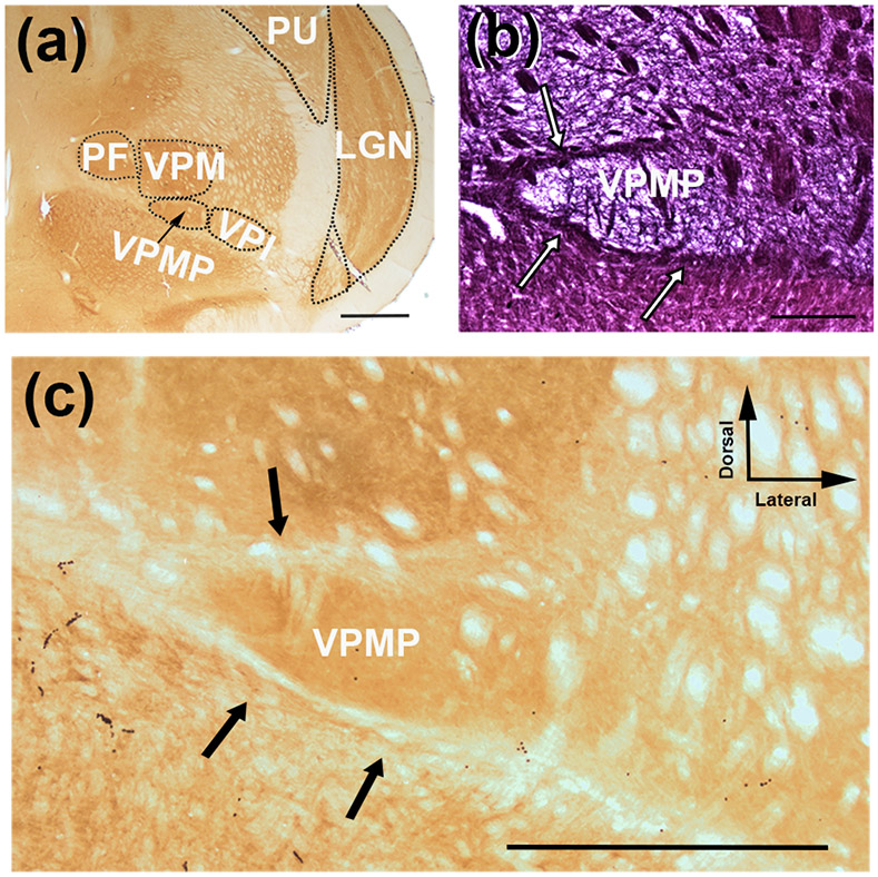
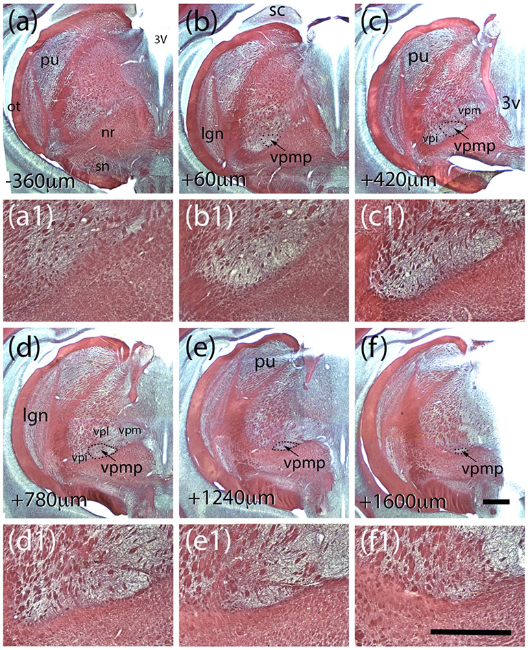
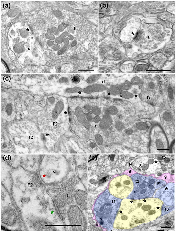
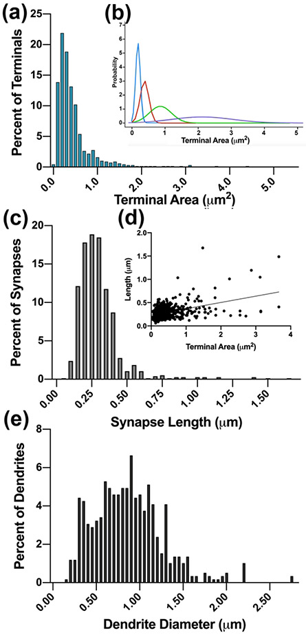
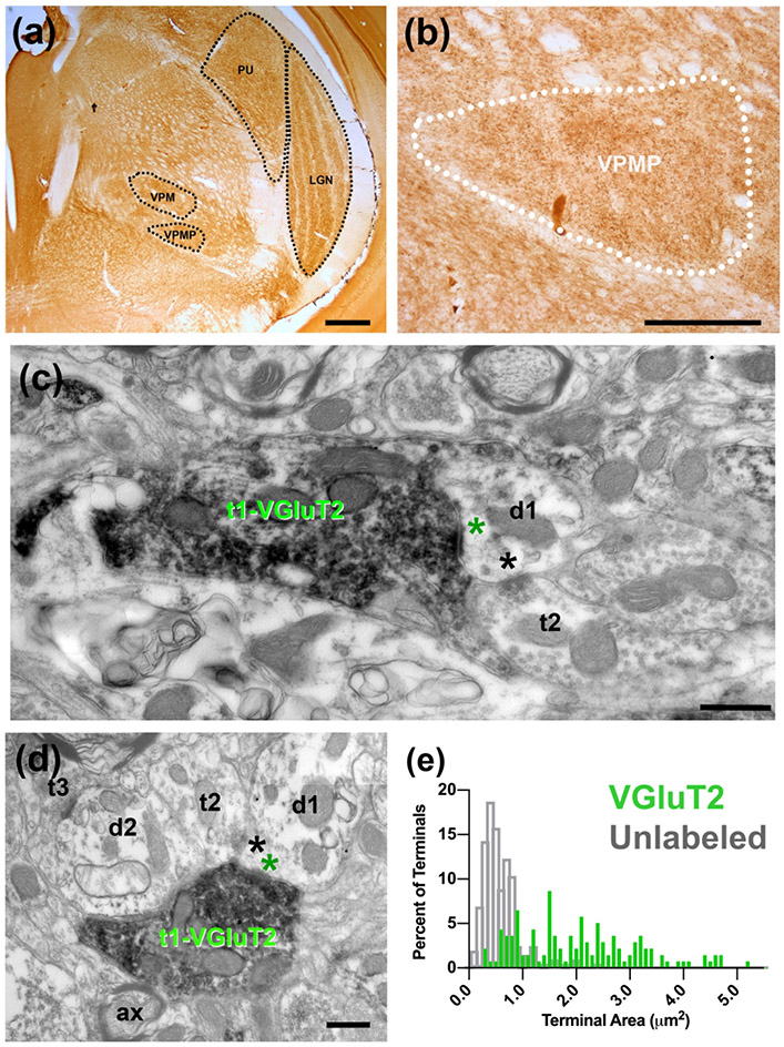
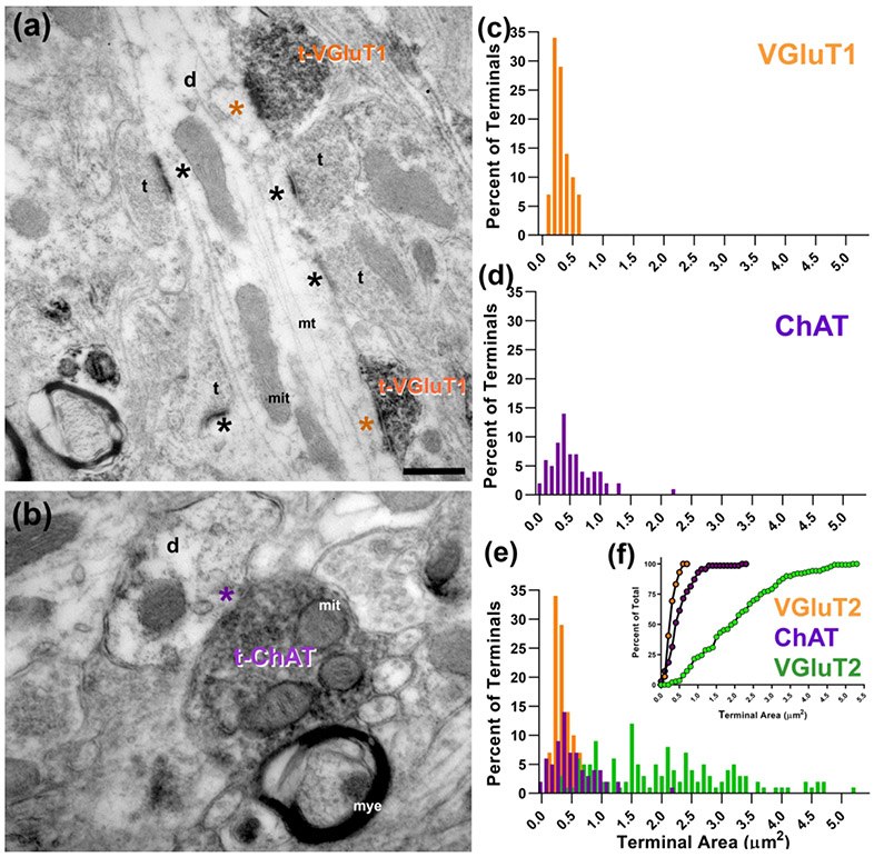
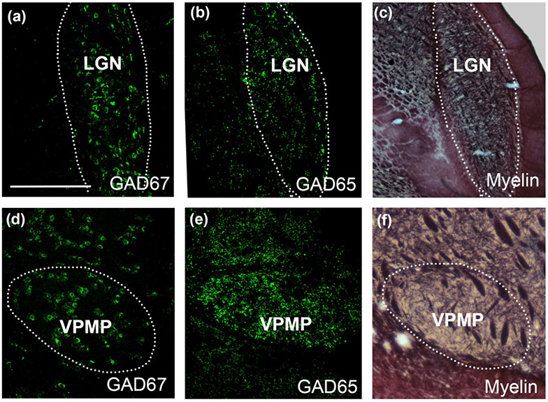
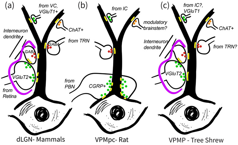
Ventroposterior medialis parvocellularis (VPMP) nucleus of the primate thalamus receives direct input from the nucleus of the solitary tract, whereas the homologous thalamic structure in the rodent does not. To reveal whether the synaptic circuitries in these nuclei lend evidence for conservation of design principles in the taste thalamus across species or across sensory thalamus in general, we characterized the ultrastructural and molecular properties of the VPMP in a close relative of primates, the tree shrew (Tupaia belangeri), and compared these to known properties of the taste thalamus in rodent, and the visual thalamus in mammals. Electron microscopy analysis to categorize the synaptic inputs in the VPMP revealed that the largest-size terminals contained many vesicles and formed large synaptic zones with thick postsynaptic density on multiple, medium-caliber dendrite segments. Some formed triads within glomerular arrangements. Smaller-sized terminals contained dark mitochondria; most formed a single asymmetric or symmetric synapse on small-diameter dendrites. Immuno-EM experiments revealed that the large-size terminals contained VGLUT2, whereas the small-size terminal populations contained VGLUT1 or ChAT. These findings provide evidence that the morphological and molecular characteristics of synaptic circuitry in the tree shrew VPMP are similar to that in nonchemical sensory thalamic nuclei. Furthermore, the results indicate that all primary sensory nuclei of the thalamus in higher mammals share a structural template for processing thalamocortical sensory information. In contrast, substantial morphological and molecular differences in rodent versus tree shrew taste nuclei suggest a fundamental divergence in cellular processing mechanisms of taste input in these two species.

Keywords: ChAT; GAD65; GAD67; VGLUT1; VGLUT2; electron microscopy; gustatory system; synaptic circuitry; ventral posteromedial parvocellular nucleus (VPMP).


Figures

Figure 1.
Figure 1.
Coronal sections of the VPMP in the tree shrew stained for cytochrome oxidase (a,c) and myelin (b). VPMP, similar to other sensory nuclei, displays high cytochrome oxidase activity (a,c). Myelin histochemistry reveals myelinated axon bundles that demarcate VPMP at the dorsal, medial and ventral aspects (b; white arrows), which are lightly stained with cytochrome oxidase (c, black arrows). The tree shrew VPMP is ventral to the VPM, lateral to the PF and medial to the VPI (a). Scale bar = 1000 μm in a and c; and 600 μm in b.
Figure 2.
Figure 2.
Myelin-stained coronal sections of tree shrew brain reveal the landmarks and relative coordinates for the tree shrew VPMP. The sections in panels a - f are 360 μm apart, and they are organized from posterior to anterior. The distances from the first section that displayed VPMP borders are indicated at lower left corners. The panels a1-f1 are higher magnification views of the VPMP from the corresponding panels above. Abbrv: 3V: third ventricle; lgn: dorsal lateral geniculate nucleus; nr: red nucleus; ot: optic tract; pu:pulvinar; sc: superior colliculus; sn: substantia nigra; vpi:n. ventral posterior inferior n.; vpmp: n. ventralis posterior medialis pars parvocellularis; vpm: n. ventralis postrior medialis; vpl: n. ventralis posterior lateralis. Scale bar in panel f = 1mm and applies to a-f. Scale bar in panel f1 = 1mm and applies to a1-f1.
Figure 3.
Figure 3.
Electron micrographs of terminals in the tree shrew VPMP. (a) A large terminal (t) filled with round vesicles and many mitochondria makes a synapse onto a large dendrite (d). The asterisk (*) marks the synapse at the postsynaptic side, here and in all subsequent panels. (b) A small terminal (t) with sparse vesicles synapses onto a dendrite. (c) A large terminal (t1) filled with round vesicles and many mitochondria makes a synapse onto a dendrite (d) and a vesicle filled profile (F2; classified as such because it contains pleomorphic-vesicles and it is postsynaptic to another terminal). Note that both synapses display multiple active zones (*). Another terminal (t2) with sparse pleomorphic vesicles (presumed inhibitory) synapses onto the same F2 profile, representing an inhibitory-to- inhibitory interaction. A third terminal (t3) that is notably small compared to t1, synapses onto the large dendrite. (d) A terminal (t) with round vesicles synapses onto a presynaptic dendrite (F2). The green asterisk marks the putative excitatory synapse. The presynaptic dendrite (F2), in turn, forms a symmetrical, presumed inhibitory synapse (red asterisk) onto another dendrite (d). (e) In a glomerular structure, three terminals (t1, t2, t3) that are pseudo-colored in blue form synapses onto a large dendrite (yellow). The synaptic arrangement is encased in an astrocytic sheath (g, pink). The synapses are indicated by asterisks at the postsynaptic side. Scale bars= 500 nm.
Figure 4.
Figure 4.
(a). The frequency distribution histogram of terminal cross-section areas reveals multimodal population. (b). Four subpopulations revealed by a BIC analysis (R-MClust) are fitted as curves and plotted in different colors. (c). The frequency distribution histogram of synapse lengths. (d). A pairwise plot of terminal cross-section areas and the length of the synapses formed by each terminal (•), and the best fit linear correlation line. (e) The frequency distribution histogram of the calibers of dendrites that are postsynaptic to terminals in the dataset.
Figure 5.
Figure 5.
(a) Immunolabeling with VGLUT2 antibody delineates nuclei of the tree shrew thalamus (black outlines), including the dLGN, PU, VPM, and VPMP. (b) VGluT2 labeling of tree shrew VPMP (white outline) reveals dark puncta labeling throughout the VPMP. (c) Electron micrograph of immunolabeling with VGLUT2 antibody in terminals in the tree shrew VPMP. A large terminal (t1-VGluT2) with several many mitochondria displays VGLUT2 label primarily around vesicles; it makes an excitatory synapse (green asterisk) onto a dendrite (d1). An unlabeled terminal (t2) is synapsing (black asterisk) onto the same dendrite segment. (d) Triadic arrangement of terminals in the tree shrew VPMP. A VGluT2 positive terminal (t1-VGluT2) synapses (green asterisk) onto a presumed inhibitory terminal (t2) and a presumed relay cell dendrite (d1). The inhibitory terminal then synapses (black asterisk) on the relay cell dendrite. (e) The distribution histograms of terminal bouton area of VGluT2 labeled terminals (green) and unlabeled terminals (grey) in the tree shrew VPMP. Scale bar = 500 μm in panels (a) and (b); 500nm in panels (c ) and (d).
Figure 6.
Figure 6.
(a) VGLUT1 immunochemistry revealed labeled terminals (t-VGluT1) in the tree shrew VPMP that were often seen synapsing (orange asterisk) on medium or small caliber dendrites (d) that also receive other labeled inputs and other small terminals (t; black asterisk). (b) Cholinergic terminals (t-ChAT) also make synapses (purple asterisk) onto small or medium sized dendrites (d). (c) The size distribution histogram of VGluT1 labeled terminals in the tree shrew VPMP. (d) The size distribution histogram of ChAT labeled terminals in the tree shrew VPMP. (e, f) Terminal size distribution (E) and cumulative probability (F) of VGluT2 (green), VGluT1 (orange), and ChAT (purple) labeled terminals in the tree shrew VPMP compare the distinct size characteristics of each terminal type. Scale bars = 200 nm; mit: mitochondria; mye: myelin sheath.
Figure 7:
Figure 7:
Immunolabeling with GAD67 reveals cell bodies in the dLGN (a) and the VPMP (d). Similarly, GAD65 immunolabeling reveals large puncta throughout in the dLGN (b) and VPMP (e) of the tree shrew thalamus. The myelin-stained sections (c,f) are useful to confirm the borders of thalamic nuclei imaged at the confocal microscope. The sections that are used to outline the dLGN and VPMP in panels (c) and (f) are adjacent to sections in panels (b) and (d), respectively. Scale Bar in a = 1 mm and applies to all panels.
Figure 8:
Figure 8:
Schematic comparison of synaptic inputs onto relay cell dendrites in the dLGN of various mammals (a), in the VPMpc, thalamic taste nucleus of the rat (b), and in the VPMP, taste nucleus of the tree shrew (c). A primary characteristics of the mammalian dLGN circuitry, that is the glia (purple) encased glomerular triads involving primary sensory inputs and pre-synaptic interneuron dendrites, is a common feature of the tree shrew VPMP, but they are not found in the rat VPMpc. Instead, the axons that bring the primary sensory axons to rat VPMP target soma and dendrite emergence regions and do not engage in triads. While large-sized glutamatergic terminals both in mammalian dLGN and the tree shrew VPMP contain VGluT2, the rat VPMpc does not display any prominent VGluT2 label. Instead, the large terminals in rat VPMpc uniquely contain CGRP peptide. Similar to mammalian dLGN and unlike rat VPMpc, tree shrew VPMP contains GABAergic cells. Also similar to in mammalian dLGN, the tree shrew VPMP prominently contains ChAT+ terminals. In all three structures, the small-size terminals including those from primary visual cortex or the insular cortex, target small caliber, distal dendrites, and these are VGluT1+ in the mammalian dLGN and the tree shrew VPMP.

References

    1. Agarwala S, Güniük AE, May JG, & Petry HM (1992). Immunohistochemical organization of the ventral lateral geniculate nucleus in the tree shrew. Journal of Comparative Neurology, 318(3), 267–276. 10.1002/cne.903180304 - DOI - PubMed
    1. Agca S, Houen G, & Trier NH (2014). Characterization of continuous B-cell epitopes in the N-terminus of glutamate decarboxylase67 using monoclonal antibodies. Journal of Peptide Science, 20(12), 928–934. 10.1002/psc.2703 - DOI - PubMed
    1. Allen GV, Saper CB, Hurley KM, & Cechetto DF (1991). Organization of visceral and limbic connections in the insular cortex of the rat. Journal of Comparative Neurology, 311(1), 1–16. 10.1002/cne.903110102 - DOI - PubMed
    1. Arcelli P, Frassoni C, Regondi MC, De Biasi S, & Spreafico R (1997). GABAergic neurons in mammalian thalamus: a marker of thalamic complexity? Brain Research Bulletin, 42(1), 27–37. 10.1016/S0361-9230(96)00107-4 - DOI - PubMed
    1. Balaram P, Isaamullah M, Petry HM, Bickford ME, & Kaas JH (2015). Distributions of vesicular glutamate transporters 1 and 2 in the visual system of tree shrews (Tupaia belangeri). Journal of Comparative Neurology, 523(12), 1792–1808. 10.1002/cne.23727 - DOI - PMC - PubMed

Publication types

MeSH terms