Review
. 2021 Mar;54(3):e12993.
doi: 10.1111/cpr.12993. Epub 2021 Jan 17.

Exosomes from adipose-derived stem cells and application to skin wound healing

Affiliations
Review

Exosomes from adipose-derived stem cells and application to skin wound healing

Yang An et al. Cell Prolif. 2021 Mar.

Abstract

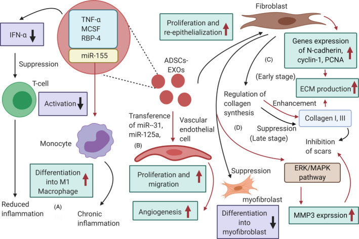
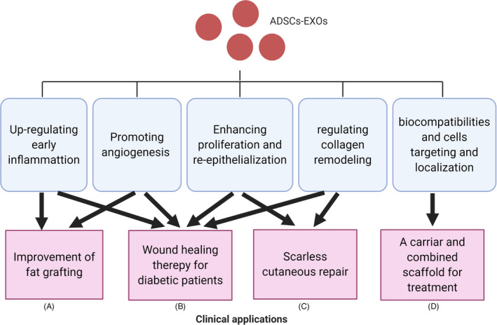
Skin wound healing is an intractable problem that represents an urgent clinical need. To solve this problem, a large number of studies have focused on the use of exosomes (EXOs) derived from adipose-derived stem cells (ADSCs). This review describes the mechanisms whereby ADSCs-EXOs regulate wound healing and their clinical application. In the wound, ADSCs-EXOs modulate immune responses and inflammation. They also promote angiogenesis, accelerate proliferation and re-epithelization of skin cells, and regulate collagen remodelling which inhibits scar hyperplasia. Compared with ADSCs therapeutics, ADSCs-EXOs have highly stability and are easily stored. Additionally, they are not rejected by the immune system and have a homing effect and their dosage can be easily controlled. ADSCs-EXOs can improve fat grafting and promote wound healing in patients with diabetes mellitus. They can also act as a carrier and combined scaffold for treatment, leading to scarless cutaneous repair. Overall, ADSCs-EXOs have the potential to be used in the clinic to promote wound healing.

Keywords: adipose-derived stem cells; angiogenesis; exosomes; inflammation; skin wound healing.


Conflict of interest statement

The authors declare no conflict of interest.

Figures

FIGURE 1
FIGURE 1
EXOs bud from endosome and plasma membranes. Exosome biogenesis may use three mechanisms: (1) vesicle budding into discrete endosomes that mature into multivesicular bodies, releasing EXOs upon plasma membrane fusion; (2) direct budding from plasma membrane and (3) delayed release by budding at intracellular plasma membrane–connected compartments (IPMCs) followed by deconstriction of IPMC neck(s). We note that this is not a comprehensive list and it is just to illustrate some of the mechanisms. Abbreviations: ECM, extracellular matrix; ERM, ezrin‐radixin‐moesin; ESCRTs, endosomal sorting complexes required for transport; MHC, major histocompatibility complex; IGSF8, immunoglobulin superfamily member 8; ICAM‐1, intercellular adhesion molecule‐1; SDC1, syndecans 1; HSPs, heat shock proteins
FIGURE 2
FIGURE 2
Mechanisms by which ADSCs‐EXOs may promote wound healing. (A) adipose‐derived stem cells (ADSCs)‐EXOs contain immunoregulatory proteins and reduce the secretion of IFN‐α, subsequently inhibiting activation of T cells, resulting in reduced inflammation. Additionally, miR‐155 in ADSCs‐EXOs can induce monocyte differentiation into M1 macrophages, causing chronic inflammation; (B) ADSCs‐EXOs can transfer miRNA‐125a and miRNA‐31 to vascular endothelial cells, stimulating proliferation and migration to promote angiogenesis; (C) In the early stages, ADSCs‐EXOs may promote proliferation, migration and collagen synthesis in fibroblasts, stimulating N‐cadherin, cyclin‐1, PCNA and collagen I, III expression and increasing ECM production; (D) in late stages, ADSCs‐EXOs prevent the differentiation of fibroblasts into myofibroblasts, and reduce scarring by inhibition of the formation of collagen and activation the ERK/MAPK pathway to increase MMP3 expression. Abbreviations: ECM, extracellular matrix; MCSF, macrophage colony‐stimulating factor
FIGURE 3
FIGURE 3
Potential clinical applications of adipose‐derived stem cells (ADSCs)‐EXOs: improvement of fat grafting (A), wound healing therapy for diabetic patients (B), scarless repair (C) and carrier and combined scaffold for treatment (D)

References

    1. Blakytny R, Jude E. The molecular biology of chronic wounds and delayed healing in diabetes. Diabet Med J Br Diabet Assoc. 2006;23(6):594‐608. 10.1111/j.1464-5491.2006.01773.x - DOI - PubMed
    1. Hamed S, Bennett CL, Demiot C, Ullmann Y, Teot L, Desmoulière A. Erythropoietin, a novel repurposed drug: an innovative treatment for wound healing in patients with diabetes mellitus. Wound Repair Regen Off Publ Wound Heal Soc Eur Tissue Repair Soc. 2014;22(1):23‐33. 10.1111/wrr.12135 - DOI - PubMed
    1. Goodarzi P, Alavi‐Moghadam S, Sarvari M, et al. Adipose tissue‐derived stromal cells for wound healing. Adv Exp Med Biol. 2018;1119:133‐149. 10.1007/5584_2018_220 - DOI - PubMed
    1. Orgill DP, Ogawa R. Discussion: the embrace device significantly decreases scarring following scar revision surgery in a randomized controlled trial. Plast Reconstr Surg. 2014;133(2):406‐407. - PubMed
    1. Turner NJ, Badylak SF. The use of biologic scaffolds in the treatment of chronic nonhealing wounds. Adv Wound Care. 2015;4(8):490‐500. - PMC - PubMed