

Rare case of granular cell tumor of perianal region: a case report and literature review
- PMID: 33459105
- PMCID: PMC7816531
- DOI: 10.1177/0300060520982689
Rare case of granular cell tumor of perianal region: a case report and literature review
Abstract
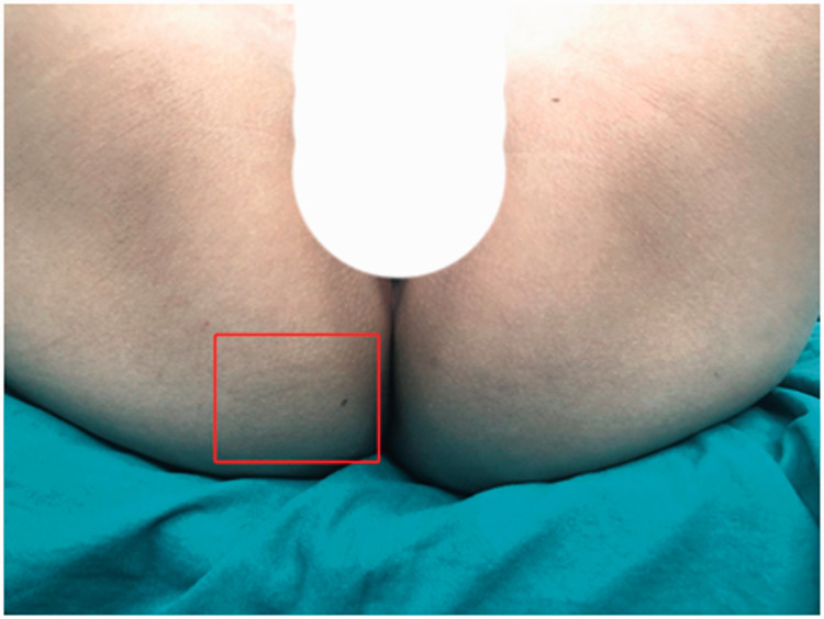
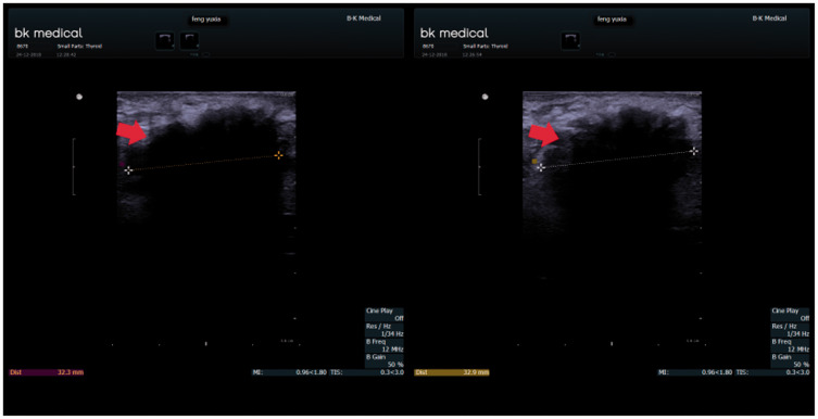
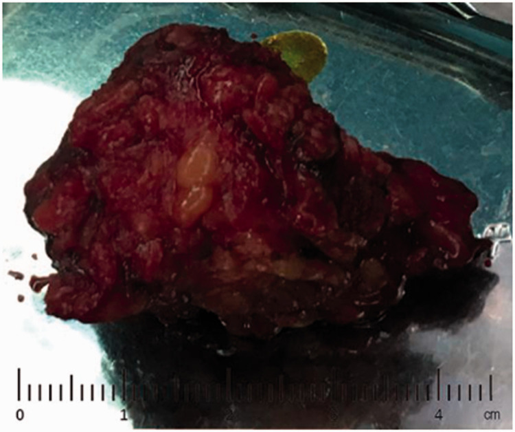
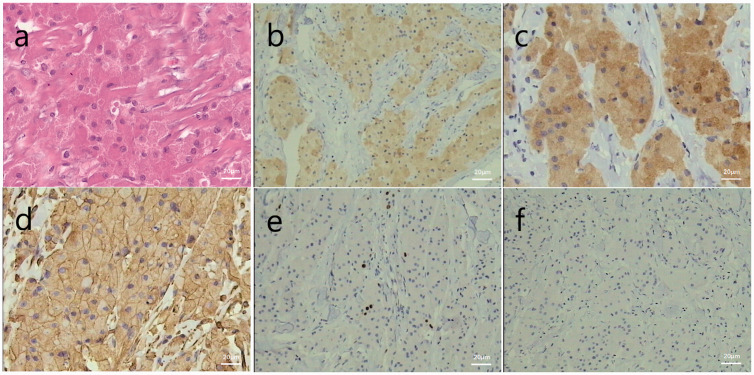
Granular cell tumors (GCTs) are rare submucosal neoplasms, with tumors in the oral mucosa accounting for about a third of all cases. In contrast, GCT is a rare anal neoplasm, with fewer than 30 cases of perianal GCT reported in the literature. We report the case of a 36-year-old woman with a perianal lump with no obvious local lesion as the main clinical complaint. The tumor was completely resected and histologically confirmed as a GCT. The patient remained under continuous follow-up. GCT is difficult for surgeons and pathologists to diagnose, and biopsy and immunohistochemical analysis are prerequisites for an accurate diagnosis. An integrated understanding of GCT in terms of its differential diagnosis will contribute to better identification and more appropriate treatment of this disease.
Keywords: Granular cell tumor; anal neoplasm; biopsy; differential diagnosis; immunohistochemistry; perianal region.
Conflict of interest statement
Figures
Similar articles
-
A rare case of perianal granular cell tumor: case report and literature review.J Surg Case Rep. 2017 Jun 6;2017(6):rjw186. doi: 10.1093/jscr/rjw186. eCollection 2017 Jun. J Surg Case Rep. 2017. PMID: 28603602 Free PMC article.
-
Unexpected Presentation of Perianal Granular Cell Tumor in a Young Woman.Curr Probl Diagn Radiol. 2017 Nov-Dec;46(6):452-454. doi: 10.1067/j.cpradiol.2017.01.004. Epub 2017 Jan 14. Curr Probl Diagn Radiol. 2017. PMID: 28284459
-
Granular cell tumor on perianal region: a case report.Acta Med Iran. 2013 Aug 7;51(7):509-11. Acta Med Iran. 2013. PMID: 23945899
-
Perianal granular cell tumor: report of a case and review of the literature.Tumori. 2009 Jul-Aug;95(4):538-41. doi: 10.1177/030089160909500424. Tumori. 2009. PMID: 19856672 Review.
-
Intramuscular granular cell tumor in the sternocleidomastoid muscle: A case report and literature review.Ear Nose Throat J. 2025 Mar;104(1_suppl):307S-312S. doi: 10.1177/01455613221143357. Epub 2022 Dec 5. Ear Nose Throat J. 2025. PMID: 36468452 Review.
Cited by
-
Imaging of perianal granular cell tumor with lung metastasis: A case report and literature review.Radiol Case Rep. 2021 Nov 26;17(2):314-319. doi: 10.1016/j.radcr.2021.10.048. eCollection 2022 Feb. Radiol Case Rep. 2021. PMID: 34876957 Free PMC article.
-
A Rare Case of Granular Cell Tumor in the Groin: Importance of Excision With Negative Margins.Cureus. 2025 May 22;17(5):e84637. doi: 10.7759/cureus.84637. eCollection 2025 May. Cureus. 2025. PMID: 40546484 Free PMC article.
-
A Rare Case of Granular Cell Tumor of the Anus.Clin Case Rep. 2024 Dec 5;12(12):e9688. doi: 10.1002/ccr3.9688. eCollection 2024 Dec. Clin Case Rep. 2024. PMID: 39649516 Free PMC article.
References
-
- Carinci F, Piattelli A, Rubini C, et al. Genetic profiling of granular cell myoblastoma. J Craniofac Surg 2004; 15: 824–834. - PubMed
-
- Wan XY, Hu B, Zhou ZY, et al. Recurrent granular cell tumor of the anal-perianal region: how much anal sphincter can be resected? Tech Coloproctol 2014; 18: 597–600. - PubMed
-
- Yang SW, Hong SW, Cho MY, et al. Malignant granular cell tumor at the retrotracheal space. Yonsei Med J 1999; 40: 76–79. - PubMed
-
- Souto GR, Caldeira PC, Johann AC, et al. Evaluation of GLUT-1 in the granular cell tumour and congenital granular cell epulis. J Oral Pathol Med 2013; 42: 450–453. - PubMed
Publication types
MeSH terms
LinkOut - more resources
Full Text Sources
Other Literature Sources
