

The prevalence of Vickers' ligament in Madelung's deformity: a retrospective multicentre study of 75 surgical cases
- PMID: 33459142
- PMCID: PMC8056710
- DOI: 10.1177/1753193420981522
The prevalence of Vickers' ligament in Madelung's deformity: a retrospective multicentre study of 75 surgical cases
Erratum in
-
Erratum to: The prevalence of Vickers' ligament in Madelung's deformity: a retrospective multicentre study of 75 surgical cases.J Hand Surg Eur Vol. 2021 May;46(4):NP1. doi: 10.1177/1753193421993305. Epub 2021 Jan 27. J Hand Surg Eur Vol. 2021. PMID: 33502285 Free PMC article. No abstract available.
Abstract
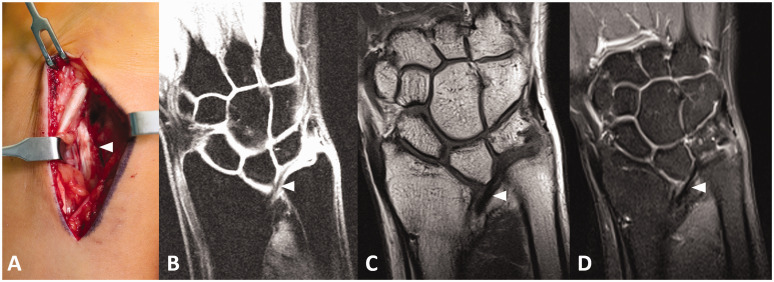
In a retrospective multicentre study, we aimed to correlate clinical factors and findings on plain radiographs and MRI with the intraoperative presence of Vickers' ligament in Madelung's deformity. We screened the records, in which the absence or presence of Vickers' ligament was affirmatively indicated, of 75 consecutive operated extremities in 58 patients. In 83% a Vickers' ligament was observed intraoperatively. The whole bone Madelung type (as opposed to the distal type) and a distal radial notch were independent, significant predictors for the presence of the ligament. The correct Vickers detection rate using MRI was 85% of the 27 cases for which MRI was available. Thus, the MRI was a good but not perfectly reliable modality. We conclude that Vickers' ligament is present in the majority but not all cases with Madelung deformity. We advise that patients with a more severe type of Madelung's deformity and a distal radial notch should be monitored closely.Level of evidence: IV.
Keywords: Madelung deformity; Vickers ligament; Zebala type; lunate subsidence; radius deformity; radius notch; ulnar tilt.
Conflict of interest statement
Figures

References
-
- Ali S, Zlotolow DA. (2015) Madelung deformity and Madelung-type deformities: a review of the clinical and radiological characteristics. Pediatr Radiol 45: 1856–63. - PubMed
-
- Del Core M, Beckwith T, Phillips L, et al. (2020) Long-term outcomes following Vickers ligament release and growth modulation for the treatment of Madelung deformity. J Pediatr Orthop 40: e306–11. - PubMed
-
- Farr S, Bae DS. (2015) Inter- and intrarater reliability of ulna variance versus lunate subsidence measurements in Madelung deformity. J Hand Surg Am 40: 62–6. - PubMed
-
- Firl M, Wünsch L. (2004) Measurement of bowing of the radius. J Bone Joint Surg Br 86: 1047–9. - PubMed
Publication types
MeSH terms
Supplementary concepts
LinkOut - more resources
Full Text Sources
Other Literature Sources
