. 2021 Apr;39(4):467-481.
doi: 10.1002/stem.3322. Epub 2021 Jan 18.

Cartilage endplate stem cells inhibit intervertebral disc degeneration by releasing exosomes to nucleus pulposus cells to activate Akt/autophagy

Affiliations

Cartilage endplate stem cells inhibit intervertebral disc degeneration by releasing exosomes to nucleus pulposus cells to activate Akt/autophagy

Liwen Luo et al. Stem Cells. 2021 Apr.

Erratum in

Abstract

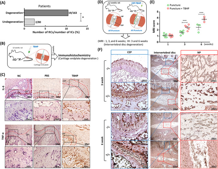
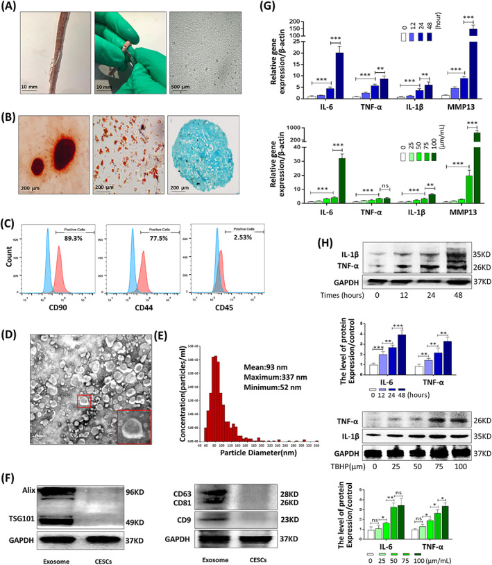
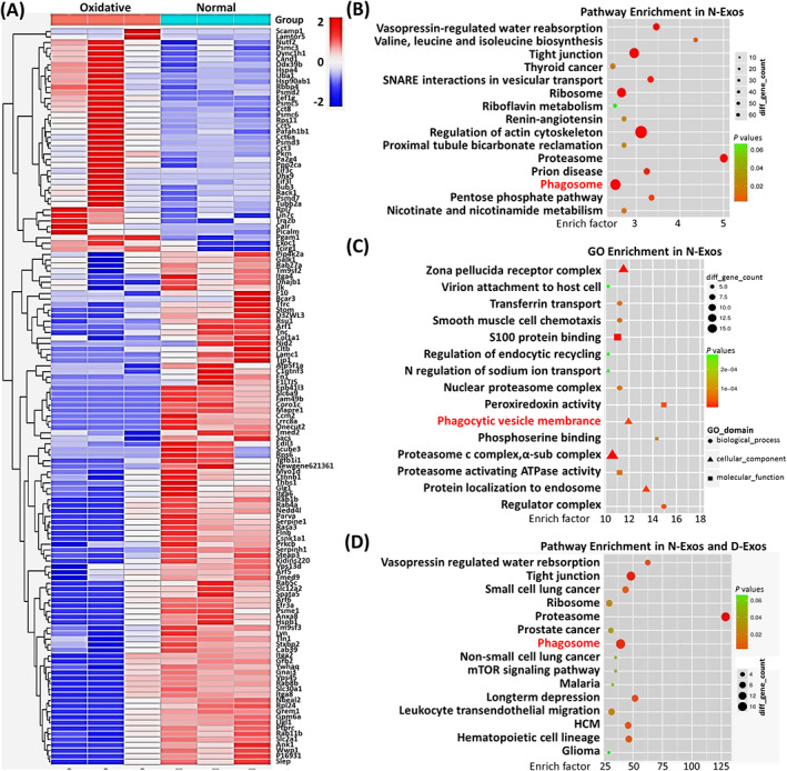
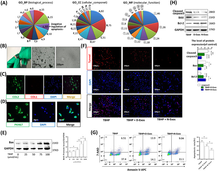
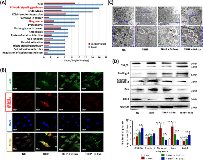
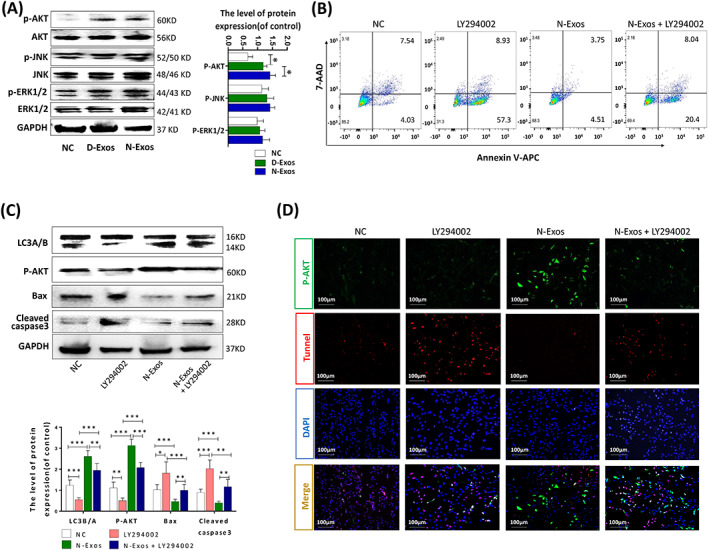
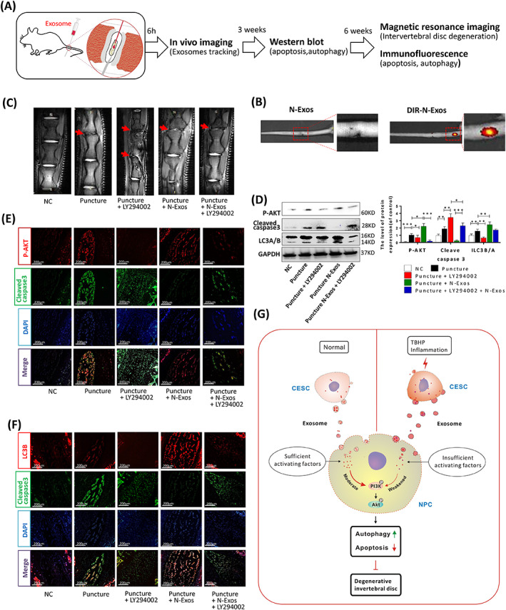
Degeneration of the cartilage endplate (CEP) induces intervertebral disc degeneration (IVDD). Nucleus pulposus cell (NPC) apoptosis is also an important exacerbating factor in IVDD, but the cascade mechanism in IVDD is not clear. We investigated the apoptosis of NPCs and IVDD when stimulated by normal cartilage endplate stem cell (CESC)-derived exosomes (N-Exos) and degenerated CESC-derived exosomes (D-Exos) in vitro and in vivo. Tert-butyl hydroperoxide (TBHP) was used to induce inflammation of CESCs. The bioinformatics differences between N-Exos and D-Exos were analyzed using mass spectrometry, heat map, and Kyoto Encyclopedia of Genes and Genomes (KEGG) enrichment analysis. NPC apoptosis was examined using TUNEL staining. The involvement of the AKT and autophagy signaling pathways was investigated using the signaling inhibitor LY294002. Magnetic resonance imaging, Western blotting, and immunofluorescence staining were used to evaluate the therapeutic effects of N-Exos in rats with IVDD. TBHP effectively induced inflammation and the degeneration of CEP in rat. N-Exos were more conducive to autophagy activation than D-Exos. The apoptotic rate of NPCs decreased obviously after treatment with N-Exos compared to D-Exos. N-Exos inhibited NPCs apoptosis and attenuated IVDD in rat via activation of the AKT and autophagy pathways. These results are the first findings to confirm that CEP delayed the progression of IVDD via exosomes. The therapeutic effects of N-Exos on NPC apoptosis inhibition and the slowing of IVDD progression were more effective than D-Exos due to activation of the PI3K/AKT/autophagy pathway, which explained the increase in the incidence of IVDD after inflammation of the CEP.

Keywords: apoptosis; autophagy; cartilage endplate stem cells; exosome; intervertebral disc degeneration.


Conflict of interest statement

The authors declared no potential conflicts of interest.

Figures

FIGURE 1
FIGURE 1
Degeneration of cartilage endplate (CEP) accelerates intervertebral disc degeneration (IVDD) in humans and rats. A, Clinical statistics on the recurrence rate of 243 patients with lumbar disc herniation with (163 cases) or without (80 cases) CEP degeneration after surgery. B, Tert‐butyl hydroperoxide (TBHP)‐induced model diagram of CEP degeneration in rat. C, Representative immunohistochemical staining of IL‐6 and TNF‐α in CEP under different treatments with microsyringe (NC, PBS [50 μL] or TBHP [50 μL,100 μmoL/mL]). D, The degeneration model diagram of puncture‐induced IVDD with or without TBHP‐induced CEP inflammation. AF:Puncture, puncture into annulus fibrosus; CEP:TBHP, TBHP was injected into CEP); IH, immunohistochemistry; MRI, magnetic resonance imaging; NP, nucleus pulposus. E, The IVDD of rats was evaluated using Pfirrmann grading according to the T2‐weighted images at 1, 3, and 6 weeks after the puncture or TBHP+puncture. F, Representative immunohistochemical staining of IL‐6 in CEP and NP 3 and 6 weeks after puncture or TBHP+puncture treatment (n = 5 per group; *P < .05). −/+, only puncture; +/+, TBHP and puncture; AF, annulus fibrosus; NC, normal control; ns: P > .05; *P < .05; **P < .01; ***P < .001
FIGURE 2
FIGURE 2
Identification of exosomes derived from rat cartilage end plate stem cells (CESCs‐Exos) and tert‐butyl hydroperoxide (TBHP)‐induced CESC degeneration. A, Horizontal views of rat cartilage endplate (CEP) and morphology of P3 CESCs at 100% confluence. B, After osteogenic induction for 14 days (left panel), adipogenic induction for 15 days (middle panel), and chondrogenic induction for 21 days (right panel), the ability of CESCs to differentiate into different cell lines was confirmed using Alizarin Red staining, Oil red O staining, and Alcian blue staining, respectively. C, Cell surface markers (CD90, CD44, and CD45) of CESCs was detected using flow cytometric analysis. The red curves represent the fluorescence intensity of CESCs stained with the corresponding antibodies. D, TEM images were used to identify the morphology of CESCs‐Exos. E, Nanoparticle trafficking analysis (NTA) was used to analyze the particle size distribution of CESCs‐Exos. F, Representative Western blots of Alix and TSG101 in CESCs‐Exos and CESCs. G, The gene expression of IL‐6, TNF‐α, IL‐1β, and MMP13 in the CESCs treated with different concentrations of TBHP (0, 25, 50, 75, or 100 μm) for 48 hours or TBHP (100 μm) for the indicated time points (0, 12, 24, or 48 hours). H, The Western blotting and quantitative protein levels of IL‐1β and TNF‐α in the CESCs as treated above. ns: P > .05; *P < .05; **P < .01; ***P < .001
FIGURE 3
FIGURE 3
Bioinformatics analysis between normal cartilage end plate stem cell (CESC)‐derived exosomes (N‐Exos) and degenerated CESC‐derived exosomes (D‐Exos). A, Heat map analysis of differential proteins between N‐Exos and D‐Exos. B,C, KEGG enrichment analysis and gene ontology (GO) data analysis of proteins contained in N‐Exos. D, KEGG enrichment analysis of the top 50 quantitative differential proteins between N‐Exos and D‐Exos
FIGURE 4
FIGURE 4
N‐Exos more effectively inhibited apoptosis compared to D‐Exos. A, Gene ontology (GO) analysis of all differential proteins carried in the normal cartilage end plate stem cell (CESC)‐derived exosomes (N‐Exos) and degenerated CESC‐derived exosomes (D‐Exos). a: small GTPase mediated signal transduction; b: protein transport; c: negative regulation of apoptotic process; d: response to drug; e: cell‐cell adhesion; f: cell adhesion; g: exocytosis; h: positive regulation of gene expression; i: cell‐matrix adhesion; j: protein folding; k: vesicle‐mediated transport; l: positive regulation of cell migration; m: substrate adhesion‐dependent cell spreading; n: protein heterooligomerization; o: positive regulation of angiogenesis. A: extracellular exosome; B: cytoplasm; C: membrane; D: plasma membrane; E: nucleus; F: Golgi apparatus; G: cytosol; H: focal adhesion; I: extracellular matrix; J: perinuclear region of cytoplasm; K: intracellular; L: cell‐cell adherens junction; M: protein complex; N: cell surface; O: centrosome. ①: protein binding, ②: poly(A) RNA binding, ③: ATP binding, ④: GTP binding, ⑤: cadherin binding involved in cell‐cell adhesion, ⑥: protein kinase binding, ⑦: protein domain specific binding, ⑧: identical protein binding, ⑨: integrin binding, ⑩: GTPase activity, ⑪: protein complex binding, ⑫: GDP binding, ⑬: unfolded protein binding, ⑭: ion channel binding, ⑮: glycoprotein binding. B, Morphological observations of rat NP tissue (left panel), P1 NPCs at 50% confluence (middle panel) and P3 NPCs at 100% confluence (middle panel). C, Double immunofluorescence staining of collagen II (green) and collagen I (red) in NPCs. D, Representative images of NPCs incubated with PBS or PKH67‐labeled N‐Exos for 24 hours. E, Western blotting and quantitative levels of apoptotic protein Bax in the NPCs treated with different concentrations of TBHP (0, 25, 50, 75, or 100 μmoL/mL) for 48 hours. F‐H, TUNEL staining, flow cytometry, representative Western blots, and quantification data of cleaved caspase3, Bax, and Bcl‐2 in NPCs treated with TBHP (100 μmoL/mL), TBHP (100 μmoL/mL) + D‐Exos (40 μg/mL), and TBHP (100 μmoL/mL) + N‐Exos (40 μg/mL). ns: P > .05; *P < .05; **P < .01; ***P < .001
FIGURE 5
FIGURE 5
N‐Exos more effectively promoted nucleus pulposus cell (NPC) autophagy and apoptosis inhibition than D‐Exos. A, KEGG enrichment analysis of all differential proteins carried in the normal CESC‐derived exosomes (N‐Exos) and degenerated CESC‐derived exosomes (D‐Exos). B, Double immunofluorescence staining of LC3‐B (green) and cleaved caspase3 (red) in NPCs treated with NC (0 μg/mL), TBHP (100 μmoL/mL), TBHP (100 μmoL/mL) + D‐Exos (40 μg/mL), or TBHP (100 μmoL/mL) + N‐Exos (40 μg/mL). C, Autophagosomes (black arrow: autophagosome) were examined using TEM after NPCs were treated as described above. D, Representative Western blots and quantitative data of LC3A/B, Beclin‐1, cleaved caspase3, Bax, and Bcl‐2 expression in NPCs treated with NC, TBHP (100 μmoL/mL), TBHP (100 μmoL/mL) + D‐Exos (40 μg/mL), or TBHP (100 μmoL/mL) + N‐Exos (40 μg/mL). NC, normal control; ns: P > .05; *P < .05; **P < .01; ***P < .001
FIGURE 6
FIGURE 6
N‐Exos inhibited nucleus pulposus cells (NPCs) apoptosis by activating the PI3K/AKT/autophagy signaling pathway. A, Western blot analysis and quantitative data of p‐AKT, AKT, p‐JNK, JNK p‐ERK1/2, and ERK1/2 expression in NPCs treated with NC (0 μg/mL), D‐Exos (40 μg/mL), and N‐Exos (40 μg/mL). B, Flow cytometry was used to detect apoptosis of NPCs treated with NC (0 μg/mL), LY294002 (20 μmoL/mL), N‐Exo (40 μg/mL), and LY294002 (20 μmoL/mL) + N‐Exos (40 μg/mL). C,D, Representative Western blots and quantitative data of LC3A/B, p‐AKT, cleaved caspase3, and Bax expression and double immunofluorescence staining of p‐AKT (green) and TUNEL (red) in NPCs treated as described above. NC, normal control; ns: P > .05; *P < .05; **P < .01; ***P < .001
FIGURE 7
FIGURE 7
N‐Exos alleviated disc degeneration via activation of the PI3K/AKT/autophagy pathway in rat. A, Exosome and reagent treatment via microsyringe in the intervertebral disc degeneration (IVDD) and the subsequent experimental steps. B, in vivo imaging of rat IVD and the vertebral segments treated with unlabeled N‐Exos or DIR‐labeled N‐Exos (DIR‐N‐Exos). C, The representative images of MRI of rat intervertebral disc treated with NC, Puncture, Puncture+LY294002 (20 μmoL/mL), Puncture+ N‐Exos (40 μg/mL), and Puncture+ N‐Exos (40 μg/mL) + LY294002 (20 μmoL/mL). D, The Western blotting and quantification data of LC3A/B, p‐AKT, capase3, and Bax in the rat intervertebral disc 3 weeks after the above‐listed treatments. E,F, Representative double immunofluorescence of p‐AKT (red) and cleaved caspase3 (green) images and LC3B (red) and cleaved caspase3 (green) images of rat discs in each group (n = 5 per group; *P < .05). G, Graphical abstract of the mechanism of CEP inflammation acceleration of the progression of IVDD. Normal cartilage end plate stem cell (CESC)‐derived exosomes (N‐Exos) more effectively inhibit NPC apoptosis than degenerated CEPC‐derived exosomes (D‐Exos) due to the decrease in anti‐apoptotic proteins carried by exosomes after CEP degeneration. N‐Exos also better activated the PI3K/AKT signaling pathway in NPCs compared to D‐Exos, enhanced autophagy, alleviated NPC apoptosis in vitro, and ameliorated IVDD in vivo. NC, normal control; ns: P > .05; *P < .05; **P < .01; ***P < .001

Similar articles

Cited by

References

    1. Andersson GB. Epidemiological features of chronic low‐back pain. Lancet. 1999;354:581‐585. - PubMed
    1. Urban JP, Roberts S. Degeneration of the intervertebral disc. Arthritis Res Ther. 2003;5:120‐130. - PMC - PubMed
    1. Wang F, Gao ZX, Cai F, et al. Formation, function, and exhaustion of notochordal cytoplasmic vacuoles within intervertebral disc: current understanding and speculation. Oncotarget. 2017;8:57800‐57812. - PMC - PubMed
    1. Wong J, Sampson SL, Bell‐Briones H, et al. Nutrient supply and nucleus pulposus cell function: effects of the transport properties of the cartilage endplate and potential implications for intradiscal biologic therapy. Osteoarthr Cartil. 2019;27:956‐964. - PMC - PubMed
    1. Li FC, Zhang N, Chen WS, Chen QX. Endplate degeneration may be the origination of the vacuum phenomenon in intervertebral discs. Med Hypotheses. 2010;75:169‐171. - PubMed

Publication types

MeSH terms