Effect of injectable platelet-rich fibrin (i-PRF) on the rate of tooth movement
- PMID: 33459765
- PMCID: PMC8084468
- DOI: 10.2319/060320-508.1
Effect of injectable platelet-rich fibrin (i-PRF) on the rate of tooth movement
Abstract
Objectives: To evaluate the efficiency of injectable platelet-rich fibrin (i-PRF) in accelerating canine tooth movement and to examine levels of the matrix metalloproteinase-8 (MMP-8), interleukin-1β (IL-1β), receptor activator of nuclear factor kappa-light-chain-enhancer of activated B cells ligand (RANKL), and osteoprotegerin (OPG) in the gingival crevicular fluid during orthodontic treatment.
Materials and methods: Twenty patients (mean age = 21.4 ± 2.9 years) with Class II Division 1 malocclusion were included in a split-mouth study. The treatment plan for all patients was extraction of maxillary first premolars followed by canine distalization with closed-coil springs using 150 g of force on each side. The study group received i-PRF two times, with a 2-week interval, on one side of the maxilla. The contralateral side served as the control and did not receive i-PRF. Maxillary canine tooth movement was measured at five time points (T1-T5) on each side. Also, the activity of inflammatory cytokines was evaluated at three time points in the gingival crevicular fluid samples.
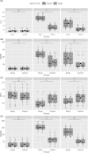
Results: There was a significant difference in canine tooth movement between the two groups (P < .001). i-PRF significantly increased the rate of tooth movement, and stimulation in the levels of inflammatory cytokines supported this result (P < .001). The levels of cytokines changed in both groups between T1 and T2. The IL-1β, MMP8, and RANKL values were significantly increased in the study group compared with the control group, while the OPG values were significantly decreased.
Conclusions: i-PRF-facilitated orthodontics is an effective and safe treatment modality to accelerate tooth movement, and this method can help shorten orthodontic treatment duration.
Keywords: Injectable platelet-rich fibrin; Rate of tooth movement; i-PRF.
© 2021 by The EH Angle Education and Research Foundation, Inc.
Figures


Comment in
-
Letter to the Editor.Angle Orthod. 2022 Mar 1;92(2):301. doi: 10.2319/1945-7103-92.2.301. Angle Orthod. 2022. PMID: 35168257 Free PMC article. No abstract available.
-
Response to the Letter.Angle Orthod. 2022 Mar 1;92(2):300. doi: 10.2319/1945-7103-92.2.300. Angle Orthod. 2022. PMID: 35168258 Free PMC article. No abstract available.
-
Letter to the Editor.Angle Orthod. 2022 Mar 1;92(2):299. doi: 10.2319/1945-7103-92.2.299. Angle Orthod. 2022. PMID: 35168259 Free PMC article. No abstract available.
-
Response to the Letter.Angle Orthod. 2022 Mar 1;92(2):302. doi: 10.2319/1945-7103-92.2.302. Angle Orthod. 2022. PMID: 35168260 Free PMC article. No abstract available.
-
Response to the Letter.Angle Orthod. 2022 Mar 1;92(2):297-298. doi: 10.2319/1945-7103-92.2.297. Angle Orthod. 2022. PMID: 35168261 Free PMC article. No abstract available.
-
Letter to the Editor.Angle Orthod. 2022 Mar 1;92(2):296. doi: 10.2319/1945-7103-92.2.296. Angle Orthod. 2022. PMID: 35168262 Free PMC article. No abstract available.
References
-
- Skidmore KJ, Brook KJ, Thomson WM, Harding WJ. Factors influencing treatment time in orthodontic patients. Am J Orthod Dentofacial Orthop. 2006;129:230–238. - PubMed
-
- Mavreas D, Athanasiou AE. Factors affecting the duration of orthodontic treatment: a systematic review. Eur J Orthod. 2008;30:386–395. - PubMed
-
- Alikhani M, Raptis M, Zoldan B, Sangsuwon C, Lee YB, Alyami B, et al. Effect of micro-osteoperforations on the rate of tooth movement. Am J Orthod Dentofacial Orthop. 2013;144:639–648. - PubMed
-
- Güleç A, Bakkalbaşı BÇ, Cumbul A, Uslu Ü, Alev B, Yarat A. Effects of local platelet-rich plasma injection on the rate of orthodontic tooth movement in a rat model: a histomorphometric study. Am J Orthod Dentofacial Orthop. 2017;151:92–104. - PubMed
-
- Rody WJ, Jr, King GJ, Gu G. Osteoclast recruitment to sites of compression in orthodontic tooth movement. Am J Orthod Dentofacial Orthop. 2001;120:477–489. - PubMed
MeSH terms
LinkOut - more resources
Full Text Sources
Other Literature Sources
Medical

