Review
. 2021 Apr;110(4):479-506.
doi: 10.1007/s00392-020-01799-3. Epub 2021 Jan 18.

Diagnosis and treatment of cardiac amyloidosis: position statement of the German Cardiac Society (DGK)

Affiliations
Review

Diagnosis and treatment of cardiac amyloidosis: position statement of the German Cardiac Society (DGK)

A Yilmaz et al. Clin Res Cardiol. 2021 Apr.

Abstract

Systemic forms of amyloidosis affecting the heart are mostly light-chain (AL) and transthyretin (ATTR) amyloidoses. The latter is caused by deposition of misfolded transthyretin, either in wild-type (ATTRwt) or mutant (ATTRv) conformation. For diagnostics, specific serum biomarkers and modern non-invasive imaging techniques, such as cardiovascular magnetic resonance imaging (CMR) and scintigraphic methods, are available today. These imaging techniques do not only complement conventional echocardiography, but also allow for accurate assessment of the extent of cardiac involvement, in addition to diagnosing cardiac amyloidosis. Endomyocardial biopsy still plays a major role in the histopathological diagnosis and subtyping of cardiac amyloidosis. The main objective of the diagnostic algorithm outlined in this position statement is to detect cardiac amyloidosis as reliably and early as possible, to accurately determine its extent, and to reliably identify the underlying subtype of amyloidosis, thereby enabling subsequent targeted treatment.

Keywords: Amyloidosis; Endomyocardial biopsy; Magnetic resonance imaging; Myocardium; Scintigraphy.


Conflict of interest statement

Ali Yilmaz: speaker and consultant activities for Alnylam, Pfizer Pharma GmbH and Akcea Therapeutics; research cooperation with Philips. Johann Bauersachs: speaker and consultant activities for Pfizer Pharma GmbH. Frank Bengel: speaker and consultant activities for Siemens Healthineers, GE Healthcare, Alnylam and Pfizer Pharma GmbH. Ronny Büchel: speaker honoraria from Pfizer Pharma GmbH and GE Healthcare. Ingrid Kindermann: speaker honoraria from Akcea Therapeutics, Alnylam and Pfizer Pharma GmbH. Karin Klingel: speaker honoraria from Akcea Therapeutics, Alnylam and Pfizer Pharma GmbH. Fabian Knebel: speaker and consultant activities for Akcea Therapeutics, Alnylam and Pfizer Pharma GmbH. Benjamin Meder and Fabian aus dem Siepen: speaker honoraria from Akcea Therapeutics, Pfizer Pharma GmbH and Alnylam as well as travel grants from Pfizer Pharma GmbH. Caroline Morbach: research cooperation with the University of Würzburg and Tomtec Imaging Systems funded by a research grant from the Bavarian Ministry of Economic Affairs, Regional Development and Energy, Germany; speakers honorarium and travel grants from Amgen, Tomtec, Orion Pharma, and Alnylam; participation in Advisory and Patient Eligibility Boards sponsored by Akcea, Alnylam, Pfizer, and EBR Systems; principal investigator in trials sponsored by Alnylam and AstraZeneca. Eike Nagel: speaker honoraria from Bayer AG and Neosoft; research cooperation with Bayer AG. Eric Schulze-Bahr: None. Norbert Frey: None.

Figures

Fig. 1
Fig. 1
Transthoracic echocardiography with measurement of longitudinal strain and detection of a characteristic “apical sparing” phenomenon in a patient with cardiac amyloidosis
Fig. 2
Fig. 2
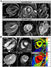
a Cardiovascular magnetic resonance (CMR) images of a patient with cardiac amyloidosis and a DDD pacemaker. In the upper row, cine images are shown in a 4-chamber view, in the short axis, and in a 3-chamber view. In the lower series, the corresponding contrast images (“late gadolinium enhancement” [LGE]) are shown with evidence of diffuse LGE in all heart cavities and myocardial sections. b In addition to the cine and LGE images, T1 mapping and ECV (extracellular volume fraction) images are shown in the right column. Particularly in the basal septum, measured values of T1 (native) = 1300 ms and ECV = 70% were significantly increased and characteristic for cardiac amyloidosis
Fig. 3
Fig. 3
Planar 99mTc-DPD scintigraphies of four different patients with varying degrees of cardiac radionuclide uptake: No cardiac uptake (Perugini score 0, a). Light cardiac uptake with preserved delineation of bone tissue (Perugini score 1, b). Strong cardiac uptake above that of bone tissue and increased soft-tissue uptake, particularly in the shoulder, abdominal wall, and gluteal region (Perugini score 2, c). Strong cardiac and soft-tissue uptake with no discernable bone-tissue uptake, suggesting diffuse amyloid soft-tissue infiltration (Perugini score 3, d). In case of myocardial uptake on planar scintigraphy, SPECT imaging should be performed, which allows for a detailed assessment of radionuclide distribution within the left-ventricular myocardium (e, short axis, vertical long axis, and horizontal long axis slices at the top and the corresponding polar plot at the bottom; white/yellow indicates high uptake; blue/black indicates little or no uptake)
Fig. 4
Fig. 4
Schematic illustration of the target regions of endomyocardial biopsy of the right (RV) or left ventricle (LV). While in case of RV biopsy, the right-ventricular parts of the septum can be reached very well, an LV biopsy allows sampling of tissue in the area of the left-ventricular lateral or inferolateral wall easily. In advanced cardiac amyloidosis, the risk for a sampling error is quite low for both RV and LV biopsy due to diffuse amyloid deposits
Fig. 5
Fig. 5
a Electron microscopic detection of amyloid protein fibrils (in an endomyocardial tissue sample) that show a characteristic structure of antiparallel beta strands with a diameter of 7–13 nm. b Histopathological studies including immunohistochemical stainings allow an accurate classification of the amyloid subtype at an early stage of disease. c Typically, in cardiac AL amyloidosis, a rather reticular pattern is found, whereas in ATTR amyloidosis, focal amyloid deposits predominate
Fig. 6
Fig. 6
Recommended path for the diagnostic work-up of suspected cardiac amyloidosis. The main objective of this diagnostic path is to detect the presence of cardiac amyloidosis as reliably and early as possible, to characterize the extent of cardiac amyloidosis as precisely as possible, to reliably identify the underlying type of amyloidosis, and to subsequently enable targeted treatment (including the possibility of monitoring the success of therapy). * = If cardiac amyloidosis is suspected, further clarification by means of free light chains and immunofixation for the determination of light chains is recommended promptly and without waiting for additional imaging results

Similar articles

Cited by

References

    1. Vaxman I, Gertz M. Recent advances in the diagnosis, risk stratification, and management of systemic light-chain amyloidosis. Acta Haematol. 2019;141(2):93–106. - PubMed
    1. Ihne S, Morbach C, Obici L, Palladini G, Störk S. Amyloidosis in heart failure. Curr Heart Fail Rep. 2019;16(6):285–303. - PubMed
    1. Palladini G, Russo P, Bosoni T, Verga L, Sarais G, Lavatelli F, Nuvolone M, Obici L, Casarini S, Donadei S, Albertini R, Righetti G, Marini M, Graziani MS, Melzi D’Eril GV, Moratti R, Merlini G. Identification of amyloidogenic light chains requires the combination of serum-free light chain assay with immunofixation of serum and urine. Clin Chem. 2009;55(3):499–504. doi: 10.1373/clinchem.2008.117143. - DOI - PubMed
    1. Ablasser K, Verheyen N, Glantschnig T, Agnetti G, Rainer PP. Unfolding cardiac amyloidosis—from pathophysiology to cure. Curr Med Chem. 2019;26(16):2865–2878. doi: 10.2174/0929867325666180104153338. - DOI - PubMed
    1. Hammarstrom P, Jiang X, Hurshman AR, Powers ET, Kelly JW. Sequence-dependent denaturation energetics: a major determinant in amyloid disease diversity. Proc Natl Acad Sci USA. 2002;99(Suppl 4):16427–16432. - PMC - PubMed