. 2021 Aug;70(8):2179-2195.
doi: 10.1007/s00262-020-02792-5. Epub 2021 Jan 18.

Tumor-draining lymph nodes are survival niches that support T cell priming against lymphatic transported tumor antigen and effects of immune checkpoint blockade in TNBC

Affiliations

Tumor-draining lymph nodes are survival niches that support T cell priming against lymphatic transported tumor antigen and effects of immune checkpoint blockade in TNBC

Meghan J O'Melia et al. Cancer Immunol Immunother. 2021 Aug.

Abstract

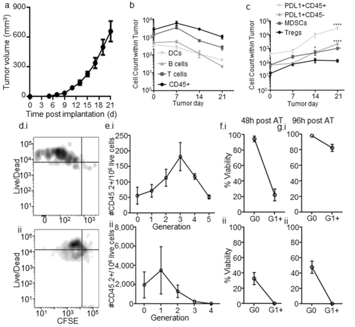
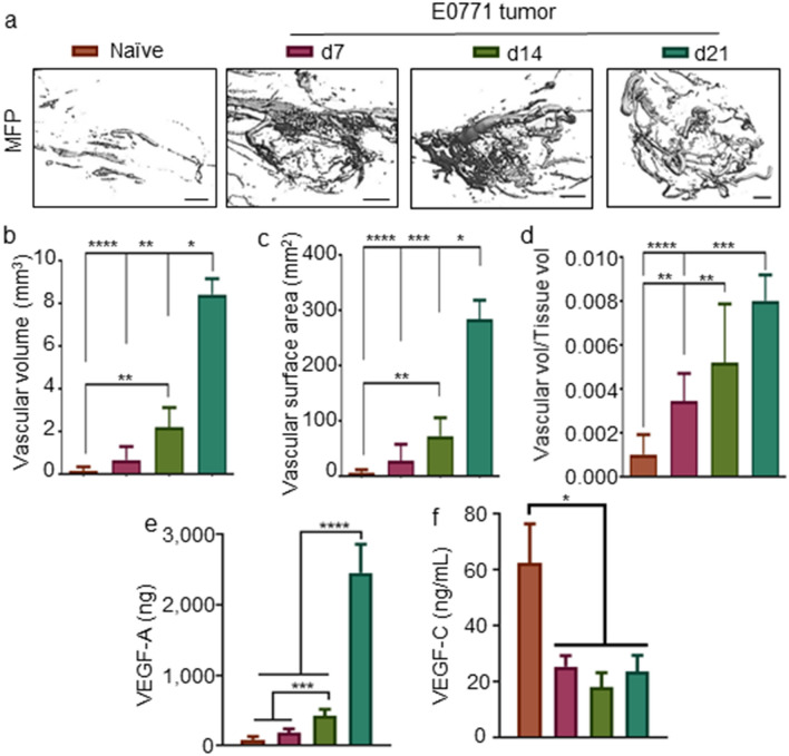
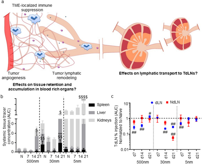
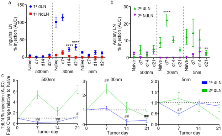
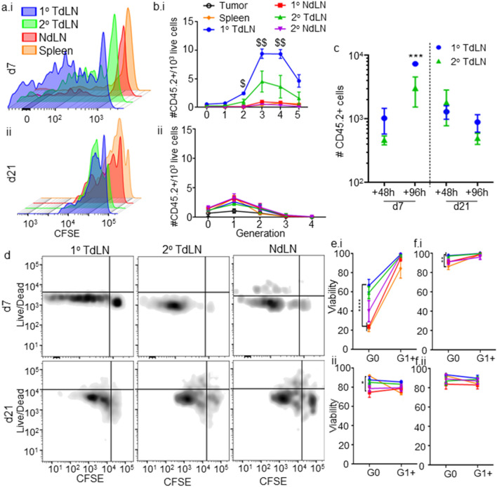
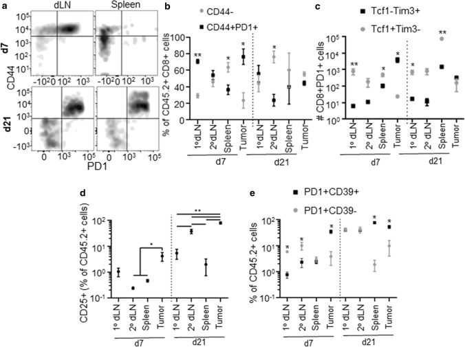
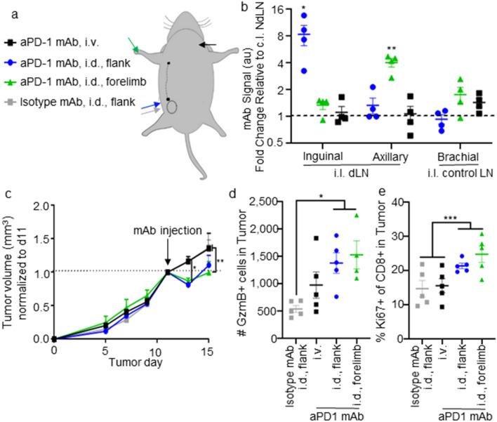
Triple negative breast cancer (TNBC) is a significant clinical problem to which immunotherapeutic strategies have been applied with limited success. Using the syngeneic E0771 TNBC mouse model, this work explores the potential for antitumor CD8+ T cell immunity to be primed extratumorally in lymphoid tissues and therapeutically leveraged. CD8+ T cell viability and responses within the tumor microenvironment (TME) were found to be severely impaired, effects coincident with local immunosuppression that is recapitulated in lymphoid tissues in late stage disease. Prior to onset of a locally suppressed immune microenvironment, however, CD8+ T cell priming within lymph nodes (LN) that depended on tumor lymphatic drainage remained intact. These results demonstrate tumor-draining LNs (TdLN) to be lymphoid tissue niches that support the survival and antigenic priming of CD8+ T lymphocytes against lymph-draining antigen. The therapeutic effects of and CD8+ T cells response to immune checkpoint blockade were furthermore improved when directed to LNs within the tumor-draining lymphatic basin. Therefore, TdLNs represent a unique potential tumor immunity reservoir in TNBC for which strategies may be developed to improve the effects of ICB immunotherapy.

Keywords: Drug delivery; Immune checkpoint blockade; Lymph nodes; T cell priming; Triple-negative breast cancer; Tumor dissemination.


Conflict of interest statement

Not applicable.

Figures

Fig. 1
Fig. 1
The tumor immune microenvironment remodels throughout E0771 disease progression and supports poor priming and viability of tumor antigen-specific CD8 + T cells. a E0771 tumor growth after implantation in the 4th mammary fatpad. b, c Number of major [CD45+; DC, CD45+CD11c+B220−CD3−; B cells (CD45+B220+CD11c−CD3−); T cells (CD45+CD3+B220−)] (b) and immunosuppressive [PDL1+CD45+; PDL1+CD45−; MDSC (CD45+CD11b+Gr1+); Tregs (CD45+CD3+B220−CD4+CD8−FoxP3+CD25+)] (c) immune cell subsets in TME. Representative CFSE versus live/dead staining (d) and quantification of number of live (e) and % viable (fg) CD45.2+CD3+CD8+ donor T cells adoptively transferred OT-I cells within TME in each proliferative generation in d7 (i) or 21 (ii) CD45.1 E0771-OVA tumor-bearing mice 48 (df) or 96 (g) h post-transfer. *Significance by two-way ANOVA with Tukey comparison; #Significance against theoretical value of 1.0 by one-sample t test; n = 5–6 mice
Fig. 2
Fig. 2
The tumor vasculature remodels throughout E0771 disease progression. Representative images (a) and quantification of mean vascular volume (b), vascular surface area (c), and vessel diameter (d) of micro-computed tomography 3D reconstructions of the mammary fatpad or E0771 tumors. VEGF-A (e) and VEGF-C (f) levels in the naïve or tumor-bearing mammary fatpad throughout disease progression. *Significance by two-way ANOVA with Tukey comparison; #Significance against theoretical value of 1.0 by one-sample t test; n = 5–6 mice; scale bar indicates 1 mm
Fig. 3
Fig. 3
Changes in lymphoid tissue immune microenvironments during E0771 progression. Fold change in the relative number of immune [CD45+; DC, CD45+CD11c+B220−CD3−; B cells (CD45+B220+CD11c−CD3−); T cells (CD45+CD3+B220−)] (a, b) and immunosuppressive [PDL1+CD45+; PDL1+CD45−; MDSC (CD45+CD11b+Gr1+); Tregs (CD45+CD3+B220−CD4+CD8−FoxP3+CD25+)] (d–f) cell subsets in the dLN (a, d), spleen (b, e), and NdLN (c, f) relative to naïve tissue; IFN-γ concentration in the tumor (g) and dLN (h) microenvironments throughout disease progression. *Significance by two-way ANOVA with Tukey comparison (af) or one-way ANOVA with Tukey comparison (gh); n = 5–6 mice
Fig. 4
Fig. 4
Molecular dissemination from breast tumors is altered by E0771 disease and progression. a Vascular and lymphatic remodeling of the TME may impact molecular dissemination into systemic tissues versus dLN. b Tracer accumulation within systemic tissues (spleen, liver, and kidneys) of naïve (N) and d 7, 14, and 21 tumor-bearing animals over 72 h post-injection as quantified as concentration AUC. c Relative change in levels of tracer accumulation within the dLN and NdLN over 72 h post-injection quantified as % injection AUC. *Significance relative to naïve, $ indicates significance relative to d7, and ^significance relative to d14 by two-way ANOVA with Tukey’s post hoc test; #significance relative to theoretical value of 1.0 by one-sample t test; n = 5–6 mice
Fig. 5
Fig. 5
E0771 disease progression effects on accumulation of intra-fatpad injected tracers within inguinal and axillary LNs. Levels of tracer accumulation within the inguinal (1°) (a) and axillary (2°) (b) draining and NdLNs over 72 h post-injection during tumor development quantified as % injection AUC. (c) Fold change in tracer accumulation within TdLNs over 72 h post-injection in the mammary fatpad (% injection AUC) relative to naïve animals. *Significance relative to naïve by two-way ANOVA with Tukey comparison; #significance relative to theoretical value of 1.0 using one-sample t test; n = 5–6 mice
Fig. 6
Fig. 6
Tumor antigen-specific T cell priming occurs in E0771 TdLNs. Representative CFSE dilution plots (a) and relative density of each proliferative generation per 103 cells (b) of OT-I CD8 + CD45.2 + donor T cells 48 h post-adoptive transfer into d 7 (i) and 21 (ii) E0771-OVA tumor-bearing CD45.1 animals within lymphoid tissues and TME. Total number (c), representative live/dead and CFSE staining flow cytometry plots (d), and viability (ef) of tumor antigen-specific (OT-I) CD8+CD45.2+ T cells 48 (ce) or 96 h (f) post-adoptive transfer in 1° (inguinal) and 2° (axial) dLNs (cf), NdLNs (df), spleens (ef), or TME (ef) of d7 or 21 E0771-OVA tumor bearing animals. *Significance by two-way ANOVA with Tukey comparison; $significance against all other groups (including tumor) by RM ANOVA; n = 5–6 mice
Fig. 7
Fig. 7
Quality of endogenous CD8+ T cells within lymphoid tissues and TME changes during E0771 disease progression. Representative CD44 and PD1 staining flow cytometry staining (a), fraction of CD44- versus CD44+PD1+ of total CD8+ T cells (b), number of Tcf1−Tim3+ or Tcf1+Tim3− PD1+CD8+ T cells (c), fraction of CD44+CD25+ cells of CD45.2+ cells (d), and fraction of PD1+CD39− versus PD1+CD39+ of CD45.2+CD44+ cells (e) in lymphoid tissues (ae) or TME (be) of d7 or 21 E0771 tumor bearing animals. *Significance by Mann–Whitney test; n = 5–6 mice
Fig. 8
Fig. 8
Locoregional administration of mAb directed to either 1o or 2o TdLN improves therapeutic and immunomodulatory effects of aPD1 ICB. a Schematic depicting routes of i.d. injection targeting 1o (blue, gray) or 2o (green) dLNs versus or systemic administration (i.v., black). b Fluorescently labelled mAb signal in inguinal, axial, or brachial (as control) LNs 24 h after i.d. injection in the flank (targeting 1o TdLN), i.d. in the forelimb (targeting 2o), or i.v. in the jugular vein relative to LNs contralateral to the tumor (NdLN). c Relative tumor size in response to treatment with 100 ug aPD1 mAb on d11. d–e Number of granzyme B (GzmB)- (d) and Ki67- (e) expressing CD8+ T cells in the TME in animals d15 after tumor implantation. * indicates significance by two-way (a) or one-way (b–c) ANOVA with Tukey comparison; n=5-6 animals.

References

    1. Siegel RL, Miller KD, Jemal A. Cancer statistics, 2019. CA Cancer J Clin. 2019;69:7–34. doi: 10.3322/caac.21551. - DOI - PubMed
    1. Ferlay J, Soerjomataram I, Dikshit R, et al. Cancer incidence and mortality worldwide: sources, methods and major patterns in GLOBOCAN 2012. Int J Cancer. 2015;136:E359–E386. doi: 10.1002/ijc.29210. - DOI - PubMed
    1. Foulkes WD, Smith IE, Reis-Filho JS. Triple-negative breast cancer. N Engl J Med. 2010;363:1938–1948. doi: 10.1056/NEJMra1001389. - DOI - PubMed
    1. Seidel JA, Otsuka A, Kabashima K. Anti-PD-1 and anti-CTLA-4 therapies in cancer: mechanisms of action, efficacy, and limitations. Front Oncol. 2018;8:86. doi: 10.3389/fonc.2018.00086. - DOI - PMC - PubMed
    1. Brahmer JR, Tykodi SS, Chow LQM, et al. Safety and activity of anti-PD-L1 antibody in patients with advanced cancer. N Engl J Med. 2012;366:2455–2465. doi: 10.1056/NEJMoa1200694. - DOI - PMC - PubMed

Substances

LinkOut - more resources