

Efficacy of echocardiography for differential diagnosis of left ventricular hypertrophy: special focus on speckle-tracking longitudinal strain
- PMID: 33460030
- PMCID: PMC8154763
- DOI: 10.1007/s12574-020-00508-3
Efficacy of echocardiography for differential diagnosis of left ventricular hypertrophy: special focus on speckle-tracking longitudinal strain
Abstract
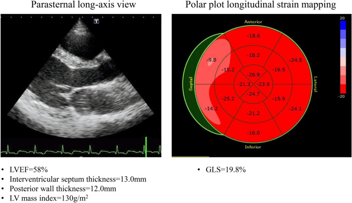
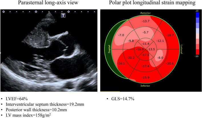
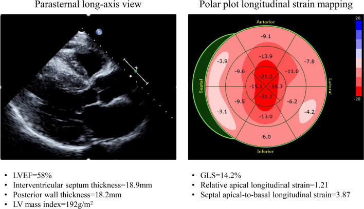
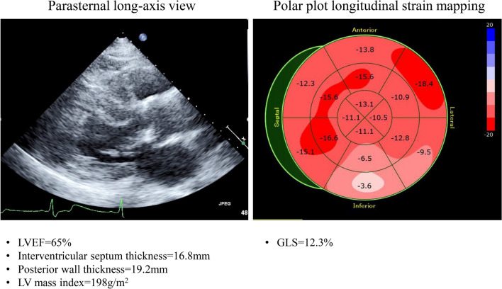
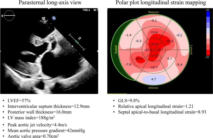
Left ventricular (LV) hypertrophy (LVH) is a frequent imaging finding in daily clinical practice, and its presence is associated with poor outcomes and ventricular arrhythmias. It is commonly detected in athletes, arterial hypertension, aortic stenosis, hypertrophic cardiomyopathy, cardiac amyloidosis, Fabry disease, or Friedreich's ataxia. Echocardiography plays an important role in detecting LVH and underlying causes in current clinical practice. While echocardiography is essential for the quantification and early detection of LV structural findings for various cardiovascular diseases, it has been reported that speckle-tracking echocardiographic parameters are also useful for the detection of early LV structural abnormalities. In particular, global longitudinal strain (GLS) assessed by two-dimensional speckle-tracking echocardiography is reportedly a sensitive marker for early subtle abnormalities of LV myocardial performance, helpful for the prediction of outcomes for various cardiac diseases, and superior to conventional echocardiographic indices. GLS is determined as the averaged peak longitudinal strain of 18 LV segments from standard apical views and can be assessed as a polar plot. This polar plot longitudinal strain mapping offers an intuitive visual overview of the global and regional LV longitudinal myocardial function status of various cardiomyopathies with LVH. This mapping is clinically practicable and the plot patterns obtainable as the result of further development of this technique for clinical practice provide clues to the etiology of cardiomyopathies. This article reviews the efficacy of echocardiography for differential diagnosis of LVH, with a special focus on the utility of speckle-tracking longitudinal strain.
Keywords: Echocardiography; Global longitudinal strain; Left ventricular hypertrophy; Speckle-tracking strain.
Conflict of interest statement
Hidekazu Tanaka has received a speaker honorarium from Boehringer Ingelheim GmbH, Sumitomo Dainippon Pharma, AstraZeneca PLC, Mitsubishi Tanabe Pharma Corporation, Taisho Pharma Co., Ltd., TOA EIYO LTD, Bayer Yakuhin, Ltd, ONO PHARMACEUTICAL CO., LTD., General Electric Company, Daiichi Sankyo Company, Limited, Novartis International AG, Teijin Limited, Merck & Co., Inc., Sekisui Medical Company, Limited, and Philips Medical Systems.
Figures
Similar articles
-
Left ventricular dyssynchrony and 2D and 3D global longitudinal strain for differentiating physiological and pathological left ventricular hypertrophy.Arch Cardiovasc Dis. 2017 Jun-Jul;110(6-7):403-412. doi: 10.1016/j.acvd.2016.11.003. Epub 2017 Jan 3. Arch Cardiovasc Dis. 2017. PMID: 28065448
-
Longitudinal strain of left ventricular basal segments and E/e' ratio differentiate primary cardiac amyloidosis at presentation from hypertensive hypertrophy: an automated function imaging study.Echocardiography. 2016 Sep;33(9):1335-43. doi: 10.1111/echo.13278. Epub 2016 Jun 9. Echocardiography. 2016. PMID: 27277827
-
CT-derived left ventricular global strain: a head-to-head comparison with speckle tracking echocardiography.Int J Cardiovasc Imaging. 2019 Sep;35(9):1701-1707. doi: 10.1007/s10554-019-01596-8. Epub 2019 Apr 5. Int J Cardiovasc Imaging. 2019. PMID: 30953252
-
Left ventricular hypertrophy or storage disease? the incremental value of speckle tracking strain bull's-eye.Echocardiography. 2017 May;34(5):746-759. doi: 10.1111/echo.13506. Epub 2017 Mar 19. Echocardiography. 2017. PMID: 28317158 Review.
-
Longitudinal strain bull's eye plot patterns in patients with cardiomyopathy and concentric left ventricular hypertrophy.Eur J Med Res. 2016 May 10;21(1):21. doi: 10.1186/s40001-016-0216-y. Eur J Med Res. 2016. PMID: 27165726 Free PMC article. Review.
Cited by
-
Echocardiographic findings of patients with transthyretin amyloid cardiomyopathy.J Echocardiogr. 2025 Mar;23(1):1-9. doi: 10.1007/s12574-024-00672-w. Epub 2024 Dec 27. J Echocardiogr. 2025. PMID: 39729212 Review.
-
Impact of antihypertensive treatment on myocardial mechanics in elderly hypertensive patients with different left ventricular geometry patterns: a two-dimensional speckle-tracking echocardiography study.Quant Imaging Med Surg. 2025 Mar 3;15(3):1862-1872. doi: 10.21037/qims-24-1419. Epub 2025 Feb 26. Quant Imaging Med Surg. 2025. PMID: 40160600 Free PMC article.
-
Higher Physical Activity Is Associated with Improved Ventricular-Arterial Coupling: Assessment Using the cfPWV/GLS Ratio in Primary Care-A Pilot Study.J Cardiovasc Dev Dis. 2025 May 30;12(6):208. doi: 10.3390/jcdd12060208. J Cardiovasc Dev Dis. 2025. PMID: 40558643 Free PMC article.
-
Advance of echocardiography in cardiac amyloidosis.Heart Fail Rev. 2023 Nov;28(6):1345-1356. doi: 10.1007/s10741-023-10332-3. Epub 2023 Aug 10. Heart Fail Rev. 2023. PMID: 37558934 Free PMC article. Review.
-
Evaluation of patients with high burden of premature ventricular contractions by comprehensive transthoracic echocardiography.Int J Cardiol Heart Vasc. 2022 Sep 15;42:101124. doi: 10.1016/j.ijcha.2022.101124. eCollection 2022 Oct. Int J Cardiol Heart Vasc. 2022. PMID: 36161233 Free PMC article.
References
-
- Verdecchia P, Carini G, Circo A, et al. Left ventricular mass and cardiovascular morbidity in essential hypertension: the MAVI study. J Am Coll Cardiol. 2001;38:1829–1835. - PubMed
-
- Draper TS, Jr, Silver JS. Gaasch WH adverse structural remodeling of the left ventricle and ventricular arrhythmias in patients with depressed ejection fraction. J Card Fail. 2015;21:97–102. - PubMed
-
- Weidemann F, Niemann M, Ertl G, et al. The different faces of echocardiographic left ventricular hypertrophy: clues to the etiology. J Am Soc Echocardiogr. 2010;23:793–801. - PubMed
-
- Cikes M, Sutherland GR, Anderson LJ, et al. The role of echocardiographic deformation imaging in hypertrophic myopathies. Nat Rev Cardiol. 2010;7:384–396. - PubMed
-
- Ponikowski P, Voors AA, Anker SD, et al. 2016 ESC Guidelines for the diagnosis and treatment of acute and chronic heart failure: the Task Force for the diagnosis and treatment of acute and chronic heart failure of the European Society of Cardiology (ESC) Developed with the special contribution of the Heart Failure Association (HFA) of the ESC. Eur Heart J. 2016;37:2129–2200. - PubMed
Publication types
MeSH terms
LinkOut - more resources
Full Text Sources
Other Literature Sources
Miscellaneous
