Review
. 2021 Jul;41(4):1965-1998.
doi: 10.1002/med.21783. Epub 2021 Jan 18.

What coronavirus 3C-like protease tells us: From structure, substrate selectivity, to inhibitor design

Affiliations
Review

What coronavirus 3C-like protease tells us: From structure, substrate selectivity, to inhibitor design

Muya Xiong et al. Med Res Rev. 2021 Jul.

Abstract

The emergence of a variety of coronaviruses (CoVs) in the last decades has posed huge threats to human health. Especially, the ongoing pandemic of coronavirus disease 2019 (COVID-19) caused by severe acute respiratory syndrome coronavirus 2 (SARS-CoV-2) has led to more than 70 million infections and over 1.6 million of deaths worldwide in the past few months. None of the efficacious antiviral agents against human CoVs have been approved yet. 3C-like protease (3CLpro ) is an attractive target for antiviral intervention due to its essential role in processing polyproteins translated from viral RNA, and its conserved structural feature and substrate specificity among CoVs in spite of the sequence variation. This review focuses on all available crystal structures of 12 CoV 3CLpro s and their inhibitors, and intends to provide a comprehensive understanding of this protease from multiple aspects including its structural features, substrate specificity, inhibitor binding modes, and more importantly, to recapitulate the similarity and diversity among different CoV 3CLpro s and the structure-activity relationship of various types of inhibitors. Such an attempt could gain a deep insight into the inhibition mechanisms and drive future structure-based drug discovery targeting 3CLpro s.

Keywords: 3C-like protease; binding modes; coronavirus; inhibitors; structure and function.


Figures

Figure 1
Figure 1
Structure and genome organization of coronaviruses. In panel A, structural proteins of coronaviruses are marked. In panel B, cleavage positions of PLpro and 3CLpro are indicated by scissors colored blue and orange, respectively [Color figure can be viewed at wileyonlinelibrary.com]
Figure 2
Figure 2
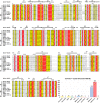
Sequence alignment of 12 CoV 3CLpros by ESPript and the number of crystal structures of 3CLpro in PDB [Color figure can be viewed at wileyonlinelibrary.com]
Figure 3
Figure 3
3D structures of 3CLpro. (A) Protomer, (B) homo‐dimer (PDB ID: 2AMQ), and (C) domain‐swapped dimer of homo‐octamer (PDB ID: 3IWM). The bound inhibitors are indicated by sticks and the N‐finger is colored magenta. Two catalytic residues are colored orange and were shown as spheres [Color figure can be viewed at wileyonlinelibrary.com]
Figure 4
Figure 4
Characterization of the substrate‐binding site of 3CLpro. (A) Expanded view of the substrate‐binding site of SARS‐CoV 3CLpro in its inactive and active forms (PDB ID: 1UJ1 and 2AMQ). (B) Detailed configurations of all residues involved in the binding site. Conserved residues are highlighted in red. (C,D) Residue configurations of each subsite of SARS‐CoV‐2 3CLpro (PDB ID: 6LU7). (E) The sequence logo, representation of the conservation of residues forming the binding site. The color coding is defined according to the chemical properties of amino acids (polar or neutral: black, basic: blue, acidic: red, and hydrophobic: orange) and the subsite legend is given under each amino acid [Color figure can be viewed at wileyonlinelibrary.com]
Figure 5
Figure 5
Schematic view of the catalytic mechanism underlying natural substrate hydrolysis by 3CLpro [Color figure can be viewed at wileyonlinelibrary.com]
Figure 6
Figure 6
Sequence logos indicating the amino acid specificity in the cleavage site of substrates for 12 CoV 3CLpros [Color figure can be viewed at wileyonlinelibrary.com]
Figure 7
Figure 7
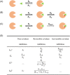
A schematic representation of mode of action of 3CLpro inhibitors: (A) noncovalent, (B) irreversible covalent, and (C) reversible covalent inhibitors. The green triangle, orange sector, and red bold line represent the inhibitor, 3CLpro, and covalent bond, respectively. (D) Kinetics parameters for inhibitors binding with 3CLpro. (a k on : the ligand binding rate constant, b k off : the ligand disassociate rate constant) [Color figure can be viewed at wileyonlinelibrary.com]
Figure 8
Figure 8
Structures of representative 3CLpro peptidomimetic inhibitors with the highlights on the P1 position (blue) and warheads (red) [Color figure can be viewed at wileyonlinelibrary.com]
Figure 9
Figure 9
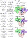
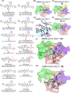
Chloromethyl ketone (CMK) and aza‐peptide epoxide (APE) inhibitors and their complex structures with 3CLpro. (A) Chemical structures of CMK and APE, and the proposed molecular mechanism of 3CLpro inhibited by APE. Under each chemical structure are shown the reported activity data and the available PDB ID of the crystal structure(s) with 3CLpro(s). The CoV type is given in the bracket. (B–D) Detailed configuration of the inhibitors interacting with the S1ʹ‐S4 subsites of 3CLpro in the crystal structures. The subsites are colored purple (S1ʹ), pink (S1), yellow (S2), and green (S4) here and in the figures hereinafter. The 3CLpro residues involved in the interactions are shown in sticks and colored according to their belonging subsites. Hydrogen bonds are shown with dashed lines [Color figure can be viewed at wileyonlinelibrary.com]
Figure 10
Figure 10
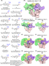
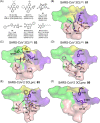
(A,B) Chemical structures of inhibitors containing Michael acceptors and the molecular mechanism of 3CLpro inhibited by these inhibitors. (C–E) Binding modes of the representative inhibitors with a Michael acceptor with 3CLpro. The superposed inhibitors are distinguished by different colors [Color figure can be viewed at wileyonlinelibrary.com]
Figure 11
Figure 11
Peptides with an aldehyde: (A) Chemical structures and (B–F) ligand binding modes with various 3CLpros [Color figure can be viewed at wileyonlinelibrary.com]
Figure 12
Figure 12
(A) Chemical structures of peptides containing a phthalhydrazido group. (B) A thioacyl‐like covalent bond formed in the SARS‐CoV 3CLpro:41 complex. (C) An episulfidel cation and a Tl2‐like configuration seen in the SARS‐CoV 3CLpro:36 complex. (D) A proposed mechanism for the reaction of the episulfidel cation and the generation of the tetrahedral intermediate in the SARS‐CoV 3CLpro:36 complex [Color figure can be viewed at wileyonlinelibrary.com]
Figure 13
Figure 13
Peptides with a nitrile group: (A) chemical structures and (B) the binding mode of the representative compound (47) with the SARS‐CoV 3CLpro[Color figure can be viewed at wileyonlinelibrary.com]
Figure 14
Figure 14
Peptides with an aldehyde bisulfite adduct: (A) chemical structures and (B, C) the ligand binding modes with 3CLpro[Color figure can be viewed at wileyonlinelibrary.com]
Figure 15
Figure 15
Peptides with an alpha‐ketoamide: (A) chemical structures and (B–G) the ligand binding modes with various 3CLpros [Color figure can be viewed at wileyonlinelibrary.com]
Figure 16
Figure 16
Benzotriazole‐based inhibitors: (A) chemical structures and (B) the proposed mechanism for the benzotriazole ester of inhibitors reacted with the protease. (C–E) The binding modes of the representative benzotriazole‐based inhibitors with various 3CLpros [Color figure can be viewed at wileyonlinelibrary.com]
Figure 17
Figure 17
Pyridyl‐based and other small molecule inhibitors: (A) chemical structures and (B–E) the binding modes of these small molecule inhibitors with various 3CLpros [Color figure can be viewed at wileyonlinelibrary.com]
Figure 18
Figure 18
Chemical structures of 3CLpro inhibitors in preclinical studies [Color figure can be viewed at wileyonlinelibrary.com]

References

    1. Bailey OT, Pappenheimer AM, Cheever FS, Daniels JB. A murine virus (JHM) causing disseminated encephalomyelitis with extensive destruction of myelin: II pathology. J Exp Med. 1949;90(3):195‐212. - PMC - PubMed
    1. Hamre D, Procknow JJ. A new virus isolated from the human respiratory tract. Proc Soc Exp Biol Med. 1966;121(1):190‐193. - PubMed
    1. McIntosh K, Dees JH, Becker WB, Kapikian AZ, Chanock RM. Recovery in tracheal organ cultures of novel viruses from patients with respiratory disease. Proc Natl Acad Sci USA. 1967;57(4):933‐940. - PMC - PubMed
    1. Bradburne AF, Bynoe ML, Tyrrell DA. Effects of a "new" human respiratory virus in volunteers. Br Med J. 1967;3(5568):767‐769. - PMC - PubMed
    1. Bradburne AF, Somerset BA. Coronative antibody tires in sera of healthy adults and experimentally infected volunteers. J Hyg. 1972;70(2):235‐244. - PMC - PubMed

Publication types

LinkOut - more resources